Feeds:
Posts
Comments

Posts Tagged ‘brugada’

Recently , a young celebrity lost his life during gym session suddenly . The media erupted as expected .Every TV channel became a temporary schools of advanced cardiology . It seemed anchors knew more cardiology than us.

Among the peers, so many hypothesis were going around. The loudest one was prolonged QT. We are discussing a relatively new, (rather les popular ) entity for the potential cause of SCD. What is it ? DID is the new buzzword in electrophysiology . Next to Long QT, Brugada, DID is looked upon as a new marker for SCD in young as well as elderly .

What is DID ?

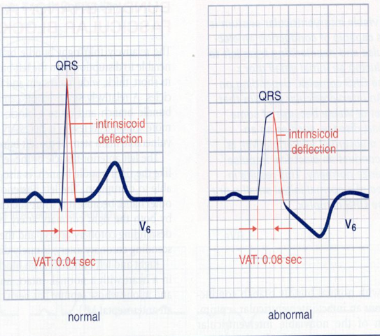
Delayed Intrinsicoid deflection. (DID) Mind you, Intrinsicoid deflection ( ID ) is nearly 100 year old concept, being rediscovered. ID is the time it takes for the electrical impulse to travel from the endocardium to the epicardium directly beneath the recording electrode.

Macleod, A. G., Wilson, F. N., & Barker, P. S. (1930). The form of the electrocardiogram; intrinsicoid electrocardiographic deflections in animals and man. Proceedings of the Society for Experimental Biology
and Medicine. 27(6), 586–587


1.How to measure, what is the normal ?

Variable in each lead.

2. Which lead it is measure ? Is ID relevant in Limb leads ?

V5, V6. Normal less than 50 ms

3.When do you call Intrinsicoid deflection as prolonged ?

>50ms

4.What is the Ionic basis of ID ?

Sodium

5.Is there both congenital and acquired forms?

Yes

7.Is it different over RV vs LV ?

Yes

6.How does a DID trigger an arrhythmia ?

Not clear

Two more questions

7.Why it is called Intrinsicoid deflection rather than Intrinsic ?

Intrinsic was the original term coined by Sir Thomas Lewis in 1914, To get this “intrinsic” measurement, an electrode had to be placed in direct contact with the epicardium . Measuring it from surface ECG leds make it Intrinsicoid.

8. Is Intrinsicoid deflection and ventricular activation time (VAT) both are same ?

Yes. Both are used inter changeably , though one denotes time other the wave as such. While VAT is the same time , specifically look at time to R wave peak. However , ID can be applied to the leads where there is no R wave. Then it becomes time to peak of Q

9.Is it true, in every LBBB, ID is prolonged by default ? Then what is the risk of SCD in LBBB ?

For the first part of the question, the answer is Yes. The second part , we don’t know the true risk yet.

10. …………………………………………………………………..(Question left for the readers to ask)

Final message

As cardiologists, we have given disproportionate importance to the QT interval and gets the blame for many SCD. It is time, the 100 year old ECG parameter Intrinsicoid deflection (ID,) seems to be equally important. Much of the secret ionic codes, for many SCDs are believed to hide behind this eerie deflection.

Reference

Here is an important review

1.Aiken AV, Goldhaber JI, Chugh SS. Delayed intrinsicoid deflection: Electrocardiographic harbinger of heart disease. Ann Noninvasive Electrocardiol. 2022 May;27(3):e12940. doi: 10.1111/anec.12940. Epub 2022 Feb 17. PMID: 35176188; PMCID: PMC9107081.

Read Full Post »

None exposes the expertise and stretches the electrophysiological acumen of a cardiologist, more than a strip of wide QRS tachycardia. Here is a patient who comes with palpitations, with mild hypotension , still comfortably entering the OPD with a non-emergency appointment.

How many diagnosis are running through your mind when you see this ECG . How much it got narrowed after applying criteria like Brugada, Vereckei  etc ( VT, FT, OTVT, Mahaim Tachycardia, AVRT /AVNRT with aberrancy etc)

Now let me reveal the age of the patient .He is a 13 year old boy. Does this help you to narrow down the list of possibilities ? Logically, It should , but for some it can widen the list as well .( Brugada, Anderson, Long QT, Naxos all runs through the mind)

What happed next ?

As the cardiologist was wondering how to tackle this arrhythmia . Adenosine? Amiodarone or DC shock, thankfully, the boy got to spontaneous sinus rhythm .

Click here to see the ECG after restored sinus rhythm & the diagnosis is Instant .

Yes it is a WPW substrate. Localization of pathway is a must and a headache for the fellows. .For me, It looks like a posterior pathway in the para-spetal area. 90% of AVRT are narrow QRS . Here it is wide. Why ?

It’s simple. The tachycardia is anti-dromic.

Is it ? Look at the strip again and confirm is this antidromic ?

I am not sure , I still think it is still antidromic .

I am saying No. This is Ortho-dromic wide QRS tachycardia .

How do you say so ?

Antidromic will still more wide and rapid and won’t terminate spontaneously .Of course one problem here aberrancy usually take an RBBB morphology.

How are you so sure . Let us send this to an EP guy.

*What did the EP say? Well, he didn’t commit to anything as I expected. He said until he measures the refractory period of the accessory path, he won’t comment anything about ortho or antidromic query of AVRT. He asked us to send the case for ablation immediately,


Ok. Let us analyse the reason for orthodromic wide QRS AVRT

1.Rate dependent aberrancy

2.Functional abberancy.

3.Preexisting BBB

4.If patients are on drugs that can prolong his Purkinje conduction


Final message

Two lessons may be learnt.

Lesson 1: Trying to decode a wide QRS tachycardia, without knowing clinical background, should be forbidden. This topic is intentionally made complicated by a flawed teaching methodology of wide QRS tachycardia for over 4 decades. Maybe the single lead AVR algorithm promises to be a quick remedy. If AVR is positive, it is VT; cannot be SVT.

(For the curious readers , please go through Dr. Masood Akhtar’s article from Wisconsin on how to differentiate VT & SVT without even looking at the ECG by law of statistics that beat the invasive EP studies).

Lesson 2 : Wide QRS AVRT is not always antidromic . In fact, orthodromic wide QRS AVRT is 2 to 3 times more common. Many of us don’t realise this bias in our learning.

Related topic

How common is orthodromic wide QRS atrial fibrillation ?

Reference

1.Vereckei A. Current algorithms for the diagnosis of wide QRS complex tachycardias. Curr Cardiol Rev. 2014 Aug;10(3):262-76. doi: 10.2174/1573403×10666140514103309. PMID: 24827795; PMCID:

2.Vereckei A, Duray G, Szénási G, Altemose GT, Miller JM. New algorithm using only lead aVR for differential diagnosis of wide QRS complex tachycardia. Heart Rhythm. 2008 Jan;5(1):89-98. doi: 10.1016/j.hrthm.2007.09.020. Epub 2007 Sep 20. PMID: 18180024.

3.Akhtar M, Shenasa M, Jazayeri M, Caceres J, Tchou PJ. Wide QRS complex tachycardia. Reappraisal of a common clinical problem. Ann Intern Med. 1988 Dec 1;109(11):905-12. doi: 10.7326/0003-4819-109-11-905. PMID: 3190044.

4.Tchou P, Young P, Mahmud R, Denker S, Jazayeri M, Akhtar M. Useful clinical criteria for the diagnosis of ventricular tachycardia. Am J Med. 1988 Jan;84(1):53-6. doi: 10.1016/0002-9343(88)90008-3. PMID: 3337132.

Acknowledgemt

The ECG is posted with courtesy of my colleague Dr. C.Moorthy, Cardiologist, Chennai.

Read Full Post »