Interventricular septum, is the common shared wall between LV and RV . For various biological and hemodynamic reasons , this sharing never follows the law of equity.. It has a bias toward the bigger brother LV. Still, it never lets the RV down in contributing to RV function.
Let us see, how the IVS behaves when it comes to responding, to RV pressure overload .It is clear ,from what we know so far, IVS behavior rarely follows a pattern. We know it resists the pressure and transforms to a D shape. Does the D shaped LV really trigger an increased IVS thickness ?
IVS hypertrophy in RVH : A mixed mystery pheonmenon
In pulmonary hypertension (PHT) or any right ventricular hypertrophy (RVH), the interventricular septum (IVS) does not hypertrophy primarily due to its unique hemodynamic positioning, as IVS is anatomically and functionally linked with left ventricular (LV) mechanics. Unlike LV, the chronic RV pressure overload which is required for septal myocyte growth is rarely sustained because RV tends to dilate as well, in the process interrupting hypertrophy.
RVH occurs with elevated pulmonary artery pressures , but the pressure distibution is uneven. It is more on RV free wall and outflow tract rather than the septum. The pressure distribution concentrated at the RV free wall (infundibulum and body) .Also, the trabeculae sparing effect on IVS from direct overload .
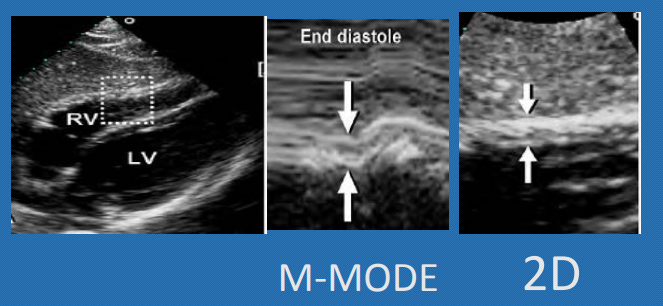
RV free wall hypertrophy defined as thickness exceeding 5 mm on echo in subcostal view in end diastole, ideally on inspiratory phase when it is maximally filled. ( MRI is a still more reliable index of RVH severity) correlating well with RV function and prognosis.
Final message
IVS rarely hypertrophies in RVH in most pathological RV pressure overload conditions. This is due to the complex shape of the RV as well as the non-uniform pressure distribution of RV intracavitary pressure. Unlike LVH, there is no strict concentric RVH. RV free wall hypertrophy is the best index for accurate identification and quantification of RVH.
A note of caution
Congenital heart diseases like isolated valvular PS, TOF can cause severe IVS hypertrophy. Similarly some Inherited or acquired infiltrative diseases can cause disproportionate RVH .We should be cautious , not to mis-classify IVS hypertrophy as LV pathology in these situations.
Reference


