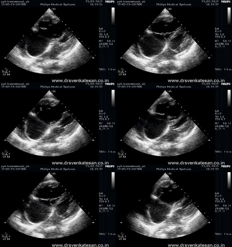
Right atrium and right ventricle are significantly
Home | Aim of this blog | Archives
April 13, 2012 3:11 pm
Echocardiogram in pulmonary HT has many aims .
Echo helps us to confirm the valvular, myocardial or congenital heart diseases in the evaluation of PAH. Apart from these etiological diagnosis of PAH predominantly lies in a systematic medical work up .(Read Dana Point classification )
2-D features
* For some reason RVH does not occur commonly in pulmonary hypertension , while LV hypertrophies promptly in systemic hypertension .
Doppler
Other Echo findings
At what pressure RV begins to dilate in PAH ?
It is not known .It is highly variable . But most will dilate their RV at a systolic pressure > 50mmg.
It is also possible the onset of TR and the magnitude of TR has a major say in the timing of RV enlargement .
We know RV is more sensitive to volume overload than pressure overload .
Paradoxically , it is often observed acute elevation in RV pressure dilate the RV faster than chronic ones.
Right atrium and right ventricle are significantly
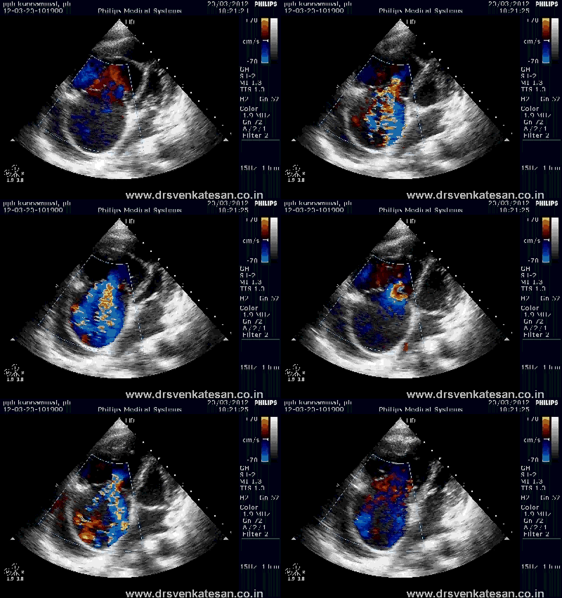
The tricuspid annulus is dilated .Note the severe TR with twin jet morphology.
Estimating Pulmonary artery pressure
PA systolic pressure : TR jet + 10 mmhg
PA diastolic pressure : PR end diastolic jet + 10
PA mean pressure : Peak PR gradient
Other complex methods to arrive ar PAP in the absence of TR or PR
The Dabestani -Mahan ‘s equation* – The mean PA pressure = 90 – (0.62 X acceleration time).
It is popular for calculating PAP by measuring pulmonary artery Doppler acceleration time .
Many believe it is neither sensitive nor practical in real clinical setting.
*Even though Dabestani is the first author of this paper Mahan got the full credits for the simple reason his name is easily remembered !
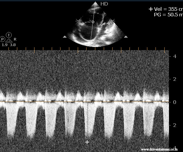
Note the peak TR jet is around 50mmhg and predicted RVSP would be 60mmhg.One would have expected still higher RV pressure but since the RV is dysfunctional the true PAP may be underestimated.
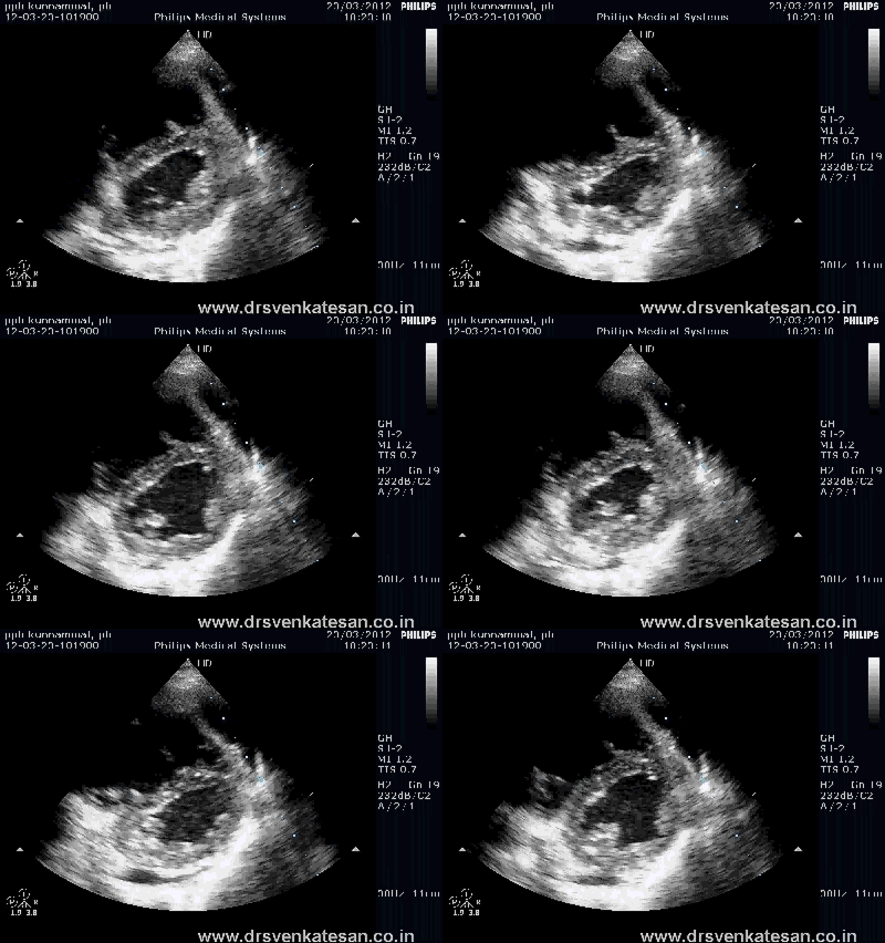
The classical D shaped IVS during systole . D shape indicates RV pressure during systole is almost equal or even higher than LV. ( Please recall D shape occurs in Volume overload also but the timing is in diastole !)
Pulmonary valve M-Mode
According to Wyeman the following M mode signs are useful in diagnosing PAH.
Currently , one may consider M-Mode echo to be an obsolete , but still the foundations help us understand the hemo-dynamics.
The most important principle in the motion of pulmonary valve , is the relationship between pulmonary “a” wave and right atrial “a” wave
Normally atrial contraction produce an inward movement* on the closing pulmonary leaflet . This happens because the MPA end diastolic pressure is usually lower than right atrial a wave .In severe PAH the elevated pulmonary diastolic pressure does not allow the atrial contraction to intend the pulmonary leaflet in pre-systolic atrial contraction .Hence pulmonary valve a wave in m -mode is diminished or even absent .
In PAH even premature closure of pulmonary valve may occur resulting in mid systolic notch .This is referred to as flying “W” -Mid systolic notch. (See below)
* The motion we see in short axis M-Mode is that of left pulmonary cusp that moves posteriorly.
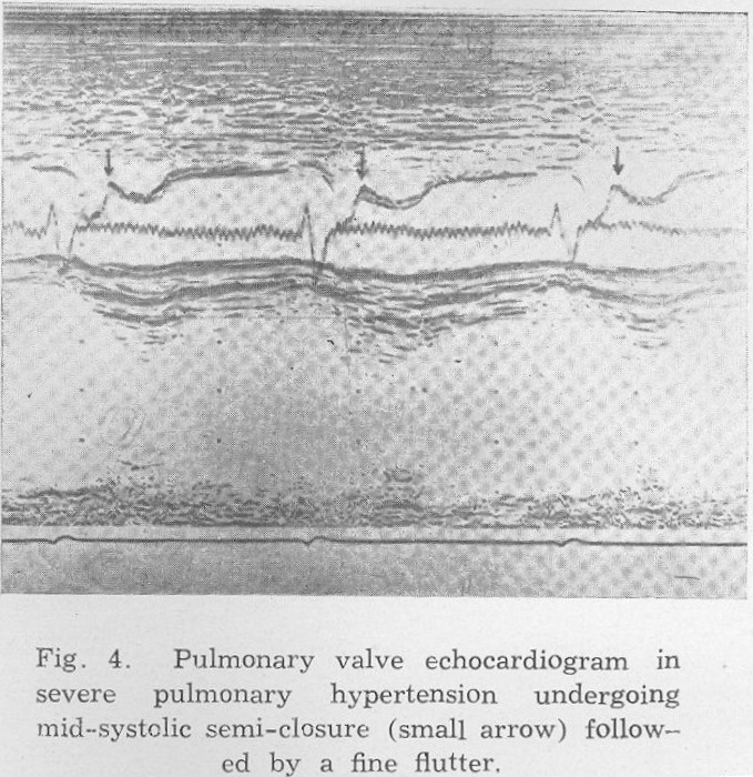
Image source : Karmarkar SG. Pulmonary valve echocardiography. J Postgrad Med 1979;25:219-23
Absence of a dip is a hemo-dyanmicaly important sign pf PAH but with one important caveat .This absence of a dip is valid only until RV failure occur.In th presence of elevated RVEDP a begin to appear again
Reference

1 Karmarkar SG. Pulmonary valve echocardiography. J Postgrad Med 1979;25:219-23
2.http://circ.ahajournals.org/content/50/5/905.full.pdf
4.Kitabatake A, Inoue M, Asao M, Masuyama T, Tanouchi J, Morita T. et al. Noninvasive evaluation of pulmonary
hypertension by a pulsed Doppler technique. Circulation. 1983; 68(2): 302-9.
5.Stevenson JG. et al, Comparison of several noninvasive methods for estimation of pulmonary artery pressure. J Am
Soc Echocardiogr. 1989; 2: 157-71.
Posted by dr s venkatesan
Categories: Cardiology - Clinical, Echo library and gallery, echocardiography, pulmonary hypertension
Tags: a dip in pulmonary valve, berger equation, d sahped ivs in pah, echocardiogram in pah, flying w sign, m mode in pulmoanry, mahan equation, mid ssytolic notch in pulmonary, pa ssytolic pressure, preamture pulmonary valve closure, priamry pulmoanry hypertension, pulmonary acceleration time, pulmonary valve m mode echo, The Dabestani Mahan equation, tr jet pap
Mobile Site | Full Site
Get a free blog at WordPress.com Theme: WordPress Mobile Edition by Alex King.
with PHT the RV hypertrophy initially but the RV contractility deteriorate rapidly bec. thinner wall RV in comprasion to LV so the RV dilate earlier.
By moamin on April 22, 2012 at 1:18 am