Feeds:
Posts
Comments

Posts Tagged ‘mitra clip’

Preamble : The Lubs & Dubs

The lubs and dubs, along with some added sounds are the only language, the heart can speak in health and distress. It’s a worrying story altogether, gradually many of us are becoming “cardiac illiterates” as we struggle to read , its gentle communication. it is not our fault. Stethoscopes are reduced to become a social marker of being a doctor. We may excuse ourselves, even if we can’t differentiate a systolic from diastolic murmur, after all, hand held echo machines, instantly tell the diagnosis.

( After reading this article, fellows are expected to understand why the first heart sound in MR (ie the lubs,) are mostly soft,  some times normal or even loud in certain conditions)

Now, let us go to the mitral valve dynamics

How many of us are aware, there is a big science of physics and biology operating when the mitral valve perfectly closes at the level of the annulus, with each systole , balancing different sets of known and unknown forces.

In this article, we will see how these two sets of forces mitral valve tethering and closing forces balance out each other to seal the mitral valve and what happens when the forces begin to fight each other.

Balance of Tethering and Closing Forces in Mitral Valve Coaptation

The mitral valve (MV) coaptation refers to the edge-to-edge apposition of the anterior and posterior leaflets during systole, ensuring a competent seal to prevent regurgitation. This process is governed by a delicate balance between tethering forces (which restrain leaflet motion to prevent prolapse into the left atrium) and closing forces (which approximate the leaflets for sealing).

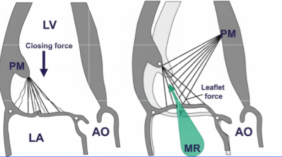
  • Tethering forces: These are primarily transmitted through the chordae tendineae from the papillary muscles (PMs) to the leaflet free edges and bellies, pulling the leaflets apically and laterally toward the left ventricular (LV) apex. They arise from:
  • Closing forces: These are driven by the transmitral pressure gradient during systole, where rising LV pressure (generated by LV contraction) exceeds left atrial (LA) pressure, pushing the leaflets together. The force is proportional to the LV dP/dt (rate of pressure rise) and peaks in midsystole.
  • Balancing mechanism: Coaptation occurs when closing forces overcome tethering, enabling leaflets to meet with sufficient overlap (coaptation length >8 mm typically). Imbalance favors regurgitation: excessive tethering (e.g., from PM displacement) causes apical tenting and incomplete closure; insufficient closing (e.g., low LV contractility) fails to seal the orifice. In health, the forces are synchronized with systole, with closing forces dominating midsystole to minimize the effective regurgitant orifice area (EROA).

Paradoxes in the Balancing Mechanism

MV mechanics exhibit several counterintuitive paradoxes, where adaptive or dysfunctional responses lead to outcomes opposite to expectations. These highlight the interplay of geometry, contractility, and force transmission:

  1. Paradoxical systolic PM elongation: Normally, PMs shorten during systole (1 cm) to offset annular descent and maintain annulopapillary balance. Post-myocardial infarction (MI), scarred or ischemic PMs paradoxically elongate driven by transmitral pressure tension. This decreases annulopapillary distance, attenuates tethering, and reduces MR severity—contrary to the intuition that PM weakness worsens regurgitation. However, extreme elongation risks leaflet prolapse, flipping the paradox to increased MR.
  2. PM dysfunction attenuating ischemic MR: In isolated dysfunction, reduced PM contraction intuitively increases slack chordae and prolapse risk. Yet, in localized basal inferior LV remodeling, PM dysfunction (measured as reduced longitudinal systolic strain) inversely correlates with MR fraction attenuating MR by limiting excessive tethering. This holds only with certain level of remodeling . Gross and asymmetrical remodeling can exaggerate tethering and increase the MR.
  3. Dynamic EROA reduction despite peak driving pressure: MR often peaks early systole (when closing forces are low and tethering dominates) but paradoxically decreases midsystole, even as LV pressure (driving force) maximizes. This occurs because rising closing forces (transmitral gradient) overcome tethering, shrinking the orifice mimicking reduced regurgitation when it should worsen.Thgis mechansim can some times seen when MR jet is bi-fid in doppler tracing.
  4. Imbalanced chordal forces causing focal prolapse: In acute ischemic MR (e.g., posterior wall ischemia), tethering redistributes unevenly: tension drops in ischemic-side chordae but rises on the nonischemic side causing focal tenting and relative prolapse on the ischemic commissure. This creates an eccentric jet despite global LV contraction.

This article clearly tells us that the forces acting on the mitral valve apparatus are so complex. The conceptual model of tethering and closing forces may be oversimplified. There are variable interactions between them. More importantly, the atrial forces also influence and intrude into these forces. Realize that MV competence is not just about force magnitude but their vectorial distribution and timing, often amplified by LV geometry changes.

Final message

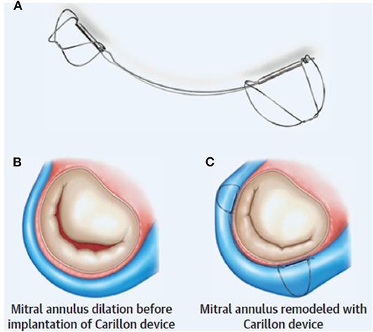
As cardiologists and surgeons, we must realize the fact, how important it is to analyze both anatomy and the physiological impact when we rush to clip, cut, or repair it with annuloplasty and subvalvular interventions.

*Sometimes, it might even be tempting to do mitral valve replacement, even when it is not indicated, because we need not bother about all these dizzy mechanics and physics of MR jet forces.

Read Full Post »

Cardiologists have always struggled to classify, assess, and grade one important valve disease, which is Mitral regurgitation. While valve replacement is the ultimate treatment, the timing of MVR is still a big debate. Apart from valve replacement, valve repair is a strong contender in selected patients. In recent times, cardiologists have made great strides to grab MR patients from cardaic surgeons. MitraClip, a percutaneous edge stitching , is possible with a varying degree of success.

Mitral valve edge-to-edge repair (MEER) is an interventional clone of Alfieri surgery that has shown conflicting results in the MITRA-FR and COAPT studies. The reason for this discrepancy in the MITRA-FR population is that they had larger-sized ventricles, which continued to pose challenges for the clip, which is focused only on the leaflets.

A new subdivision of secondary MR

Now, some of the cardiologists want to classify SMR/FMR into Proportionate vs Disproportionate MR. It may not be a great innovation, but it sub-divides secondary MR for optimal therapy. It simply says if LV dilation is significant, clipping the leaflet alone will not be sufficient; it would rather need an annular restriction either at the time of the index clip procedure or in due course. While Disproportionate MR implies, it is more of a leaflet coaptation defect, dominating over annular contribution.

Who proposed this ? What is the implication?

It is an afterthought, I think, from the makers of annular restriction device makers. MEER is found to be less effective in proportional MR.The Carillon device is a new arrival to tackle secondary MR .It is actually a wire that forcibly tightens the AV annulus inserted through coronary sinus . This modality takes advaantage of the aantomical proximity of coronary sinus to mitral annulus. Coronary sinus encircles and from a virtual wall along significant circumference of mitral annulus.

Unlike mitraclip, the Carillon device is claimed to tackle secondary MR irrespective of whether it is proportional or disproportional. It also has the potential to reduce LV dimensions in the long run. We have another device called IRIS-Millipede (to compete with Carillon).

Front. Cardiovasc. Med., 20 November 2020
Sec. Structural Interventional Cardiology
Volume 7 – 2020 | https://doi.org/10.3389/fcvm.2020.576058

Final message

We are free to have as many classifications in MR (Primary, Secondary, Functional, Atrial MR, & now Proportionate Disproportionate.) It is not the aim to bring up a rivalry between leaflet vs annular intervention. Ultimately, the most powerful component of the mitral valve apparatus, i.e., the LV muscle that matters.

I would request the esteemed researchers in MR ,not to keep EROs, regurgitation fractions, or chamber dimensions as primary markers of success of a device. Having strong clinical outcomes as the endpoint should be made mandatory, i.e., prolonging good quality of life and survival (But, the reality can bite hard. Someone told me, walking 20 meters extra in a 6-minute walk test is enough to get device approval from the authorizing entities.)

Final message

1.Grayburn PA, Sannino A, Packer M. Distinguishing Proportionate and Disproportionate Subtypes in Functional Mitral Regurgitation and Left Ventricular Systolic Dysfunction. JACC Cardiovasc Imaging. 2021 Apr;14(4):726-729. doi: 10.1016/j.jcmg.2020.05.043. Epub 2020 Aug 26. PMID: 32861653.

Postamble : A surgeons perspetive

While we debate about devices, the true benefits may lie elsewhere. A good MVR done by a mitral heart surgeon is the need of the hour. Says this paper from a top heart surgeon in India. Yadava OP. Disseminating valve repairs – a clarion call. Indian J Thorac Cardiovasc Surg. 2020 Jan;36

Read Full Post »

AV valves exist to divert blood to the outflows when ventricles are contracting. If they leak significantly, obviously there are major consequences. What happens to the net cardiac output when these valves leak?

Clincally , we know ,fatigue is sign of reduced forward cardaic out put , that is more common in MR. But, edema , congestion, muscle fatigue is eqaully if not more common with TR. One importnat diference is MR jet is mainly a hemodynamic trouble in pulmonary circuit while TR jet , has one more component hits on liver function making it metabolic consequnce as well.

Can’t escape, tell us your answer for the question

*I am afraid, anyone has done a specific hemodynamic study on this. It should be clear for everyone, that RV stroke volume, is not only the preload of LV, and it is going to bcome LV stroke volume, a few seconds later. Hence TR equally reduce * (if not more ) the forward cardiac output as does MR. In fact, the compensatory mechanism of LV with its reserve work might maintain the forward output for a longer time. RV reserve functions are less.Fellows must analyze this. It is not a difficult one. Measure LV stroke volume by echo ( LVOT area X Aortic TVI) in patients with TR vs MR pre and post-correction.

Final message

RV & LV are like conjoined twins. One’s function can’t be decoupled from the other. Now we realize, TR plays a more central role in both symptom generation and the overall outcome in cardiac failure. Hence, aggressive interventions are being attempted to plug this leak. (We will come to know whether it is truly beneficial or not only later, as arresting TR puts more burden on RV, as TR has a pressure cooker release/relief effect on RV)

Reference

Unterhuber M, Kresoja KP, Besler C, Rommel KP, Orban M, von Roeder M, Braun D, Stolz L, Massberg S, Trebicka J, Zachäus M, Hausleiter J, Thiele H, Lurz P. Cardiac output states in patients with severe functional tricuspid regurgitation: impact on treatment success and prognosis. Eur J Heart Fail. 2021 Oct;23(10):1784-1794. doi: 10.1002/ejhf.2307. Epub 2021 Jul 28. PMID: 34272792.

Rebecca T Hahn, Luigi P Badano, Philipp E Bartko, Denisa Muraru, Francesco Maisano, Jose L Zamorano, Erwan Donal, Tricuspid regurgitation: recent advances in understanding pathophysiology, severity grading and outcome, European Heart Journal – Cardiovascular Imaging, Volume 23, Issue 7, July 2022, Pages 913–929https://doi.org/10.1093/ehjci/jeac009

Read Full Post »

Mitra clip is a small metal device that is delivered percutaneously, to clip the incompletely coapting (closing) mitral valve. It was first introduced to treat degenerative mitral regurgitation. It is an interventional imitation of the famous edge to edge Alfieri stitch repair.This procedure in fact converts the single mitral valve orifice into two. In the process, curtails the regurgitation jet orifice significantly. Though the technique looks nice and simple to hear, lots of per and post-procedure issues need refinement. Conceptually it is ideal in primary disorders of the mitral valve. (Read EVEREST 2 criteria for optimal patient selection)  There have been more than 60000 Mitra clips implanted worldwide wide. Thanks to Abbot.

In secondary MR (due to LV dysfunction) Mitra clip has shown mixed results( MITRA-FR not much benefit, COAPT -Did show benefits)

Now, what about Mitra clip as a remedy for rheumatic mitral regurgitation?

This is the question everyone likes to ask. Now we have some interesting breakthroughs. Dr. Ningyan Wong from the National University of Singapore reports probably the first case (Ref 1) . The videos are reproduced with the creative commons license.


Note the classical thickened AML in rheumatic mitral regurgitation.


TEE showing severe MR


Post Mitra clip : A real surprise to note near-total abolition of regurgitation. (This really is good news for the rheumatic mitral valves )

Technical issues

  • Should be isolated MR
  • P2/A2 scallop clipping is the key to success. 
  • The thickness of the leaflet limits the success (Grasping the leaflet will be difficult)
  • Clip Induced mitral stenosis is a distinct risk.

Potential role and future

RHD forms 90 % of valvular heart disease in a country like India. The incidence of Isolated MR in both acute rheumatic fever and chronic  RHD are substantial. If only we refine the hardware and technique to suit these thickened rheumatic valves, Mitra clip is expected to make an impact in this unique group of patients where surgery can be avoided or at least postponed

Though we would very much like to do such a trial in our place, logistics has effectively precluded it. I wish some large centers like AIIMS New Delhi or PGIMER Chandigarh and others can take this concept to the next level.  


Reference

1.Ningyan Wong, Peilin Cheryl Marise Tan, Zee Pin Ding, Khung Keong Yeo, Successful MitraClip for severe rheumatic mitral regurgitation: a case report, European Heart Journal – Case Reports, Volume 3, Issue 3, September 2019

Read Full Post »

Truely a great demonstration of life saving Mitra clip procedure.

Found this from

Read Full Post »