Feeds:
Posts
Comments

Posts Tagged ‘la pressure volume loop’

A brief learning session with cardiology fellow about a possible new concept in Left heart failure.

What is the commmest cause for acute pulmonary edema?

Left sided heart failure”

“Can you be more specific , Left sided means ?

“I meant LV failure , typically DCM of any cause or Severe un-controlled HT”

“Ok .good. Does Left Heart failure include mitral valve dysfunction also ?

“Yes sir, very much. Classical mitral stenosis and Isolated MR can cause pulmonary edema. In fact, acute AR Iis also part of left heart disease”

So far, so good, now coming to the complex part of left heart

Can LA fail in isolation independent of LV , ie I mean with normal Left ventricle ?

I am not sure. Can we call new onset atrial fibrillation as a primary atrial failure that can result in pulmonary edema?

Excellent. You are absolutely right. But I am talking about mechanical atrial failure, not electrical. Are you aware that most of the time AF is a well tolerated arrhythmia , it can even be silent in many cases. This is because the pumping function of the atria contributes only 20-25% to LV filling. This can easily be compensated by augmented LV suction force , provided the baseline LV function is normal.

Have you heard about ACM. Atial cardiomyopathy?

“No sir”,

“You will hear more about it soon” (Ref 1) Scientists, especially Echo guys are closing in on this concept. We know, the atria has three functional components, namely conduit, reservoir, and pumping. Curiously, we have realized that the pumping function of LA may not be that critical from indirect observations from some land mark studies . (Rate control vs rhythm control studies in AF are a powerful proof on the atrial pumping function .(AFFIRM/RACE etc ) I don’t know, whether I am right in saying the above statement.

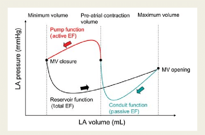
It is tempting to conclude , only if all the three functional components of atria gets affected , then only primary atrial dysfunction can be diagnosed. The concept is more complex than we realise. In diastole , pulmonary vein, LA , LV all work as single functional unit. Only in systole, we see them as different things.

Atrial DCM

Like LV systolic function, which is coupled with RV in parallel , LA function is closely knitted to LV in series during diastole. The key to suspect or diagnose this entity is to demonstrate dissociation of LVEDP with LA mean pressure & PCWP. This is not an easy job in bedside. Isolated Increase in LA volume without any reason , is one clue. LA ejection fraction is possible marker. (Kanagala P, . Int J Cardiovasc Imaging. 2020)

Final message

We are in the early days of understanding primary atrial mechanical failure, Atrial cardiomyopathy (ACM) or atrial DCM is being proposed as separate entity. It is very likely, some subsets of HFpEF might turn out to be primary atrial disease, depending on the level of investigation we do.

Reference

1.Li M, Ning Y, Tse G, Saguner AM, Wei M, Day JD, Luo G, Li G. Atrial cardiomyopathy: from cell to bedside. ESC Heart Fail. 2022 Dec;9(6):3768-3784. doi: 10.1002/ehf2.14089.

2.Patel, R.B., Lam, C.S.P., Svedlund, S. et al. Disproportionate left atrial myopathy in heart failure with preserved ejection fraction among participants of the PROMIS-HFpEF study. Sci Rep 11, 4885 (2021). https://doi.org/10.1038/s41598-021-84133-9

Read Full Post »

LA dimension and volume have become vital parameters in recent times, especially, with the entity of HFpEF is becoming so common. LA not only acts as a live barometer, reflecting all that happens in LV, but it is also a chronic marker of LV diastolic function. (Funnily referred to as HBA1c of diastolic dysfunction) 

What is normal LA dimension & volume ? 

  • Normal left atrial diameter < 4.1 cm in men or < 3.9 cm in women
  • Normal left atrial volume indexed for body surface area (BSA) is 34 ml/m2 for both women and men 

Which part of the cardiac cycle do we measure? 

Ever since Wiggers introduced the overwhelming concept of LV systole and diastole, most of us ignored the fact that atria do have a separate contraction relaxation cycle, independent of what happens in the ventricle. Of course, atria and ventricles act as a single chamber in diastole. In reality, atria lack true boundaries when it acts as a conduit. The LA dimension varies considerably during the atrial cardiac cycle. Look at the  LA pressure-volume loop, which can frighten anyone, with its horizontally lying figure of 8 pattern. During every cardiac cycle, the volume reaches atleast two troughs and one peak.

Don’t get frightened with this graph, spend some time, and you will get it right, Begin at  “3” o clock position with the onset of diastole with a downsloping green loop, that continues as the red line of atrial contraction to end up in systole. The entire black loop, that happens during ventricular systole depicts the true reservoir function. with MV closed. ,


As of now, we have a consensus, LA volume is measured typically in LV end-systolic frame. ( Rather, we measure it at maximum LA volume ) However, we have 4 different LA  volumetric components to assess, as this article excellently depicts. (Hoit BD. Left atrial size and function: role in prognosis. J Am Coll Cardiol. 2014 Feb 18;63(6):493-505.)

What could be the limitations of the traditional end-systolic measurement?

No single measurement will give an overall LA function assessment. But still, Somehow, we have measured the maximum LA  volume as a reference of true diastolic function. This happens in LV end-systolic point where atria reach the maximum size. But, here is a catch, we assess the left atrial function before its main physiological function of emptying takes place.

How about assessing  LA efficiency after it completes its job, ie end diastole? 

In LV function end-systolic dimension has pride of place as it is devoid of influence from loading condition. If applying the same logic, the “end atrial” systolic dimension(Which is the same as LV end-diastolic point/or post A ) should be perfect. It can also help measure the residual LA volume after its systole.

A potential advantage of LV end-diastolic dimension (The Heart & Soul study )

Maybe, this is less affected in the presence of MR systolic jet will spuriously elevate LA volume. In AF also this parameter is less likely to be influenced by LA preload.

Final message

Suddenly, we are debating a fundamental Issue, ie timing of LA measurement. While the end-systolic size/volume is the current standard, the LA dimension in the end diastole also provides useful info. There are at least 4 different LA volumes, at different parts of the LA cycle that need to be studied for a proper understanding of diastology (Unlike LV which has only two).

Now, we may need to ponder, if there is a mean LA volume, measured with the 3D volumetric analysis or MRI, that could be representative of the global LA function. 

Reference

Thadani SR, Shaw RE, Fang Q, Whooley MA, Schiller NB. Left Atrial End-Diastolic Volume Index as a Predictor of Cardiovascular Outcomes: The Heart and Soul Study. Circ Cardiovasc Imaging. 2020 Apr;13(4):e009746. doi: 10.1161/CIRCIMAGING.119.009746. Epub 2020 Apr 20. PMID: 32306763; PMCID: PMC8846436.


Read Full Post »