Reading X -ray chest can be as blind as a bat flying in the dark . It needs lots of Imagination . (Many times the blindness continues to cath lab as well during structural interventions is a different story !)
Yes ,its true any one can recognise a cardiomegaly in X-ray . . . but Which chamber is responsible for cardiomegaly ? and quantifying each ones contribution to the increased CTR is the critical question.
We know the 4 chambers in the heart are arranged in a complex pre-specified (Antero -superior and right to left orientation ) still , the CT ratio in X-RAY chest is based on the diameter formed by two chambers only ie right atrium and left ventricle.
However, any of the 4 chamber enlargement can increase CT ratio in pathological conditions.
- LV enlargement is the most common cause for cardiomegaly as it is the normally border forming.(DCM, Aortic valve, HT diseases)
- RV can do it when it enlarger grossly forming the left heart border(COPD, Severe pulmonary hypertension of any cause)
- RA can enlarge to both pressure and volume overload.(CHF, with RVF)
- LA is least likely to be border forming as it is midline structure .Since It tends to enlarge posteriorly and superiorly it rarely enlarges sideways. Occasionally In severe mitral stenosis it can enlarge to the right and cross the right heart border causing the classical shadow in shadow.
Since I have struggled with X ray orientation of heart chambers in my early days (Still i do sometimes!) Just thought , why we are not fusing a X-ray with a given patients echocardiogram that will help understand the chamber anatomy .
Fusion Image of X ray chest PA view with apical 4 chamber in ECHO. (Rotated to specified angle to match heart border)
Note : The Left atrium is not only left of RA , its also posterior and superior to RA.This makes the IAS not actually pure right left to relationship but also a slight infero to superior and antero posterior orientation.(This can be realised when we puncture the IAS from RA side the needle goes more of superior)
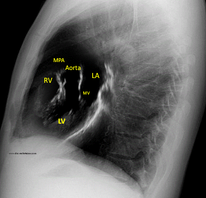
X ray chest left lateral view is fused with para- sternal long axis view. Please note this is not true anatomical correlates. The RV shown in echo is actually RVOT but in X-ray its more of RV body .
* A note of caution : The fused Images are rough attempt to co-register x-ray with echo. There is sophisticated software in some new generation cath labs to mix fluro images with live TEE data that aid in Interventions.
Postamble
A bedside Instant point of care echo is becoming a norm in clinical cardiology practice. Why bother about X-ray then ? Agreed to that point to a certain extent. But, I used to tell my (amused ) students that technology based lazy learning doesn’t help build a strong scientific foundation which would ultimately threaten the patient care one day !


Leave a comment