Posted in bio ethics, Medical ethics, medical quotes, medical satistics | Tagged fraud in medical research, hippocrates oath modern science, how to interpret a study, how to read a scientific paper, jokes in science, medical ethics, medical research errors, medical statistics | Leave a Comment »
One casual question in my class led to this search for an anatomical mystery. When we were discussing why left atrial oxygen saturation never reaches 100 % ? , it was attributed to desaturated bronchial venous blood draining into pulmonary vein.
How does this bronchial vein enter pulmonary venous circulation ? How many bronchial veins are there ? What anatomical plane it runs ?
Surprisingly, even in this hi-tech era of academic excess, literature is sparse for this basic anatomical question. It is reported (In Greys anatomy ? ) Bronchial veins are two in number and both drain to Azygos and Hemiazygos veins (systemic) rather than pulmonary veins.
So is our assumption wrong ?
May not be.We realise these are only two visible and named bronchial veins .It is learnt they probably carry only about 13 % of bronchial venous blood to systemic venous circuit.

Image showing right and left bronchial veins draining to Azygos and hemiazygos veins.
It is assumed , remaining 87 % of bronchial venous blood drains to pulmonary venous circuit in an invisible fashion (By unnamed twigs ?) desaturating the LA blood by about one percent from 100 to 99 %. This is our current understanding. I haven’t come across any specific human research that quantifies the bronchial venous channels and it saturation . It’s gratifying to find one study specifically looked answer this question in sheep study .(Charan H.B et all Reference 1 )

True physiological bronchial venous drainage seems to be different from anatomical bronchial venous circuits .
Clinical implication of bronchial venous circulation.
In physiology it may not be important . However bronchial circulation (both arterial and venous) can take many anatomical tracts when pulmonary micro vascular bed is structurally and functionally altered as in COPD, , pulmonary atresia with aorto-pulmonary collaterals , congenital left to right shunts,post Fontan circulation pulmonary AV malformations,lung tumors etc .
Hemoptysis in acute pulmonary venous hypertension is thought to be due to rupture of these bronchial veins as elevated pulmonary venous pressure reflect into bronchial veins (As in mitral stenois and other conditions. ) This again would vouch for bronchial veins draining to pulmonary veins.
Final message
As on today , it can be concluded bronchial vein drainage goes both systemic and pulmonary venous circuit.Bulk of them appear to end in pulmonary veins though clear anatomical evidence is lacking.
Postamble
Exploring human anatomy appear a grossly unfinished agenda even today, especially the micro and histo-anatomy. Teachers of basic sciences should impress upon youngsters entering the medical school to pursue translational research relevant to specific clinical problems.
Students may contact <drsvenkatesans@yahoo.co.in> for specific areas of clinical cardiac anatomy topics that still requires answers.
Reference
Posted in Anatomy of heart, Bronchail arterial and venous circulation, bronchial arterial and venous circulation | Tagged bronchial circulation, bronchial vein rupture hemoptysis in mitral stenosis, bronchial venous drainage, clincial antomy research topics, research topics in basic sciences anatomy, where does bronchial veins drain ? | 2 Comments »
We know, atrial fibrillation is the commonest clinical cardiac arrhythmia , that is extensively studied , subjected to exotic investigations and state of the art treatment strategies.Interestingly , this arrhythmia also drags the economics of cardiology practice of a community in a big way with heavy influence on drug , device and usage.We know, RF ablation of pulmonary vein is one of the modern ways to manage this arrhythmia.
Iam sharing this article from medscape by an EP specialist Dr. Jhon Mandrola , surprisingly exposes our fundemental ignorance about this arrhythmia and the near futility of certain procedures.
Posted in Uncategorized | Leave a Comment »
Scientific cardiology has forced us to believe ACS management must be catheter based and all others are inferior and those who pursue the later , carry a risk of being labelled as unethical in near future. However ,experienced cardiologists will know where the truth lies.
Now,in the interventional cardiology board rooms there is a big debate going on regarding the value of early total revascualrisation in STEMI with multivessel CAD.Suddenly , every lesion looks suspect ( Ex,current or future culprit ! ) and all stentable lesion are stented either in an emergency or semi emergency fashion (The new age post PCI dialogue goes something like this “I have tackled one culprit , other one seems to hide in LAD , we will arrest it next 48 hours or so* ? ( This is the concept of deferred or staged non-IRA stenting )
*Ironically it brings one more dubious therapeutic time window in ACS !
The recent studies like PRAMI, PRIMULTY ,CvLPRIT are trying to find out an answer to this issue and suggest acute multivessel PCI may be good strategy. Some of them advocate a FFR guided non IRA intervention , knowing fully well micro-circulatory bed is completely altered by the index acute thrombotic event.( Mind you , for FFR, we need to induce maximum hyperemia with Adenosine in a highly varying local autonomic milleu within the thrombus clogged capillary network)
Final message ( Intentionally biased !)
Till we learn or unlearn it is vital to go with conventional wisdom.Don’t pursue a random hunt for coronary culprits in acute phase of STEMI.Many of them are innocents and likely to suffer in cross fire.Tender coronary arteries need some rest,peace and time to heal thyself . Just keep away , they will definitely say big thanks with folded hands !
Reference
1.Gershlick AH, Khan J, Kelly DJ, et al. Randomized Trial of Complete Versus Lesion-Only Revascularization in Patients Undergoing Primary Percutaneous Coronary Intervention for STEMI and Multivessel Disease: The CvLPRIT Trial. J Am Coll Cardiol. 2015;65(10):963-972.
Posted in acute coronary syndrome, Cardiology -unresolved questions, Primary PCI | Tagged diferred pci for non ira, ira non ira culprit vessels, multivessel pci in stemi, primary pci | Leave a Comment »
ICDs are revolutionary devices in the management of patients at risk for electrical sudden death .Its is indeed a boon for patient’ s with a primary electrical disease with occasional risk for VT.
Unfortunately , the usefulness of ICD in patients with severe mechanical dysfunction is marginal at best as these patients succumb sooner or later inspite of ICD, especially if the episodes of arrhythmia is more.
This is understandable as electrical events are directly linked to primary mechanical problem and one begetting the other.Of late , we realised these patients require some methods to stop the arrhythmia generation in the first place rather than terminating it after it manifest.
ICD may be great devices but it simply does nothing in preventing an episode VT.It trys to battle the fire after its ignition.Not a great concept to be pride upon.At best it can be called as back up safety device.So , for long term therapy it seems we need additional support system to ICD .
This can either be RF ablation or medical therapy (Amiodarone ,Sotolol, Mexiletene).It is likely , intensive anti -arrhymic therapy is essential in most.In some patients all three modalities(ICD, RF ablation, drugs) will be required for complete protection.
The VANISH trial has added important data on this issue .
Posted in Uncategorized | Leave a Comment »
We all know to err is human , but most of us probably won’t agree medical mistakes , (bulk of which happen in the name of practicing state of the art of science ! ) could be the dominant theme in modern medical care !
BMJ exposes this well known secret with the help of most authentic data from an apex scientific body CDC , Atlanta .
Reference
Posted in bio ethics, Cardiology -guidelines, Cardiology -Patient page, cardiology -Preventive, Cardiology quotes, medical quotes, Uncategorized | Leave a Comment »
Bifurcation lesions (BFL) remain a true challenge to interventional cardiologists. For over two decades , at least a dozen strategies are being tried to conquer it without true success . . . if iam allowed to say that.
We often talk about side branch in BFLs.Ironically , the importance of side branch is largely determined by our cortical linguistic perception of the word “side”
The much famed Medina classification does little to clarify the importance of side branch with reference to left main vs non left main bifurcation lesions.
In true sense , both LAD or LCX can be side branches in left main BFL depending upon how one views it.
Commonsense would tell us, since LAD is a major vessel , LCX gets the side branch tag by default.
However, If LAD is diminutive, or its serving a infarcted , non functional zone and if LCX is really big and dominant, it has every right to reject the humiliation of being refered to as a sidekick.
Note , in non left main BFL there is no much confusion since main branch continues as main and side branch just exit.
Final message
Interventional cardiologists use the term “side and main branch ” in variety of ways .Though, it could mean vitally important things , oftentimes its simply semantics prevailing over complex coronary hemodynamics.
Posted in Uncategorized | 1 Comment »
There can be no debate to call diabetes as major cardiac risk factor . But , how about calling all diabetics to be deemed (Rather doomed ) to suffer from CAD and label them with a fanciful terminology as CAD equivalent ?
This is what happened few years ago.From the beginging it was a controversial concept. The argument in favour of it was , many diabetics will have micro or macro vascular disease process in coronary or peripheral disease which are sub-clinical .One major study from Fiinish population in (NEJM 1998 ;Ref 1 ) suggested this possibility and was dissiminated without proper scrutinty . The same Finnish group ( I need to confirm this as few authors are same in both studies !) has comeout with 18 year old data (1998-2016 ) and conclude their earlier conclusion could be wrong after all (Reference 3 )
Premature conceptualisation can be rampant and crucial time is wasted in unlearning. This emphasizes an important aspect of medical learning what I call as “discontinuing medical education” (DME) that would make sense in the future for sure !
Reference
Posted in Uncategorized | Leave a Comment »
This happened recently in one of my private ER visits. When I asked my fellow to lyse a patient with STEMI who arrived within 20 minutes after the onset of chest pain to our CCU.
He was reluctant and surprised, seemed to suggest thrombolysis is a banished indication.
I asked him , whether he is aware of any study that showed early , fast pre-hospital thrombolysis is as good as primary PCI ?
Yes sir. . but these studies clearly say it is useful only if its done prehospitally sir, not inside the hospital or coronary care units.
I told him to think CCU as an ambualnce ,consider the patient is in transit and lyse him.
He was amused , as it looked a comical concept and an unscientific uttering from a professor !
Still, he was courteous enough to follow my advice.The patient stabilised within 6 hours and the ST segment resoluted to near 100 % , No LV dysfunction.Discharged in 48 hours.
Final message
I realised in a harsh way , modern day scientists driven by evidence would struggle to regain the lost common sense ! There is a real risk for irreversible damage to our faculty of wisdom !

http://www.nejm.org/doi/full/10.1056/NEJMoa1301092
Posted in Uncategorized | Tagged STREAM Trial Nejm | Leave a Comment »
The ECG changes in ACS can be “as dynamic as” an occluding thrombus. The initial events include sudden total occlusion, early lysis, a trickle of flow, partial re-occlusion, reflow, no-flow, etc. The extent of transmural vs sub-endocardial ischemia, the competing force of re-perfusing vs necrotic wavefront, would define ECG findings. This makes the ST segment labile in the early hours of ACS. This is also the basis of some cases of STEMI evolving into NSTEMI and vice versa.
A 65-year-old man presented to with this ECG,
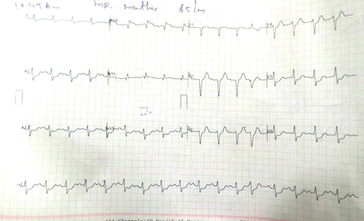
Does this ECG allow you to go ahead for thrombolysis? It actually looks like NSEACS with ST elevation in AVR suggesting left main lesion
The initial diagnosis of NSTEMI was made, and hence thrombolysis was not considered. Even as the fellows were mulling over the diagnosis, one of them could find one more ECG available taken a few hours ago in another hospital.
It had something on it ,
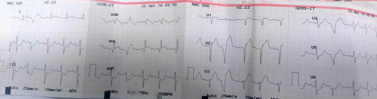
This ECG taken a few hours ago, shows ST elevation in 1 and AVL, and few VPDS in the chest leads unmasks the anterior ST elevation.
The moment we saw this ECG it was decided to go ahead with thrombolysis.The final ECG after thrombolysis with (Streptokinase) showed further stabilization. The question of thrombolysis in NSTEMI though not indicated in general, in selected situations we need to Introspect!
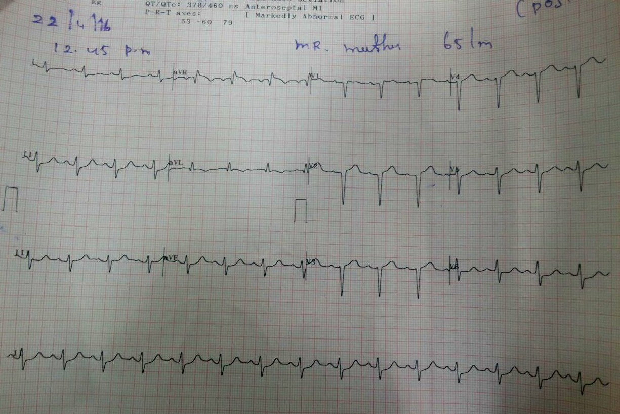
How to manage a patient who presents as NSTEMI but had STEMI a few hours ago?
Four ways to ponder!
- This patient should not be lysed as we have to treat the current event, not the past. ,(Its NSTEMI and no need for lysis) Just heparin, dual antiplatelets. That will do.
- One can go ahead with lysis as there is evidence for STEMI in prior ECG.
- There is ST elevation in AVR even in the second ECG and so you have to thrombolyse !
- “Come on guys, . . . don’t live in the primitive era of managing ACS in CCU . Forget the ECG take him to the cath lab , suck out all thrombus and deploy a stent and come out”.
* The last one , though appear practical (and most of us would love that ) is an unprofessional way of practicing cardiology. Management of ACS requires sound principles of ECG and its correlation with the Intra-coronary and myocardial pathology.
What happened to this patient?
He did well, free of angina with minimal LV dysfunction. He was discharged. Will be reviewed later, for further evaluation. This is a typical example of a patient with ACS managed successfully without entering the cath lab.(A forbidden practice and a potential coronary blasphemy )
Final message
ECG changes are as dynamic as the Intra-coronary blood flow in ACS. Multiple factors determine ST elevation or depression. While thrombolysis is reserved for STEMI, NSTEMI has little or no benefit to accrue with thrombolysis. However, this is applicable only for de-novo NSTEMI and may not apply for STEMI in transition into NSTEMI as in the above patient.
Posted in acute coronary syndrome, Cardiology -Therapeutic dilemma | Tagged ecg in acs, ecg in ccu, NSTEMI NSTACS STEMI STEACS, STEACS AND NSTEACS, transformation of nstemi to stemi and stemi to nstemi | Leave a Comment »
Categories
-

-
The contents of the this blog is being published as Kindle E book , as per the request of many of the readers. Every article will continue to be open source in this site. Again I shall reiterate the book format is not aimed at any commercial intent. It is only to facilitate learning in a single book format Here is the link to book
https://amzn.in/d/euhL5vu Archives
- March 2026 (7)
- February 2026 (8)
- January 2026 (8)
- December 2025 (11)
- November 2025 (7)
- October 2025 (8)
- September 2025 (7)
- August 2025 (9)
- July 2025 (10)
- June 2025 (8)
- May 2025 (9)
- April 2025 (7)
- March 2025 (10)
- February 2025 (4)
- January 2025 (9)
- December 2024 (11)
- November 2024 (8)
- October 2024 (10)
- September 2024 (5)
- August 2024 (5)
- July 2024 (6)
- June 2024 (5)
- May 2024 (4)
- April 2024 (7)
- March 2024 (4)
- February 2024 (8)
- January 2024 (6)
- December 2023 (8)
- November 2023 (13)
- October 2023 (14)
- September 2023 (5)
- August 2023 (6)
- July 2023 (10)
- June 2023 (5)
- May 2023 (5)
- April 2023 (4)
- March 2023 (5)
- February 2023 (2)
- January 2023 (7)
- December 2022 (3)
- November 2022 (5)
- October 2022 (5)
- September 2022 (4)
- August 2022 (3)
- July 2022 (9)
- June 2022 (2)
- May 2022 (1)
- April 2022 (2)
- March 2022 (1)
- February 2022 (3)
- January 2022 (7)
- December 2021 (3)
- November 2021 (5)
- October 2021 (8)
- September 2021 (4)
- August 2021 (6)
- July 2021 (6)
- June 2021 (7)
- May 2021 (5)
- April 2021 (4)
- March 2021 (3)
- February 2021 (6)
- January 2021 (8)
- December 2020 (4)
- November 2020 (5)
- October 2020 (7)
- September 2020 (7)
- August 2020 (10)
- July 2020 (6)
- June 2020 (9)
- May 2020 (9)
- April 2020 (5)
- March 2020 (7)
- February 2020 (3)
- January 2020 (4)
- December 2019 (4)
- November 2019 (6)
- October 2019 (3)
- September 2019 (6)
- August 2019 (3)
- July 2019 (1)
- June 2019 (3)
- May 2019 (2)
- April 2019 (2)
- March 2019 (2)
- February 2019 (4)
- January 2019 (2)
- December 2018 (2)
- November 2018 (2)
- October 2018 (2)
- September 2018 (1)
- August 2018 (2)
- July 2018 (3)
- June 2018 (1)
- May 2018 (3)
- April 2018 (1)
- March 2018 (3)
- February 2018 (3)
- January 2018 (1)
- December 2017 (3)
- November 2017 (3)
- October 2017 (3)
- September 2017 (2)
- August 2017 (2)
- July 2017 (2)
- June 2017 (2)
- May 2017 (4)
- April 2017 (3)
- March 2017 (3)
- February 2017 (5)
- January 2017 (3)
- December 2016 (2)
- November 2016 (5)
- October 2016 (4)
- September 2016 (3)
- August 2016 (5)
- July 2016 (3)
- June 2016 (4)
- May 2016 (3)
- April 2016 (6)
- March 2016 (4)
- February 2016 (3)
- January 2016 (5)
- December 2015 (6)
- November 2015 (5)
- October 2015 (8)
- September 2015 (2)
- August 2015 (5)
- July 2015 (7)
- June 2015 (4)
- May 2015 (6)
- April 2015 (5)
- March 2015 (7)
- February 2015 (15)
- January 2015 (8)
- December 2014 (5)
- November 2014 (9)
- October 2014 (7)
- September 2014 (9)
- August 2014 (5)
- July 2014 (11)
- June 2014 (5)
- May 2014 (4)
- April 2014 (5)
- March 2014 (8)
- February 2014 (8)
- January 2014 (5)
- December 2013 (7)
- November 2013 (7)
- October 2013 (14)
- September 2013 (12)
- August 2013 (15)
- July 2013 (15)
- June 2013 (15)
- May 2013 (15)
- April 2013 (15)
- March 2013 (15)
- February 2013 (15)
- January 2013 (15)
- December 2012 (15)
- November 2012 (15)
- October 2012 (15)
- September 2012 (15)
- August 2012 (15)
- July 2012 (15)
- June 2012 (15)
- May 2012 (15)
- April 2012 (15)
- March 2012 (15)
- February 2012 (15)
- January 2012 (15)
- December 2011 (15)
- November 2011 (17)
- October 2011 (17)
- September 2011 (17)
- August 2011 (21)
- July 2011 (20)
- June 2011 (17)
- May 2011 (15)
- April 2011 (17)
- March 2011 (25)
- February 2011 (20)
- January 2011 (20)
- December 2010 (18)
- November 2010 (21)
- October 2010 (21)
- September 2010 (25)
- August 2010 (20)
- July 2010 (10)
- June 2010 (11)
- May 2010 (19)
- April 2010 (16)
- March 2010 (14)
- February 2010 (22)
- January 2010 (18)
- December 2009 (20)
- November 2009 (20)
- October 2009 (3)
- September 2009 (21)
- August 2009 (19)
- July 2009 (12)
- June 2009 (12)
- May 2009 (11)
- April 2009 (15)
- March 2009 (21)
- February 2009 (4)
- January 2009 (12)
- December 2008 (13)
- November 2008 (9)
- October 2008 (22)
- September 2008 (20)
- August 2008 (16)
- July 2008 (14)
- June 2008 (7)
Blog Stats
- 6,637,761 hits
Please give your feed back .
Click below to see who is watching this website live !

- This site will never aim for profit. Still ,this donation link is added at the request of few visitors who wanted to contribute and of-course that will help make it sustainable .

Please Note

