Feeds:
Posts
Comments

Posts Tagged ‘HFpEF’

Some times I wished, I could have become a Nephrologist. This thought comes whenever we face a patient with unexplained dyspnea, normal EF, and heart size. Am I missing a HFpEF? The heart is not like the kidney, which injects the all too familiar BUN & creatinine into blood promptly (with a high degree of reliability). Even a medical neophyte can diagnose renal failure just like that.

Cardiologists don’t have that luxury. Of course, we do have NT-Pro BNP recently , which nowhere comes near to renal failure markers . Still, many of the youngsters are tuned to believe NT-Pro BNP is equivalent to creatinine for the heart. Remember, biochemical diagnosis is always an adjunct and never confirmatory This is because NT-Pro BNP is universal cardiac stretch molecule , that can elevate in big list of conditions. Similarly , to appear in the blood, there can be a time lag unlike creatinine. (Read the universal definition of heart failure below).

“The universal definition of HF , defines HF is a clinical syndrome with symptoms and/or signs caused by a structural and/or functional cardiac abnormality and corroborated by elevated natriuretic peptide levels and/or objective evidence of pulmonary or systemic congestion”

* Though , a key link word and /or is used widely, unfortunately , the definition fails to reiterate clinical signs will prevail over biochemistry at any given point of time. It has inadvertantly ? given some over wiegtage to biochemistry.

Also, the new definition has gotten rid of hemodynamic, metabolic, and chamber-wise RV, LV classification systems. I think the committee wanted to remove the clutter of terms. In a broad sense, it’s good. By using the word structural, it includes valve defects or pericardial pathology into the basket of HF.


Now getting deeper, can we differentiate differentiate HFpEF from HFrEF, based on biochemical markers ?

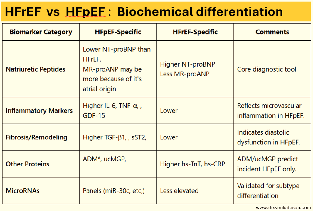
Differentiating HFpEF and HFrEF is vital but looks complex. One may say it is not really complex. Funnily , the purpose of the above chart becomes redundant if the EF of 50% has already differentiated the two conditions . what is the big deal about this table ? It is purely for prognostic and etiological purposes.

Summary

NT-proBNP is typically lower in HFpEF due to reduced ventricular wall stress, while MR-proANP which originates in the atria are expected to be more in HFpEF . Inflammatory and fibrotic markers and MicroRNA panels are high and more specific to HFpEF.

*Growth differentiation factor-15 . **ADM -Adrenomedulin a biomarker for arterial stiffness and arterial calcification . ***ucMGP refers Uncarboxylated Matrix Gla Protein. MGP to function as a calcification inhibitor,

Final message

Recognizing and confirming HFpEF, itself can be considered a partial clinical success. (Since more often it is missed) As mentioned earlier, biochemical differentiation is largely an academic exercise but gives us an idea of systemic pro-inflammatory and local fibrotic status.

There is a rough pathological rule of thumb (Note the word rough again). HFpEF is more often a systemic condition where the heart is affected. (In contrast to HFrEF where the pathology of the heart is central, and all systemic features are secondary to it.) Treatment options for heart failure have improved overall .However it is more limited in HFpEF as Inotropic agents are redundant here. Prognosis is highly variable and not necessarily better.

Reference

1.Ho JE, Lyass A, Lee DS, et al. Circulating biomarkers of incident heart failure with preserved ejection fraction: the ARIC study. JAMA Cardiol. 2018;3(5):415-423.

2.Zile MR, Desantis SM, Baldauff N, et al. Biomarker-guided risk assessment for the diagnosis and treatment of heart failure with reduced ejection fraction and preserved ejection fraction. JACC Heart Fail. 2021;9(12):893-903.

For Fellows . How do you diagnose HFpEF ?

  1. Clinical features of heart failure (Apply some criteria , ex-Framingham )
  2. Some evidence of elevated LV filling pressure at rest or exercise (Diastolic stress test)
  3. Normal LVEF > 50% (Must ensure this should not be a recovered and improved EF from HFrEF condition)
  4. NT-ProBNP -Supportive

Read Full Post »

Restrictive LV filling is an advanced form of diastolic dysfunction. The mean LA pressure is high, and LVEDP is also correspondingly elevated (need not be linear though, as LA reservoir/conduit dysfunction can independently hike the LA pressure). This clinical scenario of restrictive LV filling usually occurs as part of HFpEF, though it can occur in HFrEF as well. (25% of DCM have restrictive filling)

Image source and courtesey : Sean Haney, Denise Sur and Zijian Xu The Journal of the American Board of Family Practice May 2005, 18 (3) 189-198; DOI: https://doi.org/10.3122/jabfm.18.3.189

Pre-load reduction is the mainstay in relieving pulmonary congestion, but it has a trade-off at a particular point, as it impacts the stroke volume and forward cardiac output. Diuretic excess, ultimately worsens the symptoms, especially fatigue, though they keep the lungs dry.

So, dear fellows , remember prescription of diuretics in restrictive LV filling is a tight pharmacological rope walk.It requires continuous monitoring of symptoms and E/E” in echocardiography.

Whether to push the LA blood with more preload or bring it down to redcue pulmonary congestion is the question

Some physicians use the E-DT as a visual guide (Deceleration time of E velocity, which is inversely related to the degree of restriction). Normal is more than 150 ms. In most restrictive filling, it is 100 ms or less. Diuretic dose can be adjusted based on E-DT.

The usual daily dose of frusemide is 80 mg. There is a huge upper limit.It will be useful if the dose of frusemide is somehow indexed to the LV filling parameter.

I have tried a personal working formula for optimal diuretic dose. It can be titrated upwards ,twice the value of E-DT when it is less than 100 ms.(Eg if E-DT is 80ms Frusemide can be 160mg, but, note there is an U curve in this .If DT is too short, diuretics will worsen the hemodynamics .At 60 ms E-DT diuretics need to be reduced to 120 mg )

I keep tellling my fellows to do an authenticated study on this. Hope some one pursues(Mayo clinic guys are well equipped to do this , may be with the help Dr Jae.K OH or Sherif F Nagueh from Methodist, Houstan, the pioneers in the field )

Final message

We realise, treating restrictive LV filling is a delicate and often difficult task.There are no specific drugs to improve the lusiotropic property of LV. Further, since LV contractility is normal in HFpEF, there is no point in using LV inotropic agents. The only available parameter to manipulate is LV preload. However, It would be a stunning discovery , if some one discover a atria specific LA inotropic agent to overcome the LV restriction .

Meanwhile, it is critical to treat associated HT, CAD, or infiltrative disease like Amyloid*. We may soon have LA sensors , that can throw LAP to your iPhone . Till then, treating restrictive LV filling is essentially a hemodynamic/clinical pharmacological guess game. Ofcourse ,*We do have protein unfolders and declutters like Tafamidis & Patiseran to clear interstitial amyloidosis. Also, IAS flow regulators are new devices being tested to decompress the LA in HFpEF. (Paitazoglou et al Ther Adv Cardiovasc Dis. 2020 )

Read Full Post »

PH is an important clinical cardio-pulmonary entity , which we confront day to day. Though the prevalence of PH in a community is just 1 % (25 times less than systemic hypertension) it deserves a special place as the diagnosis is more complex and outcome is often adverse.

The defining criteria , the classification, and grading of PH has always been a difficult and dynamic academic task .Right from WHO’s 1974 definition, we have 7 global symposiums , last one happened few months ago, in July 2024 in Barcelona.

We have made rapid strides in all aspects of PH right from molecular , genetic , imaging and therpeutics. Still, there is one important issue that has been overlooked for quiet some long. The concept of fitting PH in 5 groups based on etiology, though appear to simplify things, there is a significant flaw.

The overlaps in etiology

1.The group 1 contains the famous , (now obsolete entity of primary pulmonary hypertension) Idiopathic PH , meaning that we don’t know the cause of it or we have excluded all known causes. Meanwhile, group 5 also has set of conditions of PH of unknown or unclear etiology. So, a IPH of group one can migrate to either group 4 or group 5 or vice versa.

2.PH due to congenital heart disease can be in both Group 1 and 3

3.If you take PH due to some of the connective tissue order, I am sure, it can fall into any of the 5 groups

Suggestions for the next PH working group

It is desirable that the next working group should acknowledge existence of inter and intra group overlaps of PH in a more clear manner. Either we should take away the groupism or the current definition of group 5 need to be more elaborate . It says multi-factorial. Instead we can try to find what are the groups it is likely to have an overlap. Should we need another a sixth group ? GO-PH (Group overlapping PH)

There can also be a place for combined etiological-hemodyanmic classification . (Example : Group 1 .Pre capillary .Group 1 Intra-capillary as in PVOD) . CTEPH though essentially is a precap PH, the risk factors of CTEPH and HFpEF can be shared one, making it combined pre and post cap PH a distinct possibility. )

Final message

While the problem of groupism in PH exists, the issue of highest importance in PH is something different. This is more philosophical . We need to be very clear what we mean by Idiopathic. As physicians, we must realize how relative this terminology is . What is idiopathic in your hospital, (However big you are) may turn out to be a missed case of mixed connective tissue disorder or silent CTEPH detected only by V/Q scan or a dual energy CT or a rare case of PVOD by judiciously reading a pulmonary angiogram in a dedicated PH center.

*Also we must recall, statistically up to 80% of PH is due to left heart (This HFpEF stuff has jacked this incidence still more ) and lung disease. Our efforts and resources should be used judiciously for optimal diagnosis and management of common conditions first.

Read Full Post »

A brief learning session with cardiology fellow about a possible new concept in Left heart failure.

What is the commmest cause for acute pulmonary edema?

Left sided heart failure”

“Can you be more specific , Left sided means ?

“I meant LV failure , typically DCM of any cause or Severe un-controlled HT”

“Ok .good. Does Left Heart failure include mitral valve dysfunction also ?

“Yes sir, very much. Classical mitral stenosis and Isolated MR can cause pulmonary edema. In fact, acute AR Iis also part of left heart disease”

So far, so good, now coming to the complex part of left heart

Can LA fail in isolation independent of LV , ie I mean with normal Left ventricle ?

I am not sure. Can we call new onset atrial fibrillation as a primary atrial failure that can result in pulmonary edema?

Excellent. You are absolutely right. But I am talking about mechanical atrial failure, not electrical. Are you aware that most of the time AF is a well tolerated arrhythmia , it can even be silent in many cases. This is because the pumping function of the atria contributes only 20-25% to LV filling. This can easily be compensated by augmented LV suction force , provided the baseline LV function is normal.

Have you heard about ACM. Atial cardiomyopathy?

“No sir”,

“You will hear more about it soon” (Ref 1) Scientists, especially Echo guys are closing in on this concept. We know, the atria has three functional components, namely conduit, reservoir, and pumping. Curiously, we have realized that the pumping function of LA may not be that critical from indirect observations from some land mark studies . (Rate control vs rhythm control studies in AF are a powerful proof on the atrial pumping function .(AFFIRM/RACE etc ) I don’t know, whether I am right in saying the above statement.

It is tempting to conclude , only if all the three functional components of atria gets affected , then only primary atrial dysfunction can be diagnosed. The concept is more complex than we realise. In diastole , pulmonary vein, LA , LV all work as single functional unit. Only in systole, we see them as different things.

Atrial DCM

Like LV systolic function, which is coupled with RV in parallel , LA function is closely knitted to LV in series during diastole. The key to suspect or diagnose this entity is to demonstrate dissociation of LVEDP with LA mean pressure & PCWP. This is not an easy job in bedside. Isolated Increase in LA volume without any reason , is one clue. LA ejection fraction is possible marker. (Kanagala P, . Int J Cardiovasc Imaging. 2020)

Final message

We are in the early days of understanding primary atrial mechanical failure, Atrial cardiomyopathy (ACM) or atrial DCM is being proposed as separate entity. It is very likely, some subsets of HFpEF might turn out to be primary atrial disease, depending on the level of investigation we do.

Reference

1.Li M, Ning Y, Tse G, Saguner AM, Wei M, Day JD, Luo G, Li G. Atrial cardiomyopathy: from cell to bedside. ESC Heart Fail. 2022 Dec;9(6):3768-3784. doi: 10.1002/ehf2.14089.

2.Patel, R.B., Lam, C.S.P., Svedlund, S. et al. Disproportionate left atrial myopathy in heart failure with preserved ejection fraction among participants of the PROMIS-HFpEF study. Sci Rep 11, 4885 (2021). https://doi.org/10.1038/s41598-021-84133-9

Read Full Post »

Though all of us are aware, the incidence of heart failure is increasing exponentially and is the leading cause of global disease mortality, what we fail to understand is, we still lack a good definition cardiac failure. 

Defining HF based on EF% is convenient but adds more complexity, and is less scientific too. Still, as of now, we have adopted this. I think, one of the important factors that apparently increased the incidence of HF is the creation of an entity called HFpEF. (Formerly diastolic heart failure)

Thanks to ESC, we have a consensus document, which has defined HFpEF based on functional, morphological, and biochemical features. This is a more refined model from the original Mayo clinic H2FpEF score.

Both are given below.


The problem with both these criteria is the disproportionate importance given to AF. The knowledge gap here is, AF can be initiated at any degree of increased atrial strain which can independently raise LA mean pressure without persistent elevation of LVEDP. We recognize now, left atrial obesity (fatty atrium)  is a powerful trigger of AF, still, in this situation, an Innocent LV may get blamed with a tag of HFpEF. Likewise, many HFpEF may turn out to be primary  LA dysfunction than LV failure. To make things more confusing(scientific) for diagnosing true HFpPEF, we may soon need to look into LA-EF as well. (LA-HFpEF)

Can we diagnose clinically significant HFpEF, without pulmonary hypertension?


In my understanding, the answer is No.

Looking at the two schemes (Mayo & ESC) one thing is clear. Pulmonary hypertension is the key hemodynamic expression of HFpEF. It could be either resting and persistent or exertional and transient.t is obvious the PH in HFpEF is post-capillary. (The modern term for pulmonary venous HT). Mind you, while PVH is mandatory to diagnose HFpEF, PAH (precapillary ) is also observed in most patients with significant HFpEF. This is the reason TR jet velocity is included as one of the criteria.  (To make things simple, we may need to create a new classification of HFpEF, ie resting vs exertional HFpEF.This is what the diastolic stress testing is all about.)

Final message

It is back to basics & time to dig into the fundamentals, of what exactly we mean by heart failure. Is the elevation of LV filling pressure alone sufficient? Should it happen at rest or at exertion, and whether neuroendocrine activation is necessary? Is RASS activation similar in both HFrEF and HFpEF? Try to find the answer to this. How often does HFpEF fulfill Framingham’s criteria of HF.? ( Löfström et al  ESC Heart Fail. 2019) 

Trying to understand the nuances of HFpEF, I think, we can make a statement,- HFpEF can not be diagnosed without pulmonary hypertension. It makes a lot of sense the P in the H2FpEF  scoring system denotes PH, however, It is assigned only a single point, which needs revision. In fact, there is a strong case to argue and make it an essential criterion.

Paradoxically & curiously HF with reduced ejection fraction (which is the most common form of HF) doesn’t require the presence of PAH to diagnose it. This issue may also be examined.

Reference 

1.How to Diagnose Heart Failure With Preserved Ejection Fraction: The HFA–PEFF Diagnostic Algorithm: A Consensus Recommendation From the Heart Failure Association (HFA) of the European Society of Cardiology (ESC). Eur Heart J 2019;40:3297-3317.

2.Löfström U, Hage C, Savarese G, Donal E, Daubert JC, Lund LH, Linde C. Prognostic impact of Framingham heart failure criteria in heart failure with preserved ejection fraction. ESC Heart Fail. 2019 Aug;6(4):830-839. doi: 10.1002/ehf2.12458. Epub 2019 Jun 17. PMID: 31207140; PMCID: PMC6676283.

Read Full Post »

Doppler E/A ratio reversal is probably the most reported abnormality in clinical echocardiography. We are also pleased to label it as a grade 1 diastolic dysfunction. Making a significant population who come for regular health checks anxious and worried.

Sharing a presentation from the Annual conference ECHO INDIA 2019, I participated in a symposium on Diastolic dysfunction.

Topic : Issues in diagnosing grade 1 diastolic dysfunction: Pearls and Perils

How did we get into this academic trap? Should we continue this practice?

The current ASE guidelines 2016 have a clear message. It has taken off the E/A ratio from the Initial screening for diastolic dysfunction.

Summary & Final message 

Are we ready for the change? By understanding a simple concept, one can reduce the incidence of indiscriminate diagnosis of grade 1 diastolic dysfunction.

  • E/A ratio apparently has a no role in diagnosing diastolic dysfunction in the normal population who have normal EF %.
  • Hence, never report E/A ratio in Isolation as grade 1 diastolic dysfunction.
  • However, in patients with HFrEF it does help in triaging diastolic dysfunction.
  • Always look for symptoms and 2D features  (Unexplained dyspnea, LA enlargement, LVH ) before considering diastolic dysfunction.

*For advanced readers and researchers grade 1 diastolic dysfunction does have a deep meaning and always continues to puzzle.

Patient corner

For all those anxious patients who ramble around with a report of grade 1 diastolic dysfunction, I can assure you this. Please realize, 9/10 times, this is just a decorative echocardiography abnormality meant to add some spice to the report  does not have any significance.

*Will post a PPT presentation shortly.

Read Full Post »

Heart failure has been classified in many ways, with prevailing levels of our knowledge and ignorance. It is based on a variety of factors like rapidity of onset, etiology, chambers involved, hemodynamics, etc. 

  • Forward vs backward failure
  • Acute vs chronic failure
  • RV/LV or Biventicular failure 
  • Systolic vs diastolic heart failure
  • High output vs low out failure
  • Ischemic vs non-ischemic failure 
  • Reversible vs Refractory HF 

None of them have really helped at the bedside though it helped us understand the condition. Now, in the last decade, we have crash-landed on our favorite obsession to classify HF ie based on Ejection fraction. We believe we have found an exciting new classification. (HFrEF/HFpEF/HFmrEF).We embraced it, even after recognizing EF as a battered LV functional parameter due to its high load-dependence with a dubious reproducibility.  

If we rely too much on echo, there can be a few more classifications for HF 

  1. HF failure with preserved diastolic function(25% of all DCMs with HFrEF )
  2. HF with preserved mitral valve function
  3. HF with preserved Global longitudinal strain(Still normal EF%)
  4. HF with preserved RV function
  5. HF with preserved Torsion and Twist.
  6. Finally, HF with normal Heart (Anemia/CKD etc)  In anemia heart never fails in true sense. In fact, it works at peak capacity.(More of a Success than failure). Similarly isn’t odd to put primary CKD/CRF in the CHF basket.

Probably the most important and practical classification  could be

  1. Primary vs secondary HF (Primary means all muscle diseases under MOGES system ) 
  2. Valvular vs non-valvular failure (Surgically correctable MVR/DVR/Mitral valve repair)
  3. Revascularisable  or Non-revascularisable HF (STICH study responders)
  4. ICD/CRT eligible HF vs Non-eligible HF ( Rule out DANISH study non-responders)
  5. Refractory failure -Novel drugs/ Assist device/TAH/ Transplant suited 

Final message

 Dr Thomas Lewis said over 100 years ago, the essence of the practice of cardiology is to recognize HF early. Looking back at the literature, there will be no dearth of classification for HF. It will come and go according to academic and Imaging whims. Of course, that may aid in ruling out primary cardiac conditions. But, we must always emphasize to the next-generation that HF is often due to systemic*(reversible too) conditions in substantial numbers. Here the heart is just a bystander watching helplessly, trying to adapt to a remote systemic comorbid problem. Such hearts don’t require cowboy aggression but gentle care by concerned physicians.(One study reveals weight reduction and systematic exercise program adds more life to HF than drugs and devices. Will link the reference/ or try google)

*Eg: Anemia is the commonest cause of HFpEF on a global scale. .CKD, undiagnosed autoimmune disorders, malignancy, are other classical examples. Let us be first a physician then a cardiologist, that will ensure our we don’t miss important treatable conditions with our short-sighted definition of heart failure based on EF%.

 Reference 

1.Y. Juilliere, J.N. Trochu, P. de Groote, et al.Heart failure with preserved systolic function: a diagnostic algorithm for a pragmatic definition  Arch Mal Coeur Vaiss, 99 (2006), pp. 279-286  View Record in ScopusGoogle Scholar
 

Read Full Post »

Assessment of LV diastolic function primarily depends on the Doppler flow profile across the mitral valve and also to be noted are the 2D features of LA and LV for associated abnormality like LVH, LAE etc.

Why diastolic dysfunction assessment difficult in AF ?

Since most diastolic doppler mitral inflow parameters involve analysis of atrial contraction A wave, atrial fibrillation makes it difficult to assess diastolic dysfunction. Since we have only early diastolic velocity to assess, the changes confined to this E velocity is of paramount importance. This E velocity again is subjected to cycle length dependent alteration in both its acceleration and deceleration time , making things still more complex.

However, the following features help diagnose diastolic dysfunction in AF

  1. Lack of significant  E velocity variation (<20%)  Inspite of significant RR interval change.(This implies mean LAP is kept high irrespective of cycle length suggesting elevated baseline LAP)
  2. E deceleration time (<140ms) (In long cycle)
  3. Propagation velocity in color M Mode(Vp)  <45cm/sec might help (RR interval dependent, measure in the long cycle)
  4. E/e” in a single beat by dual doppler probe (Ref 1)  > 10 indicate diastolic dysfunction that correlate with PCWP> 15mmhg (Ref 1)
  5. Finally (and curiously ) presence of AF by itself may imply significant LV diastolic dysfunction. It could be due to an increase in atrial strain and afterload of LA (ie pre A-LVEDP) (Of course, It should be in the absence of mitral valve disease)
  6. LA dimension in AF*

*LA dimension is a very good sign of chronic elevation of LAP and diastolic dysfunction in the absence of mitral valve disease. However, AF can dilate the LA making it a less useful parameter. But, it should be noted in AF both RA and LA dilate together.So,  a disproportionate LA>RA (or if RA is normal size ) could still be a marker of baseline LV diastolic dysfunction.


Reference

  1. Kusunose K.Yamada H.,  Nishio S.et al.  Clinical utility of single-beat E/e′ obtained by simultaneous recording of flow and tissue Doppler velocities in atrial fibrillation with preserved systolic functionJ Am Coll Cardiol Img 2009 2:11471156


Read Full Post »