Yes, It’s not a humiliation, to get branded as a new generation cardiologist. In fact, the opposite is true. Sorry, no-blaming any one. We can’t avoid it as well. It is the wages, we are foreced to pay for sensationalised technological sins , that is Imploding in the world of medical science.
Coronary bloodflow is primarily known, to occcur as a diastolic circulation. Does that in any way mean coronary artery diastolic pressure, can exceed the systolic pressure ?
A. No. diastolic BP can never exceed systolic BP in side the coronary artery.
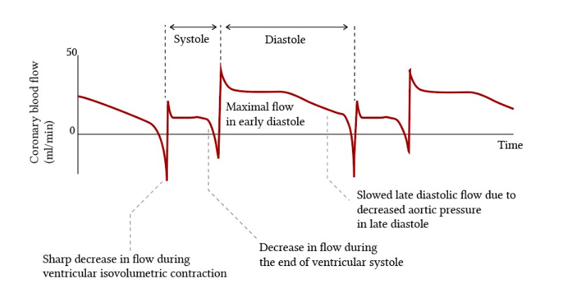
B. Yes. Coronary diastolic BP is higher than systolic, since there is little blood flow during systole due to myocardial compression.
C. There is not much difference between systolic and diastolic pressures, within the coronary artery . We need to bother only about mean perfusion pressure.
D.It is true, the coronary blood flow is compromised in systole and primarily occur in diastole .Still, the epicardial coronary arterial compresssion is not that significant. Hence systolic pressure blunting is negligible. This is called the pressure -flow paradox.
Answer : D (Ref Image 2)
What is the normal intra-coronary arterial pressure in systole and diastole? I could not get a clear answer to this question. Logically it should be sane as in radialartery 120/80mmhg. Surprisingly, most literature discusses only coronary blood flow, which primarily takes place in diastole. (Does that mean the pressure would be less in diastole, so that blood flows easily?) The complexity in understanding intra-coronary pressure , is because, we don’t know the exact blood volume, flow vs pressure relation in this dynamic organ.Further, mechanical force/pressure exerted by the muscle ,can it be recorded , within the lumen , and quntify it sepearately ?
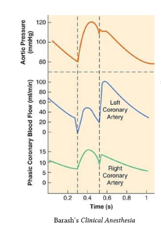
The classical illustrations that are found in cardiac physiology literature about the dominance of coronary blood flow during diastole (Image source Ref 2)
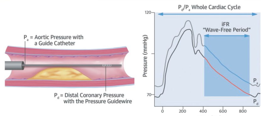
During FFR studies the Intracoronary presssure curves almost mimic radial pulse. No where we could see the effect of mechanical compression . It is likely , the epicardial coronary artery do not get compressed that much , only the micro circulation gets squeezed.
We realise ,coronary perfusion pressure, mean coronary arterial pressure, and coronary arterial wedge pressure are more important than systolic and diastolic pressures . The mean coronary artery pressure is around 45 to 60 mmHg backed up with good autoregulatory mechanism. We are not clear how this autoregulation is modified by lesion tightness. Documentation of true coronary arterial systolic BP in physiology and various pathologies is an important academic vacuum that youngsters can explore.
1.Clinical Implication : Does LV dysfunction has a favorable efffect on coronary perfusion ?
If LV contraction interferes with coronary blood flow, patients with severe LV dysfunction, may gain some advantage as systolic blood flow can happen more easily, and myocardium is perfused better, provided the aortic systolic pressure not too low enough.
2.How common is angina in DCM ? and Why ?
Angina in DCM is an exception despite elevated LVEDP. Is the above logic explain why very few dilated cardiomyopathy patients experience angina? Even in ischemic cardiomyopathy, once it sets in, Intensity of angina is mitigated or completley eliminated.(of course at the cost of failure). Is it nature’s response to prevent angina?
3.Why systemic hypertension is a weak coronary risk factor ?
Unlike the brain, where stroke risk is directly related to systolic BP, fortunately sudden systolic spikes rarely get a chance to attack the coronary endothelium as much of the coronary lumen is relatively closed (? to be confirmed , atleast during rapid ejection phase of systole) In this context, we can also be happy there is no risk of myocardial hemorrhage due to HT. However, there is evidence that diastolic BP carries much risk for CAD, as do Isometric exercises when DBP exceeds out of proportion to systolic BP.
4.Differential intra coronary pressure , RCA VS LCA is well knwon asthe RV contraction is not good enough to compress the RCA.This adds a new hemodynamic concepts in RCA CAD.(We have done a study where we found thrombolysis was more effective in RCA apparently due to bi-modal continuous delivery of the lytic drugs, unlike the left system)
5.During CPR , what would be coronary hemodynamics of chest compression ?
When we compress, it is systemic systole, and when we release it becomes coronary diastole. In fact there is now evidence to suggest , too rapid and hurried contractions reduce the success rate of CPR. The inter compression time is to be atleast 4 or even 5 full seconds, to enable coronary perfusion.The mean pressure during CPR is to be atleast 40mmhg. (Yannopoulos D et al , Resuscitation. 2005)
Final message
It is surprising why we are not recording intra-coronary pressure directly and trying to understand this. We need to go 100 years back for that Wiggers article in search of truth. (Ref 1). This is an area of good research for cardiology fellows. Also, next time,when you do FFR or IFR, ask this question : Why proximal reference pressure is taken at the aortic root instead of just before the lesion ?
OCT, the magical intraluminal coronary vision, has been a great innovation that helps us to decode many uncertainties in the morphology, behavior and vulnerability of coronary plaques. It is used widely in pre- or post-PCI or even asssit during the implantation of stents. The role of OCT/IVUS is sometimes deemed critical in dealing with left main and bifurcation lesions.
Of course, there were some overuse of OCT as well, as many centers did it for some academic fun, even in some innocuous lesions. Meanwhile, there is a striking miss. We probably failed to accrue the benefits of this revolutionary imaging in the graft evaluation. Its real role could be in LIMA grafts including anastomotic site or SVG lesions in the immediate postoperative or at long-term follow-up lesions. As far as I understand, it is very rare for cardiologists to attempt imaging these sophisticated tools in LIMA or SVG.
Published data on OCT IN LIMA
Here is a paper from a stalwart in coronary interventions, Dr. Patrick Serruys and his team from the Netherlands (Published in 2009 ,but surprised to find not many takers)
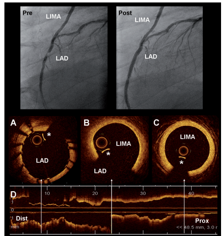
Image source & Courtesey : Ref 1 Optical coherence tomography (OCT) visualisation of left internal mammary artery (LIMA). The superior panel shows the angiogram of a patient with a graft of LIMA to the LAD.
Why OCT/IVUS is less popular in graft assessment ?
*Graft follow-up often falls under surgeon’s domain. They don’t call for check angio often, unless the patient is really, really symptomatic. (CT angiogram is more popular in post CABG)
*From cardiologist’s point of view, they rarely deem it to be necessary. Reason being, it could be technical (Will the venous graft tolerate the OCT wires?)
*Lack of experience and apprehension
*Lack of publsihed data.
Final messge
It is true, doing regular graft angiogram, by itself is less than 5-10 % of all angiograms . Asking for OCT in that population is big deal .Still, OCT can be a valuable in providing crucial information in the assessment of both LIMA and SVG, at least in the former. One more purpose of OCT is, its offline use to assess the integrity of LIMA graft on table prior to CABG. It can confirm patency and possibly rule out any significant takedown injury, that is missed otherwise.
Though the left atrium is the superior most chamber of the heart , it loses its gravity-assisted LV filling advantage in a lying posture. In patients with compromised heart function, this becomes a symptom defining factor. No surprise, patients during episodes of LVF or paroxysmal nocturnal dyspnea, natural forces make them sit up by default, and bring the LA superior & over the top of LV hence its filling is augmented. One more factor that operates is that, IVC orientation, which assumes slope and reduce venous return velocity. In the process, they decongest the lungs and patient gets Immediate relief. In fact, pillows work faster than diuretics and we can technically call it low-cost LV assit devices.
Note, how the LA takes control by its superior position, when the patient assumes erect posture from supine. In fact ,the number of pillows used, by the pateint has some direct correlation with LA mean and Echo cardiographic E/e ‘ . ESCAPE study suggest a possiblity of correlation of this LVEDP with right sided JVP as well.( Drazner et al Circ Heart Fail. 2008 )
Final message
This post may not be relevant to cardiology fellows. Whenever we receive a dyspneic patient in heart failure, prop them up with few pillows. This lesson is taught right in the first-year clinical rounds. I wanted to highlight the anatomical and hemodynamic basis of the sitting-up posture and its impact on LA mean and LVEDP. By some crazy stretch of imagination, pillows can be referred to as a temporary LV assist device.
Research suggestion for fellows
Some of you can do you a study in cath lab, how much the LA mean pressure is altered with reference to posture. It could appear a flimsy study in this era of TAVR/Mitra clips. Sill, we have an good opprtunity to analyse these things as we enter all chambers of heart in routine fashion for some indication or other. This will make us understand LV filling physiology in a better way. (Recalling the days of Guyton & Rushmer when they strugggled to know computational models to measure the pressure gradients)
A question for our hemodynamic acumen?
How does the LA empty in to LV , when LV inflow conduit need to operate against gravity during head down feet up postion as in many sports like bungee jumping or in some asanas (Shirshasana) . Has any one attempted, to know , how would be the E and A velocity across the mitral valve in this posture .Wish some one take on this and report ,if no one has done it before please add some credit . (Just kidding)
Caution
Patients (even some healthy) with diastolic dysfunction especially in elderly, should never attempt to do such sports or indulge in any compromised posture that brings LA below the LV.
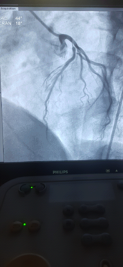
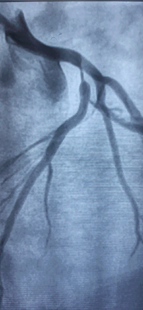
How do you explain this ? 99% occlusion still TIMI 3 flow ?
Answer
A. It could be a parallax error. Lesion may not be tight. Should be seen in other views.
B . Forcible Injection by the operator, make it an artificial TIMI -3 flow.
C .Such flows are very much possible .It Indicates a healthy distal micro-circulation a vascular bed in a fully dilated mode.
D. TIMI flow is not reliable here . We need TIMI frame count to confirm.
Follow up questions
1.How much will be the FFR ?
Likely to be less than . 8 definitely , but surprises can happpen
2.Can he be asymptomatic ?
Unlikely.
Final message
Coronary occlusions are ominpresent . While we have mastered the art of successfully taming these anatomical enemies , we are still very much ignorant what these lesions actually do, to the physiology, inspite of half a dozen flow reserve Indices we have.(FFR,iFR, rFR,qFR, dP/dT ,etc)
The question is, at what level of obstruction, it really limits the coronary bllod flow significantly ( both at rest and exertion) . One thing is clear , it is higly variable & Individualistic, the secrets of which lies deep, in the domain of invisible micro-vascular network integrity.
Counterpoint
TIMI flows may no longer be valid in non-ACS situations. The name TIMI , by itself carries flow after thrombolysis. For some unexplained (& debatable ) reasons, we are used to apply this flow grade , in every angiographic flow scenerios irrespective of underlying clinical entity.
Is primary PCI superior to thrombolysis in the first hour of STEMI ?
No, it is not. I know, most interventional cardiologists cannot accept this fact and would strongly disagree. Still, they know very well there is no clear data to back up their belief. Even the ACC and ESC sort of failed to acknowledge the low quality of available evidence in this specific issue of reperfusion in the first hour.
The fact of the matter is, at best, pPCI fights for equipoise in the first hour, but thrombolysis is a clear winner in moral, scientifc & holistic perspective, as it can be administered very early, at a fraction of cost ,independent of expertise and infrastructure. (CAPTIM). Read ref 1, 2.
Final message
However , there is a thin streak of hope, untill, the unstoppable new generation Interventional cardiologists make “pre-hospital PCI“ a reality .Till then, pPCI will trail behind pre-hospital thrombolysis in the golden hour reperfusion race both in time and probably in efficacy as well.
Peripartum cardiomyopathy (PPCM) , first reported almost a century ago (Ref 1) is getting lot of attention in recent years.Most works are trying to find out the true cause for this ubiquitous entity.The fact that it occurs in the peripartum period, we are forced to link it to hemodynamic and hormonal stress.
Advanced molecular genetic studies reveal PPCM unmasks abnormal sarcolemmal protein mutations in a random population, that express as pregnancy-related DCM. The genes most commonly associated with peripartum cardiomyopathy (PPCM) are Titin, Filamin C, Desmosome, and BAG3-Athanogene (B-Cell related). These genes are involved in the sarcomere, desmosome, intercalated discs, and autophagy.
Link between PPCM & PIH : Is real, not a speculation
Genetically vulnerable women who happen to develop PIH are obviously are at high risk for PPCM. No surprise 30 % of PPCM patients have a history of PIH. If we allow Occam’s razor to teach a few lessons in the Obstetrical suite, then simplistically, a large chunk of PPCM is just a prolonged myocardial distress to abnormal loading conditions of the heart. Afterload mismatch in PIH and preload mismatch in women without PIH. The latter term simply represents volume enhanced myocardial stretch in the postpartum period. Acute LV dilatation adds on to wall stress , thereby hiking the afterload as per Laplace law.
(*There are the molecular triggers that switch on the dormant , yet dysfunctional gap junctional proteins and stretch them beyond physiological limits, resulting in temporary loss of elastic function and precipitating PPCM, which recovers in many women , if God willing)
Hickam’s also welcome : PPCM is a likely Pituitury cardiomyopathy. Though it is a suspicious circumstantial culprit, it has become a popular hormonal model for PPCM .It is yet to accrue authentic evidence. Meanwhile, bromocrtiptine,the prolactin antagonist is being used with wide ranging efficacy. (Koenig T, Card Fail Rev. 2018 )
Final message
PPCM is essentially a form of cardiac inefficiency in handling either the afterload or preload (or both) in the peripartum period, probably influenced by the pituitary gland in those with genetically vulnerable sarcomere .The fact that it is recurring in substanial number of women in subsequent pregancy , would point out to the same hemodynamic distress story.
Counterpoint
*Some would argue , PPCM should not be diagnosed if the mother has PIH .The rule is PPCM is diagnosed only after excluding all reversible diagnosis. Many guidelines endorse that. I couldn’t understand the logic, unless we know the true reasons why some hearts struggle to handle the BP effectively. Incidence of RV dysfunction in PPCM would argue for diffuse global myocardial pathology, still it might also be poor tolerance to raised pulmonary pressure .
However, In normal hearts without LV dysfunction the risk is dramatically lower, but still not totally risk free. (The term normality can be undermined now , because, in true sense , an echocardiogram is not sufffice to rule out a structural myocardial disease. MRI has become a basic requirment to R/O concealed or localised scars , which may not alter wall motion and show up in echocardiogram)
So, do you want to say all LBBBs are risky ?
No. But we can’t say they are not risky as well. This is because the slowing of conduction, mild prolongation of the QT interval (contributed more by prolonged depolarization), and desynchrony still are the common denominators. These changes increase vulnerability to arrhythmias under specific circumstances—like electrolyte imbalances, drug effects, or extreme stress. This can trigger a VT in potentially inappropriate circumstances.
Final message
Cardiac electrical highway from SA node to Purkinje fibers is expected to be smooth flowing without any bumps and humps. Any interruptions in the electrical circuit are a potential invite for misbehavior. Fortunately, most times the adjacent cells compensate and adapt to the subtle diversions.
Restrictive LV filling is an advanced form of diastolic dysfunction. The mean LA pressure is high, and LVEDP is also correspondingly elevated (need not be linear though, as LA reservoir/conduit dysfunction can independently hike the LA pressure). This clinical scenario of restrictive LV filling usually occurs as part of HFpEF, though it can occur in HFrEF as well. (25% of DCM have restrictive filling)
Pre-load reduction is the mainstay in relieving pulmonary congestion, but it has a trade-off at a particular point, as it impacts the stroke volume and forward cardiac output. Diuretic excess, ultimately worsens the symptoms, especially fatigue, though they keep the lungs dry.
So, dear fellows , remember prescription of diuretics in restrictive LV filling is a tight pharmacological rope walk.It requires continuous monitoring of symptoms and E/E” in echocardiography.
“Whether to push the LA blood with more preload or bring it down to redcue pulmonary congestion is the question“
Some physicians use the E-DT as a visual guide (Deceleration time of E velocity, which is inversely related to the degree of restriction). Normal is more than 150 ms. In most restrictive filling, it is 100 ms or less. Diuretic dose can be adjusted based on E-DT.
The usual daily dose of frusemide is 80 mg. There is a huge upper limit.It will be useful if the dose of frusemide is somehow indexed to the LV filling parameter.
I have tried a personal working formula for optimal diuretic dose. It can be titrated upwards ,twice the value of E-DT when it is less than 100 ms.(Eg if E-DT is 80ms Frusemide can be 160mg, but, note there is an U curve in this .If DT is too short, diuretics will worsen the hemodynamics .At 60 ms E-DT diuretics need to be reduced to 120 mg )
I keep tellling my fellows to do an authenticated study on this. Hope some one pursues(Mayo clinic guys are well equipped to do this , may be with the help Dr Jae.K OH or Sherif F Nagueh from Methodist, Houstan, the pioneers in the field )
Final message
We realise, treating restrictive LV filling is a delicate and often difficult task.There are no specific drugs to improve the lusiotropic property of LV. Further, since LV contractility is normal in HFpEF, there is no point in using LV inotropic agents. The only available parameter to manipulate is LV preload. However, It would be a stunning discovery , if some one discover a atria specific LA inotropic agent to overcome the LV restriction .
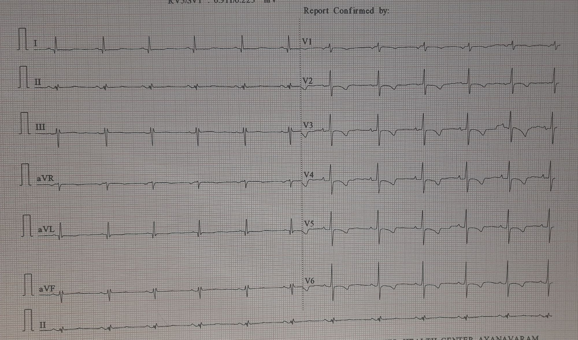
D.Primarily T wave Inversion ,with secondary ST dragging
Answer: Response C, is logical, but applying some ionic sense to the various repolarisation currents in the left shoulder region of action potential , it is the clash between late phase 2 and the premature phase 3 activity, that deforms the initial limb (forward) of T waves , dragging and effacing the ST segment mimicking ST depression. This we have proposed to call it as ST drag effect by T waves. (ST drag is generally more benign , than ST segement depression that begins at J point.
Clinical significance of such ST segment
Without knowing the symptoms or the reason for which this ECG was taken, we shouldn’t interpret this ECG. In this case, it was taken in a 36-year-old woman, routine health check and who has no specific symptom. This almost rules out an ACS or even a CCS. Firther, the fact that the heart rate is normal rules out demand side ischemia as well. Very likely, it should be LVH or anemia or some other systemic medical conditions. (Rarely, neuro-adrenergic-emotional signals from brainstem can tilt the ST segment like this. (Tansient Tako-subo equivalents)
Next step
However, we can’t leave her alone. She needs an echocardiogram to rule out any subclinical myocardial disease. TMT would seem to be a necessity, but false positivity is very likely.( A flamboyant cardiologist would order a CT angiogram either striaghtaway or a day-care radial angiogram. Nothing wrong with that, as long as the patient insists on reaching the bottom of the truth)
What will you do?
Will sit with the patient for atleast 15 minutes, listen to her daily activity ,past history and look for any subtle symptoms, and then decide. It needs lots of courage (or Ignorance) to leave her without any further Investigation. Echocardiogram is a must. (Have seen a HCM variants like this ).TMT is redunant, if her excercise capacity is excellent.
Final message
One more concept on ST segment can be extrapolated by curious observation of some of the ECGs who present at ER. . It is the secondary ST sagging by primary T wave downward forces. (Pushing ST up is also possibe , as we already know it as ERS pattern )
Postamble.
We know, the S point (Technically J ) in ST segment is well defined , while the end of ST segment is hidden in deep mystery in many clinical situations.Mind you, a flattish ST segment, with absent T wave can be an aboslute normality. Here, you can’t measure either ST segment or even the QT interval.
The contents of the this blog is being published as Kindle E book , as per the request of many of the readers. Every article will continue to be open source in this site. Again I shall reiterate the book format is not aimed at any commercial intent. It is only to facilitate learning in a single book format Here is the link to book https://amzn.in/d/euhL5vu
Click below to see who is watching this website live !
This site will never aim for profit. Still ,this donation link is added at the request of few visitors who wanted to contribute and of-course that will help make it sustainable .
Please Note
The author acknowledges all the queries posted by the readers and wishes to answer them .Due to logistic reasons only few could be responded. Inconvenience caused is regretted.