Feeds:
Posts
Comments

Archive for the ‘Infrequently asked questions in cardiology (iFAQs)’ Category

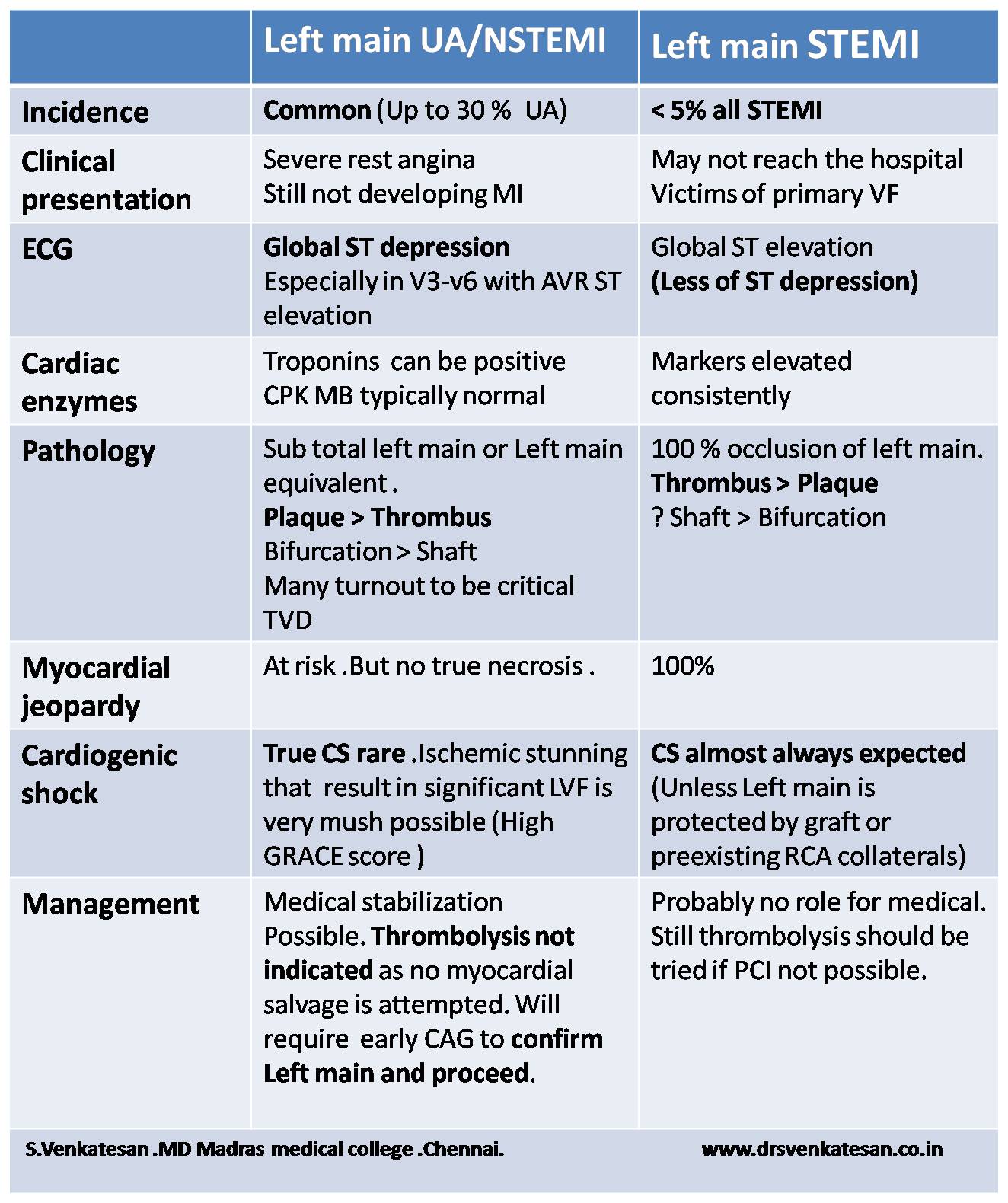
CHB with CAD is a  common combination especially in the elderly.

Which will you Intervene first ?  Is the AV block related to CAD  ?

How to differentiate Ischemic from degenerative AV block ?

Differentiating is often difficult.Even coronary angiogram may not answer the query unless it is totally normal . For AV block to occur usually  LCX / RCA lesion is required.  LAD lesion in isolation are rare to cause CHB .

How often re-vascularisation  reverses  ischemic CHB ?

Logically  you expect more reversals.In real world it rarely happens.

Therapeutic options in combined CAD and CHB

  1. PCI and pace maker in the same sitting .
  2. PCI first followed by pace-maker at a later date.
  3. Pace maker first followed by PCI at a later date if required.
  4. CABG  and epicardial pacemaker ( best option In all critical TVD and CHB)
  5. Pace maker followed by CABG later
  6. Pacemaker followed by medical management (CHB with Insignificant CAD)

Can worsening of ischemia  occur after pacemaker  ?

Very much possible . Since the patient  has been benefited by low heart rate in terms of MVO2 consumption .(Inserting a pacemaker  is  like sudden withdrawal of beta blocker !)

Rate adoptive pacing can confer chronotropic competence which  may bring back the angina.So,what was a insignificant lesion  can become hemodynamicaly relevant  and  may require  angioplasty  later.

*The above clinical issue is applicable  for sinus node dysfunction and CAD as well.

Final message

There is no  fixed rule in the management strategy in combined  CHB and CAD .

Generally , electrical  therapy  should be given preference .Symptom guided approach  may be practical.

In this scientific era , one may argue to deal both issues  together by simultaneous  PCI and pacemaker ,  still  option 3 and 6  remain clear  favorites !

If angina  occurs  even in  baseline bradycardia  it is obvious the obstructive CAD  is  significant and needs immediate fixing .

Finally , though it looks an attractive  concept , It is very rare for CHB  to get reverted by PCI or CABG.


Read Full Post »

Last week  there was a heated debate in our CCU regarding thrombolysis for  a patient with severe rest angina  and ST elevation in AVR  and ST depression in V2-V5  as it implies  Left main disease  Few argued left main disease is an exception where one can thrombolyse even with unstable angina !

One of my fellows argued ACC guidelines vouched for lysis in UA involving left main .( I do not agree )

A logical attempt to differentiate Left main NSTEMI//UA and STEMI

(In the strict sense Left main NSTEMI is misnomer as AVR shows ST elevation  isn’t ? )

left main disease

Final message

Such  patients with suspected LMD   are to be rushed to cath lab .  . . agreed . If it is not feasible , manage it as high risk unstable angina and do not thrombolyse .Let it be left main disease . Indications for lysis are clear. ST  elevation in AVR alone can not be taken as an Indication for lysis.For thromolysis to be effective there should be high thrombus burden with total occlusion . ST elevation in single lead (AVR ) is not a good  marker for left-main thrombus !

Read Full Post »

We know Nitroglycerine(NTG) as a most powerful epicardial coronary dilator  . We use it for instant relief during episodes of coronary arterial spasm in cath lab.

What will happen if we administer NTG over a stented segment ?

Does it dilate it with same vigor ? What will be the consequence  ?

A perfect setting for stent migration isn’t ?

Let us bust the myth around  NTG . NTG  rarely  show  visible coronary dilating effect except in the setting of coronary spasm .

NTG and coronary vasodilatation

Does a LAD with 3 mm diameter become 3.1 or 3.2  and so on with NTG ?

No .It won’t .It is my belief. It is well known , NTG’s action varies significantly in normal and diseased endothelium . Again , there is an irony .It seems , it can act only in normal endothelium , but  we need require it’s therapeutic action only in pathological segments.Further any stented segment would contain   clusters of  both normal and abnormal endothelium .

One more inference is that, stented segment exerts constant pressure on intima making any  pharmacological vasodilatation irrelevant .

Importance of  radial strength of a stent

This issue of vaso-dilator induced  stent migration may not arise in self expanding wall stent with high radial force.But we do not know how long these metals will carry this metallic property .Balloon delivered  stents ( currently used 99% of times ) do not have permanent radial strength .

Final message

I am yet to comprehend what nitrates are expected to do (and what it really does ?)  in a patient post PCI ? (By the way  . . . why we need to prescribe Nitrates it in the first place ? but  In real world most continue to take this for many reasons .)

We need to analyse the micro-vasomotion at the stent -coronary intimal interface.The dynamism in this  narrow space  can be critical  , and may make the difference between life and death !

After thought .

In the hind sight,  this post appears quixotic  for myself . But some one , some where , may generate a great idea  out of it , that will help our patients.

Read Full Post »

CAD is growing as an epidemic in most  parts of the globe. It  is  a major determinant of health status of any country .Great strides in diagnostic, treatment modalities of CAD  have been made in the last few decades. Still , the core principle of management of CAD resides in simple things like  risk factor reduction / optimization , life style changes and few essential cardio-protective medications  Aspirin, beta blockers and statins.

However , modern scientists have made a  firm statement that  knowing the coronary anatomy before starting the treatment is the only scientific approach . It is a huge assumption !

Is it practical ? or is it really required ?

CAD can be managed  by  means of medicines  ,  interventions or surgery. Revascularisation is required  only for  those , who have  critical , symptomatic lesions.

It is estimated , in only  a fraction of CAD patients ,  we would require to know the anatomy . We have set criteria to choose  patients  for CAG , who are  likely to have critical lesions.Physicians  are trained for that elusive wisdom to choose  such patients .Standard text books do mention clear-cut Indications for doing  CAGs. Unfortunately , it is  least respected and followed .

Cardiac physicians who  would boast  they  can’t treat a CAD without knowing  the coronary anatomy  are clinically handicapped  or poorly trained.

I am afraid such a class of  cardiologists are rapidly breeding in the country side. They are  encouraged to attend  CME on clinical  cardiology and basic principles of  clinical decision-making  .

We can’t  keep  on doing CAGs like ECG for every episode of  angina . In fact treating CAD without knowing  the anatomy remains (And it should  be ) the dominant theme contemporary  clinical practice . CAG is multi -edged sword

The most important side effect of routine  coronary angiogram  is , it  ends up in infinite number of inappropriate interventions ! 

I think , we should pray in Hippocratic  temples for sufficient wisdom  to choose our patients. We can also learn it from Neurologists , they  somehow  manage most  forms of cerebrovascular  diseases (scientifically too ! )  without asking  for angiogram of  circle of Willis !  Mind you. . . brain is equally a vital organ !

Final message

It needn’t be a crime to treat  CAD*  without knowing the coronary anatomy. Rather  . . . it would be so  , to ask for CAG indiscriminately  , in every episode of chest pain , without applying clinical sense !

* Emergencies included.

Read Full Post »

We expect LBBB in RV pacing . . .but if RBBB is recorded we are worried ! (Often times it may be neither  LBBB nor RBBB )

Is it really a panic situation ?

  • Not necessarily. The only issue is septal perforation .It is rare , can be recognised by echo or fluro .
  • In true RV apical pacing with tined leads , RBBB  is extremely uncommon .
  • If the lead  is fixed  in the septum and para hisian  area ,  there is  definite  possibility of  deviation from typical LBBB pattern . Screwing leads  that faces high septum  or outflow , RBBB  can be noted occasionally.
  • The commonest cause for RBBB pattern in RV pacing ,  is due to  screw tip going deeper into septal planes  and activating the fibers of left bundle early .
  • For LBBB pattern to occur right  bundle should be morphologically intact .In diffuse CHB  with bilateral bundle branch blocks  the relative contribution ( Impulse conduction ) will determine the QRS morphology . If right bundle is more damaged than left bundle ,RBBB pattern  may prevail  even in the midst of RV pacing !
  • In elderly men with sigmoid septum typical LBBBs are not observed.
  • Anther plausible mechanism would be , even though RV is paced , the pacemaker current’s  exit route may be from LV side .
  • Finally , always  think about coronary  sinus pacing .It is extremely common in blind temporary pacing.

What should we do if we encounter RBBB morphology after PPM ?

  • Analyse the ECG meticulosuly for capture or sensing failure .
  • Do an echocardiography in RV inflow view.
  • Screen the lead by fluroscopy
  • Check the pacing  parameters.
  • Do a holter if  you are really anxious .

If everything is fine , just forget the RBBB.Don’t split your hair for this apparent paradox. In medicine  impossibilities will always  galore !

This paper from Taiwan would vouch for this

RBBB during RV pacing safe ecg

Read Full Post »

We know LVH and SHT go together . Mind you , this is not an Intimate relationship.

Widespread utilisation  of echocardiography  has revealed  , definite  LVH occurs only in about 20% (A guess !) of  HT . (Do you know in the Famingham study the incidence of LVH  after 12 year follow up was a paltry 3 % .Will you agree with that ? Mind you , It was in 1969 when Echo was not there )

What determines LVH ?  The clear answer is elusive. It is easy to escape  from the issue by calling it  multi factorial !

Why don’t you try this question .

My guess would be ,  magnitude ( or  even duration of HT !)  is  less important than genetic predisposition  or  associated diabetes ,  renal involvement.Our analysis from  hypertension clinic reveals LVH is many fold common in secondary HT  when compared to primary HT !

I often used to provoke the students by saying if the LVH is gross in HT it can not be primary , 9/10 times  ! Invariably  we find some  other  association or reason for the HT !

Link to related topic in this site

Why-lvh-does-not-occur-in-all-patients-with-systemic-hypertension ?

How-diabetes-modifies-lvh-due-to-hypertension ?

incidence of lV left ventricular hypertrophy framingham study

Next  . . .

How does LVH regress with treatment ?

Read Full Post »

This question always creeps in any coronary care unit.

Often times , there is a significant  histo-pathological overlap between severe degrees of  Ischemia* and  myocyte necrosis . (What is called micro infarcts, lacunar infarcts,  make us over diagnose MI). It is not yet clear , whether leaky myocyte cell membrane can release free cytoplasmic enzymes without actual cell necrosis.
Clinical Implication
Fortunately , there is not much .These bio markers are primarily used as prognostication tools .Many of these patients  need to  undergo early revascularisation. However , It is unwise ,to get alarmed by  just  Troponin positivity  in an other wise comfortable ACS patient.
* Some call severe degrees of Ischemia as Injury ! It is an old thought based entirely on ECG  .There is no specific cellular equivalent of electrical injury current !

Read Full Post »

In one of my classes , this ECG  was presented .  Controversy  erupted.It was about the  basics .

What is the QRS axis  of this ECG  ?

ecg north west qrs axis  indeterminate

Not surprisingly there were  handful of answers .

  1. North west Axis
  2. Indeterminate QRS
  3. +150
  4. +180
  5. 0 degrees
  6. Extreme Right axis

Which is correct ? My guess  is ,  it should be  closer  to + 180 .  Lead 2 is equiphasic and perpendicular lead is negative limb of AVL ie + 150 .If you  plot Lead 1 and  AVF in graph and calculate  we get + 135 . (In the strict sense , we are not  supposed to take one standard lead and an augmented lead for plotting ). Finally, the strongest argument was ,  since  AVR shows  only positive forces  it  would make  north west axis more likely .

ecg qrs axis north west indeterminate

Causes of North west  QRS axis

  1. Denova North west axis
  2. Extreme Left becoming NWA*
  3. Extreme Right  becoming NWA

*Left becoming NWA is much more common than other types.

Chamber enlargement alone is not sufficient to shift the axis to NW corridor. There must be anatomical distortion of  his bundle and it’s branches to shift the axis dramatically .This usually occur in complex congenital heart disease. In acquired heart disease the  an apical VT is probably  an important cause for NWA.

One word about indeterminate qrs axis .

By the way , Indeterminate QRS axis  is not synonymous with north west axis. This term should ideally be used  if qrs complex is equiphasic  in all limb leads , when  qrs  axis  can not be truly determined .This situation commonly occurs when we encounter very very low voltage qrs  as in cardiac tamponade and severe hypothyroidism , constrictive pericardits, etc

If  the QRS is in north west  corrodor , How  to differentiate ,  whether it came from  extreme left axis  or  right axis ?

I am yet to find a correct answer for this.

  1. Pre-cardial pattern will help.
  2. A  q in V5/V6 would suggest  extreme left axis.
  3. May be we have to look the initial qrs vector in AVR lead for more clues

Traditionally , we talk about net qrs axis . We should realise net qrs axis is a combination of initial and late vectors .It can be dramatically different in different leads . QRS axis is  a two dimensional representation of three or more  (omni) dimensional electrical forces .That is the source for confusion. So,  do not unduly worry about the complexity .Blame it on the limitations of surface ECG !

Expecting some comments .

Read Full Post »

Regurgitant  lesions of cardiac valves  are often tricky for the heart . Myocardium shows “love- hate” relationship with these  leaky valves.  Some of them are  “sort of”  stress relievers for  LV . A mild MR will make the LV comfortable in terms of wall stress. When the wall stress is reduced the contractility increases and LV EF may raise a little.Hence EF is never going to help us to assess true LV function in MR .

LV end diastolic dimension(LVEDD) is  a preload dependent  parameter .A patient with 6.5cm LV EDD  may still  have good contractility  and he may reach even a  40mm LV ESD, implying an intact LV function.

LV function should be best  assessed  in systole .(After all ,  systole is the prime function of heart) .Further , it should be assessed when the LV is  free from  influence of the all  loading  conditions of heart .  (Note : The initial part of systole  depends on after load. As the systole progresses the influence  of after-load lessens .In the pressure volume loop* , the point at which loading conditions are least operative is end systole.)

* Please realise , heart is a dynamic organ there is no true load independent point in cardiac cycle  as pressure and volume are eternally coupled.

What happens in AR ?

The same rule applies for Aortic regurgitation, but the parameters worsen little later than that of MR. For same degree of regurgitant fraction MR will require early surgery than AR.The reason for better  tolerablity of  AR  is due to largely  intact LA function and compliance till very late stages of AR.(In AR- it’s single chamber volume overload , while in MR  it’s two chambers !)

Final message

LVEDD is not used in assessing MR  as it is a pre-load dependent parameter that will not reflect true myocardial  function /dysfunction. LV ESD is fairly accurate  measure of LV systolic function minus all loading factors .

Watch out  for next topic

Vasodilator therapy in MR and AR : How is it different ?

Read Full Post »

Cardiologists are  not  single organ  specialists . They are supposed to  be sincere guardians of the  the entire vascular system .Sexual dysfunction in  males  is almost synonymous with erectile dysfunction(ED) .The male sex organ is equally  dynamic organ like the heart . It demands a sudden gush of blood  to the tune of  500 ml  during complete  erection  .This  conveys an important  message . The penile macro and micro vasculature is as important  as coronary mIcrovascular bed. Atherosclerois of  LAD  can be as common as atherosclerosis  of pudendal artery .It can precede or follow the coronary lesion. Penile insufficiency is a early marker of endothelial dysfunction. All patients with CAD should be screened for  ED and vice versa.

This  is not a  sexual  intrusion in academics , but I am sure , a sustained  erection  that  completes  a normal sexual act  may very well  rule out a proximal LAD lesion 99 % of times .

Do you know , > 7  Mets  on a tread mill  will  rule out a  significant left main disease with high degree of accuracy   !  Sexual acts require more than that (One may do a study on this !)

There has been  some interesting guidelines for managing   issue of sexual dysfunction in CAD. .Princeton consensus conference is  the famous one.

References

princeton consensus conference sexual dysfunction in cardiology princeton 3 sexual dysfunction in cardiology

sexual activity in cardiovascular disease  circulation 2012

Read Full Post »

« Newer Posts - Older Posts »