Feeds:
Posts
Comments

Archive for the ‘Uncategorized’ Category

We know, LV dysfunction of any etiology can cause VPDs.This must be differentiated from VPD induced LV dysfunction. Mind you, this is not an easy job at all.

When do you suspect excess VPDs are the cause (or might cause) of LV dysfunction?

  • In young, otherwise healthy persons with “VPDs and LV dysfunction”  would suggest chronic abnormal electrical activity is the cause for subsequent LV dysfunction. (An expression of electromechanical remodeling)
  • Monomorphic VPDs more often suggest primary electrical pathology(Like OTVT, Fascicular VT)
  • VPD count more than >10000 in 24 hrs in Holter will probably Indicate an electrical defect that requires  Intervention.
  • VPD burden (>10% and usually >20%),
  • Frequent NSVT,
  • Retrograde P-wave after the PVCs,(Chronic AV desynchrony)
  • Lastly one may argue its myth as well. VVI (& mode switched DDD) is a perfect example of how our heart tackles undesired VPDS. (We see hearts living comfortably with only with VPDs from the right ventricle (Pacemaker rhythm) without any troubling LVD.

Ref 

1.Baman TS, Lange DC, Ilg KJ, et al. Relationship between burden of premature ventricular complexes and left ventricular function. Heart
Rhythm. 2010;7:865–9.
2.Ban JE, Park HC, Park JS, et al. Electrocardiographic and electrophysiological characteristics of premature ventricular complexes associated with left ventricular dysfunction in patients without structural heart disease. Europace. 2013;15:735–41

When do you suspect VPDs are because of LV dysfunction?

  • History , clinical examination is the key. If there is a known cause for myocardial pathology.

  • Here, the VPDs are more often multifocal (Rarely monomorphic)
  • QRS complex other than VPDs may show slurring or fractured abnormality 
  • More severe forms of LV dysfunction  
  • Severely scarred ventricle obviously would Indicate primary structural disease that causes VPDs

Clinical implication

  • If VPDs are the cause of LV dysfunction we may try to suppress or abolish it.
  • No point in ablating otherwise asymptomatic VPDs in cardiomyopathy. Here we have to identify the cause for LV dysfunction(CAD, Myopathy etc) 
  • Beta-blockers can be useful in both subsets.
  • The over-enthusiasm of ablating all forms of VPDs in any structurally abnormal heart is to be restricted. (Of course, Indication for RF ablation/ ICD may be appropriate in malignant forms )
  • The beenifits of CRT therapy can be negated with frequent VPDs
  • The relationship between the risk of SCD with the number of VPDs,  is never found to be linear. (We have learnt in a hard way, that It is the degree of LV dysfunction that writes the script for SCD in a given patient)

What is the mechanism of VPD induced LV dysfunction?

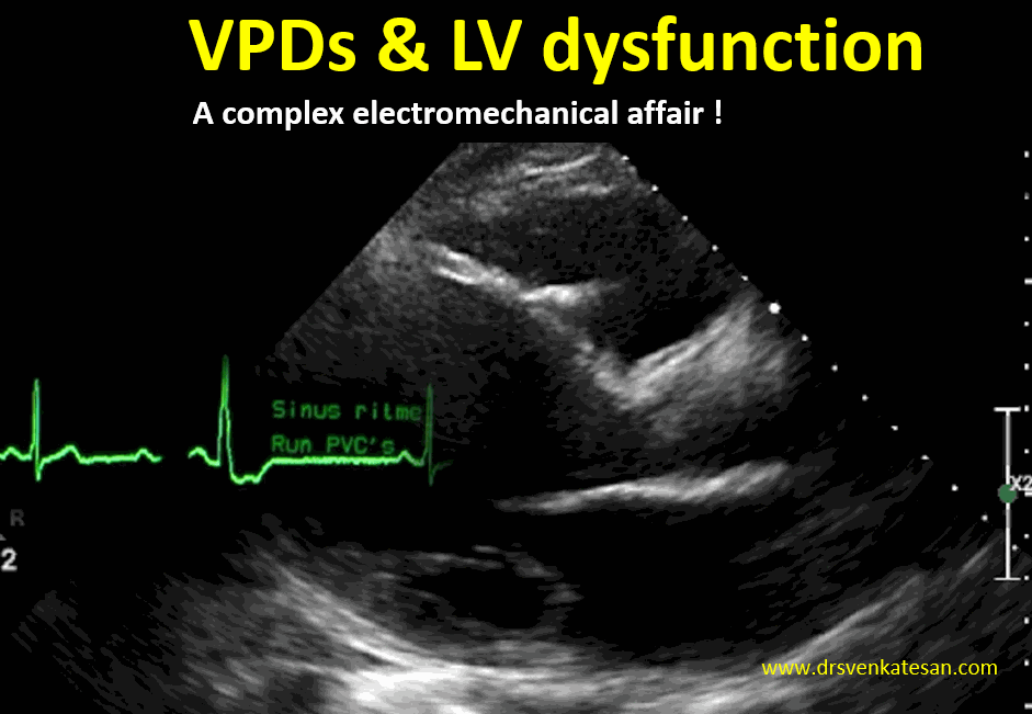
VPDs alter the way ventricle contracts by inducing wall motion defect. In fact, it is intermittent cardiac desynchrony. Generally, LV tolerates this well. When the number exceeds a critical level LV size, shape and contractility is affected.

We need to differentiate chronic tachycardia mediated LV dysfunction (Tachycardic cardiomyopathy) with VPDs per se. This differentiation we can only guess. 

* Some how cardiologists have not,  implicated RV dysfunction induced VPDS. My guess is, it is equally important. Logically,at least 30% of VPD in end-stage DCM must be attributable( and arise from) RV dysfunction.

How to confirm is it a cause or bcoz ?

The only way is to prove LV function improves to normal or near-normal with ablation of VPDs .This can happen only with primary electrical disorders.

Final message 

The link between VPDs and LV dysfunction is stuck in a complex two-way affair. Though a cause and effect component might be quantified to a certain extent, both can be coupled sequentially . “VPD promoting  LVD” &  “LVD begetting VPD”  is always a possibility. This is the reason, we are tempted to take on any VPDs,  which of course, is definitely not warranted.

  Two resources 

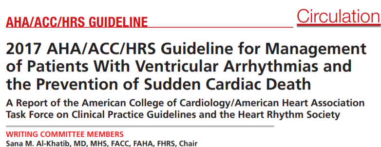
The HRS guidelines must-read for all fellows


This is a real state of the art lecture on VPDs straight from a world-renowned Dr Gacia  Courtesy : Methodist, Houston 

Read Full Post »

Surprised to see many of my colleagues, physicians and fellows are beaming with new pride even in this troubled corona times. Paradoxically, could see some fresh clinical sense in their approach to problems as well.

Each one of them has a story to tell

  • Sir, I could suddenly diagnose heart failure for the first time with my eyes and ears without NT- Pro BNP or E/E’ . I agree with you sir, textbooks seem to be right. There was indeed basal rales and JVP was elevated. I was astonished I could diagnose CHF clinically!
  • I feel proud, that I have acquired the rare expertise of giving fitness to an emergency appendectomy just by ECG.Its unbelievable, I had the courage of not asking for a pre-op echocardiogram.
  • Oh yes, it was a real flash of bedside brilliance. I could rule out Infective endocarditis, in a patient with prolonged fever, without caring to call for a bed side screening echo for vegetation. I am really proud of my acumen! I realised, Duke criteria is far more deep than our urge to have a glimpse on vegetation.
  • I can’t believe myself, yesterday, I was able to Ignore a 90% LAD lesion, first time in my life, by clinical means without FFR and QFR stuff.
  • This one is again from the echo lab. I refered a patient with aortic stenosis, for AVR, without bothering about all those low flow and high gradient conundrum. I was sure it was severe AS. The dense calcium and LVH were good enough to tell the complete story.

Finally, one of my senior colleagues, who lives half his awake time in cath lab, confessed to me. “Yes,Venkat, it’s all happening right in front of my eyes. Miracles based on absolute truths . I have since learnt the ultimate lesson in cardiology. How to treat, many of my CAD patients, without knowing coronary anatomy, that too without any major adversaries”

Postamble

After listening to these sobering stories , I got into a mid-afternoon nap, wherein, my good old professor came in my dream. He blessed me with his famous smile and hug for practicing and propagating clinical cardiology, as he taught to me.

But sir, I blinked, sorry sir, I don’t deserve your compliments. This newfound sense is going to vanish along with this dream. Blame it on the terrible corona times, which has forced us to deliver this low-quality care based on the antique clinical methods.

My mentor’s happiness was short-lived, as he realised these guys will soon be consumed again, by the glamor machines that runs medical science. He left silently, still pleading us to try our best.

Read Full Post »

I think it is an Invalid question. Whether you like it or not , medical science and philosophy are always bonded together and its relationship is eternal. It doesn’t make sense to separate them. I think we have misunderstood the meaning of philosophy. While science is presumed truths, philosophy is trying to believe in unknown truths. Philosophical truths are built-into every decision a medical professional takes.

If the expected natural history of any disease is science, unexpected deviations are philosophy. (RT PCR testing for diagnosing  Corona is science, why 90% of them are not infective and don’t transform disease is philosophy) When something is not seen or quantifiable like human immunity, it is a perfect example of concealed science or manifest philosophy.

Taking about what we think we know is science, Talking about what we really don’t know is philosophy. The term Idiopathic syndrome finds a  proud of the place in every specialty in medicine, Isn’t? 

 What will be your answer when your patient wants an assurance that a stent, you had just implanted will not get occluded in the next 6 months or so.“I don’t know, I cant assure you about that”  will be your most likely answer. (Though, we do it in style, hiding behind  the scientific hyperbole decorated with numbers,  also referred to as statistics) Please realize, this is the expression of medical philosophy in the finest form.

Final message 

My Impression is, philosophical truths should be liberally used in a regular fashion right from the first-year medical school to advanced specialty teaching. This seems essential as science in the current times suffers from too much sanctity. This has spilled over to the doctor population as well, and make them appear invincible. 

If only we realize science often trails behind the philosophical truths at least by a few decades, our patients will not be injured inappropriately and prematurely. Mixing science with philosophy in the right composition ( a perfect academic cocktail ) will bring out the best from the noble profession.   

Postamble

Can anyone guess, why scientists are given a doctorate in Philosophy degree  (PhD ) ?

Read Full Post »

Fortunately, indications for DC cardioversion in pregnancy is rare. A literature search suggests only about 50 cases are reported. I haven’t shocked electively in pregnancy but occasionally have come closer to it. In this current corona lockdown period, we had a call for a potential shock in pregnant mother with fast AF, which was again avoided by the optional  rate control measures.

Let us see, how often DC cardioversion might be necessary during pregnancy and few tips for its safety.

General principles

We know pregnancy can be pro arrhythmogenic. Most arrhythmias are non-sustained VPDs and APDs.They can be ignored if there is no structural heart disease, or at least postponed till delivery.

Drugs remain the mainstay.

The most common sustained arrhythmia in the young reproductive age group is SVT (AVNRT/AVRT) that can be managed with drugs like  Adenosine and beta-blockers. (Flecainide/Sotalol are found to be safe in pregnancy) . Though IV Verapamil is very effective, it has with some concern for fetus, so better avoided. Please, note many of the SVTs can be reverted with simple vagal maneuvres and oral beta-blocker /Verapamil. IV Digoxin has been widely used in RHD population for AF during pregnancy.

Mind you, even Injections Adenosine, Esmolol do have bradycardic potential and need to be given in monitored setting (No surprise, they are called medical cardioversion with the attendant risk) 

The universal antiarrhythmic drug Amiodarone still might come in handy in any refractory arrhythmia (Including AF) though it comes under the list of contraindication.(Safety of amiodarone in pregnancy) 

One important suggestion to make. Magnesium is a wonder antiarrhythmic drug, a membrane stabilizing agent through its indirect Ca + and K + blocking properties can be a powerful antiarrhythmic agent, especially in VT. This has a unique safety profile in pregnancy.We use it in eclampsia liberally  with the same action to suppress brain convulsions. (Cerebral tachycardia). Please consider IV magnesium  prior to considering  shock in VTs with dysfunctional ventricles as in peripartum cardiomyopathies et(Dose  to 2 g in 10mL of D5W over 1 to 2 minutes)

Consider DC shock only if there is hemodynamic instability.

Hemodynamic instability demands DC version. One practical issue is , what defines hemodynamic instability? In pregnancy, the systolic BP is already in lower normal due to systemic vasodilatory state. An HR>150 makes it further fall to around 90mmhg. This tempts us to label it as unstable. In this situation, we have to rely on patients’ symptoms to define hemodynamic instability. Never try to shock a comfortable pregnant women in whatever tachycardia she is in . (Including some VTs especially from outflow, fascicular, etc  ) Try to use drugs and get an expert opinion. to rule out subsets like cardiomyopathy, documented CAD, LV dysfunction.

When to shift to a cardiac facility?

This question crop up often. It is mainly logistic. May be in peripartum cardiomyopathy /Suspected ACS  with VT require special care.

 DC Shock checklist  & Precautions 

  • Biphasic shocks with energy levels 100 joules ( up to 200). Ideal to give single shock, ok to err on high energy  
  • Pads should be well away from the abdomen. 
  • Synchronized with QRS complex (Machine does this) 
  • In an unusual event of VF and cardiac arrest Defibrillation with 300/320 J (Here unsynchronised) 
  • Check the crash cart ready with essential drugs.
  • Keep cardiologist either on-call (Even a junior resident in labor room give immense confidence) 
  • Rule out  LV dysfunction or significant valve disease by echo (CAD can’t be ruled out though) If echo machine is not available ask the radiology or cardiology fellow to use the abdominal USG probe to document good LV contractility and gross EF% estimate.
  • If intramural thrombus is not convincingly excluded and there is AF and valvular heart disease, better to heparinse  and shock to avoid embolic events.
  • Temporary pacemaker support (Some of the cardioverters has transcutaneous pacing to tide over transient bradycardia that might occur post-shock) 
  • CPR readiness ( Extreme precaution !)
  • Fetal heart monitoring and Emergency cesarian readiness. 
  • Finally, most important consent with patient and family.

Is electrical Insulation of baby necessary or is it possible? 

 It’s not required. Fetus inherently tolerates stress better. Even if,  few joules reach the fetal heart inadvertently it may not mean much. What is, to be worried is maternal hypotension or bradycardia post-DC shock.

Impact on the fetus: Evidence?

The impact on fetal blood flow is not significant. This report from Taiwan  reassures there is no adverse effect by measuring umbilical artery flow (Yu-Chi Wang European Journal of Obstetrics & Gynecology and Reproductive Biology 126 (2006) 268–274)

While we consider DC shock during pregnancy is safe for the fetus, still, shock pads close to the abdomen, amniotic fluid being a good conductor of electricity at least one mother showed a sustained contraction of the uterus and fetal distress. This was possibly attributable to DC shock  Eleanor J. Barnes BJOG 2003 https://doi.org/10.1046/j.1471-0528.2002.02113.

Final message 

Most cardiac arrhythmias in pregnancy are carefully managed by non-electrical means. Of course, emergencies can’t afford to wait. Though two lives are at stake, it’s the mother’s heart that prevails over in drug selection and risk estimation. After all, it is her loving heart, that keeps the fetus alive.

I have seen Obstetrician anxiety (which spills over to attending cardiologist too!) can be extreme in such situations. I must admit, Obstetricians, are truly sincere warriors fighting at odd hours to protect the two delicate lives. After all, taking  responsibility brings the anxiety. Cardiologists must understand this and help them out in their difficult times.(without any super specialty ego !)

Reference

  1. Crijns HJ. Electrical cardioversion in healthy pregnant women: safe yes, but needed?. Neth Heart J. 2011;19(3):105‐106. doi:10.1007/s12471-011-0079-3


2.Finlay AY, Edmunds V. D.C. cardioversion in pregnancy. Br J Clin Pract. 1979;3:88–94

3.Oktay C, Kesapli M, Altekin E Wide-QRS complex tachycardia during pregnancy: treatment with cardioversion and review. Am J Emerg Med. 2002 Sep;20(5):492-3.

Which drugs are safe?

From BMJ 

Click to access pregnancy_heart_disease_v28_web.pdf


Further frontiers 

The DC shock from ICD experience

There have been a good number of women who got ICD for various indications (Commonly HOCM, long QT, ) who subsequently became pregnant, successfully managed during pregnancy.

(One rare study Andrea Natale et ll Circulation. 1997;96:2808–2812 documents at least 10 shock episodes documented in large series of 44 patients without any consequences)


Read Full Post »


A throbbing query …for so long

Got into this amazing lecture, that Infuses knowledge and wisdom about values in life, in just 14 minutes .Probably, It has more treaures than what we may get, in our entire life time as we search for truths.

I wonder, such thoughts can come only from God’s special messengers.Devdutt Pattanaik seems to be one. 


Read Full Post »

LA volume is one of the critical parameters that define (as well as determines the symptoms) both diastolic and systolic LV dysfunction.Still, we are tentative in the true estimates about the normal range of LA volume. (Upper limit 40ml/m²) .We are surprised to note, the difference between MRI derived and Echo measured LA volume showed a disturbing variation nearing 80 %.

How to measure  LA volume?

While the timing of LA volume measurement has not much controversy (end-systole), and the shape errors are largely eliminated by 3D echo, still why this variation?

It is a telltale error, of either including (or excluding ) the pulmonary vein ostium and complete blindness to LAA during LA border tracing. We know, LAA is physiologically, pathologically, and electrically is a critical accessory of LA. Still, we have so for excluded it from routine LA volume calculations. Is that a right-thinking in the overall evaluation of LA volume?

Normal LAA volume

Though the LAA volume is directly related to its size, shape much great confounder, since it precludes in arriving any mathematical calculation of volume from the area. Direct casts ofLAA and 3D echo to a certain extent will help measure LAA volume. MRI may also do the same.The normal LAA volume is calculated to be up to 20 ± 9 ml. Whatever be the  LAA  volume, one estimate suggests it will reach 25 % of total LA volume. This is very important to know. In fact, In significant LV dysfunction, LAA  is expected to stretch, efface, and dilate and contribute more to LA volume. Both static and dynamic LAA volume status also gives us an idea about potential thrombus formation risk.

(Ref : Measurement of Left Atrial Appendage Size by Transesophageal Echocardiography  Kazuko Yoshimoto et al. J Med Ultrason (2001). 2012 Jan.)

Are we justified to Ignore the LAA volume during routine LA function assesment ?

I don’t know. It may be wise to routinely add LAA volume to LA chamber volume to truly assess  1.Overall LA function, 2.Estimate the risk of thrombus formation and  3.Risk of developing AF. Meanwhile, its found LAA appendage volume might even approach that of  LA volume when it’s pathologically enlarged. (Left Atrial Appendage Volume as a New Predictor of Atrial Fibrillation Recurrence After Catheter AblationPedro Pinto Teixeira et al. J Interv Card Electrophysiol. 2017 August) 

LAA volume is Important for one more reason 

We are getting new data about dynamic LAA volume status during LAA closure.In fact , this particular study (JACC: Cardiovascular Interventions Volume 8, Issue 15, December 2015)  documented how LAA appendage balloons out during volume loading of LA.This study suggests we have to be careful about the hidden potential of LAA to expand and if ignored the device is likely to get dislodged with volume overloading. These observations make it clear we can’t isolate LAA volume when calculating LA volume. 

Final message

There is a strong case for measuring  LAA size & volume separately and preferably be added in the net LA volume Index. We can’t simply Ignore this vital and inherent part of LA , just because its called as an appendage. Of course, even a novice will rank LAA first,  as the pathological hot spot within the entire LA.

Reference

Everything about LAA

Giuseppe PattiVittorio PengoRossella Marcucci,The left atrial appendage: from embryology to prevention of thromboembolism  European Heart Journal, Volume 38, Issue 12, 21 March 2017, Pages 877–887, https://doi.org/10.1093/eurheartj/ehw159


Read Full Post »


If you think coronary artery ostia are exclusively related to LVOT, and they nowhere come closer to RVOT, we are seriously wrong for some hidden anatomical reasons. Though, Its a well recognised anatomical reality , we had to learn it from EP labs during the ablation of RVOT VT. The left main ostium is at equal (If not more ) at risk as we ablate across RVOT.The reason being the complex twist God conferred on the outflow tracts of heart.

The LVOT goes beneath and posteriorly travels from left to right, while the opposite happens for RVOT. This results in the net anatomical conundrum of RVOT and LVOT sharing common wall at some part.

Here is an excellent article that specifically looked into this issue of the relationship between RVOT and the coronary artery.

(VASEGHI, M., CESARIO, D.A., MAHAJAN, A., WIENER, I., BOYLE, N.G., FISHBEIN, M.C., HOROWITZ, B.N. and SHIVKUMAR, K. (2006), Catheter Ablation of Right Ventricular Outflow Tract Tachycardia: Value of Defining Coronary Anatomy. Journal of Cardiovascular Electrophysiology, 17: 632-637. doi:10.1111/j.1540-8167.2006.00483.x)

The following illustration from this paper reinforces this . The left main is just 4mm away from RVOT.(While, the distance from LVOT to left main ostium is much wider)

Final message

Never underestimate the risk of injuring the left coronary ostia when you manipulate catheters and devices across RVOT. This is especially true with RVOT ablations. There has been so many instances of injuring coronary arteries and precipitating ACS. Working within the right heart tends to give a false sense of safety, as if you are away from systemic coronary circulation. After all, both outflow tracts originate from the same embryological source (bulbus cordis) still, intertwined in adult life, not willing to leave its innate anatomical Intimacy that began and ended between 4 th and 8th week of fetal life.

  1. D. Biermann, J. Schonebeck, M. Rebel et al., “Left coronary artery occlusion after percutaneous pulmonary valve implantation,” The Annals of Thoracic Surgery, vol. 94, pp. e7–e9, 2012.View at: Google Scholar

Read Full Post »

Found a wonderful anatomy teaching resource. A succinct yet comprehensive lecture on Aortic valve .A must-read for anyone who deals with valvular Interventions. For the cardiology fellows , I can tell you can’t find anything better than this. Thanks to Prof Gregory M Scalia and the structural heart disease Australia. It starts with this one master diagram of Aorta in the center with all surrounding structures and goes on to ensure we watch this 25-minute lecture nonstop.

At the end of the lecture, you should be able to learn about.

  • The complex fusion points of LVOT with Aorta. Understanding them is critical as we deploy Aortic valve percutaneously.
  • Understand the illusion of Aortic annulus.
  • When it is an advantage to keep the aortic valve supra annularly.
  • The Implication of sharing of myocardial cells into the aortic cusp zone, and Aortic tissue into the myocardium.
  • How closely the conduction tissues located beneath the non-coronary cusp making it vulnerable with calcific spur Injury.
  • How pathology of Aortic root with its central location reaches the surrounding tissue.

These are just a few . . . It is a treasure out there.

Over to Prof Gregory Scalia

Please note ,the talk on Aortic valve begins after a brief conference Introduction.If you are not patient enough begin at 2.45mts.

Read Full Post »

Preamble  

The resting coronary blood flow (CBF) is about 5 % of cardiac output. It amounts to 250 ml /min (0.8 ml /mt/gram of myocardium ) It is estimated, blood flow across LAD is 50% . LCX and RCA share 25% each, depending upon the dominance. No need to say , the net return to coronary sinus  should match the CBF at rest or exertion.(Minus a small fraction contributed by  thebesain and vene cardia minimi flow, into the right heart chambers)

Great cardiac vein (GCV) is the venous cousin of LAD. It must receive and empty 125ml of deoxygenated blood every minute into the coronary sinus, if LAD flow is normal. When LAD microcirculation is obstructed as in severe obstruction , GCV will fill and empty  sluggishly. 

Let us move on from physiology to bedside.

Primary PCI & No -reflow in LAD 

We have painfully realized, no-reflow is almost a hemodynamic death sentence to the concerned coronary artery during  primary PCI. For practical purposes, no-reflow is common in left coronary circulation, that too in LAD.  Decades of research and experience haven’t really helped us to either overcome or treat this complication effectively. 

So, it’s worth considering experimental mechanical options.

  1.  The first option is, to forcibly open the closed microvasculature by pushing the debris across arterioles, venous capillaries and subsequently to the coronary sinus. This appears tricky as the high-pressure injection can be hazardous. This can be done either hand or even controlled pressure Injection. (It is believed few centers do this unofficially and found some success. I am not sure)


  2. The second option is again mechanical but uses negative pressure to suck from the far end of microcirculation. We can pull the debris into the coronary venous circuit by using vacuum suction deep inside the main stem of the coronary sinus or even the great cardiac vein, the venous counterpart of LAD. This appears to be more comforting to the coronaries at least theoretically. (Never under-estimate the power of vacuum as a biological Intervention.I recall tender newborns pulled out with massive vacuum pressure without any issues during my O&G days)


We have proposed this idea to scientific committee. It was rejected promptly as expected even prior to applying for ethical clearance. In fact, we were eager to try our hands on this, as a life saving modality in desperate situations. Now, It is an appeal to all those scientificaly  liberal centers to try this concept that can be made legal as a part of an official trial.

Antegrade vs retrograde aspiration

It is wiser to introspect, why most antegrade thrombus aspiration strategies in epicardial coronaries failed in STEMI .Still, what is the possiblity  it might work retrogradely? Let us hope this concept becomes a success and doesn’t add on to the long list of failed attempts to this ubiquitous hemodynamic entity.

Potential risk

Since the mechanism of MVO and no=reflo  is multifactorial, mechanical suction might help only in the clearance of thrombotic debris. Further risk of microvascular endothelial injury is real.

Final message

Many fellows write to me,  asking for some research ideas. Please acknowledge if anyone proceeds with this one (If you really believe no one has thought about this Intervention before)

Reference  for Interventions within Coronary sinus 

  1. EP guys do it , in a regular fashion with CRT.They also specifically enter the Anterior cardiac and great vein during ablation in VTs originating from LV summit .
  2. Coronary sinus aspiration done for contrast removal after angiogram to prevent AKI in renal compromised patients.(Ref Osama Ali Diab Circulation: Cardiovascular Interventions. 2017)
  3. Stem cell delivery has been done through coronary sinus (Wouter A. Gathier J Cardiovasc Transl Res. 2018)
  4. Coronary  sinus retrograde perfusion  has been tried for refractory angina (David P. Faxon Circ Cardiovasc Interv. 2015)


Read Full Post »

The Medtronic leadless Micra TPS pacemaker

In 2016, EP world saw a major breakthrough when Medtronic introduced leadless Micra TPS pacemaker. This device that looked like a small bullet, was implanted in the RV apex without the need for lead insertion and surgical pocket. It was real innovation but was not able to take off even after 4 years of marketing. The reason was simple. Though it was a smart device, it was a journey backward in time, as Micra TPS provided only non-physiological VVI pacing. (In the current era of multi-site and His bundle pacing)

Physiological pacing requires two are more leads.(Except single lead VDD, now obsolete AAI ) . Atria must be sensed for AV synchrony to happen. Atrial sensing is accomplished by

  1. A dedicated atrial lead as in AAI or DDD
  2. A floating atrial lead as in VDD mode

How does the leadless pacemaker attached to RV apex bring in AV synchrony without any add on leads ?

Medtronic, has come out with new add on to TPS ie “Micra AV” .The same gadget has been upgraded with a software to do atrial sensing. The accelerometer in the pacemaker senses the motion of the blood in early diastole followed by atrial contraction mediated S4 . This is sensed, and ventricular lead is set to fire after a programmed Interval.

Medtronic micr AV pacing 2

The initial experience appears promising. The results of the MARVEL study is published (Ref 1) However, there are important limitations. The atrial sensing function is not fully tested in real-life exertion. Further, It’s actually a form of mechanical sensing. The atrial electromechanical association has been taken for granted. The absence of electrical atrial sensing can mislead the ventricle. Currently, I guess it is a software patch that converts Micra TPS to AV . One more issue is, the soft ware consumes more energy and cut shorts the life of the pacemaker.

Reference

Read Full Post »

« Newer Posts - Older Posts »