After a hectic two years , let us pray & believe, the good old times will be back in our life.

Posted in Uncategorized on January 1, 2022|
After a hectic two years , let us pray & believe, the good old times will be back in our life.
Posted in Uncategorized on December 1, 2021|
If anyone asks to shortlist the best papers that were, ever published in clinical cardiology, I am sure, this one will reach the top ten. It was 1979, the field of cardiology is just waiting to explode. CAD was managed primarily with drugs and occasional CABGs. Coronary angiograms were an academic luxury. Both thrombolysis and PCI were unknown. Fortunately, Clinical cardiology was still alive and kicking. Dr. George Diamond and Dr. James Forrester from Cedars Sinai, New York worked together to bring this masterpiece. How and when to suspect CAD in the general population? For the first time probability was applied as a diagnostic tool.
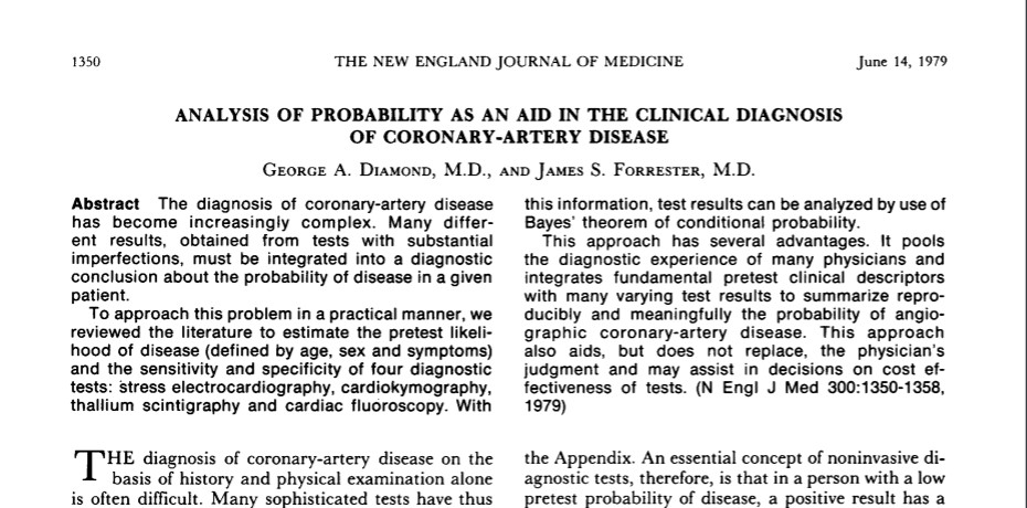
The paper begins by analysing basic clinical symptoms, risk factors, then gradually dwell deep into the population-based likelihood ratio, of CAD with the help of stress ECG, Thallium, and fluoroscopic coronary calcium. It finally ends up with a magical fusion of the Bayesian theorem into clinical medicine. It essentially taught us how to accrue scattered knowledge, clinical judgment, and diagnostic acumen among physicians in a community and aggregate them to a powerful statistical evidence base.
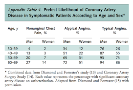
A popular Inference from the DK model still asked in cardiology boards
It’s more than four decades since this paper was published. There have been some concerns about DF classification in the current era. It was compared with the new Duke risk and found to be less valuable in the low-risk CAD population.(Wasfy MM, et all AJC 2011) The concept of pre-test probability deciding the diagnostic value of screening tests is very much valid. We need to recalibrate the DF scale for the current population and new generation screening methods like MDCT etc..(Gibbons et al Jamanetwork 2021 )
Forget the pros and cons, DF study told us the importance of clinical judgment in the decision making process. Now, we are living in a glamorous new world of cardiology. Cath labs have become our 24/7 office suits, always in hot pursuit for instant fix solutions. Still, we often find ourselves desperately blinking at the doors of EBM, for the elusive answers to some critical queries. Why the same intervention seems to work in one large study and totally go wanting in another? (MITRA-FR vs COAPT)
Where are we erring?
The problem is the way evidence is created. It is often made up of data collected from poorly framed questions and methods, which are incompletely collected or wrongly interpreted. I wish, Bayesian theorem derivatives also address the probability of how pure is the pre-test (research) evidence base available in a scientific community. The core of truth in statistical science lies in, how we understand and define the number needed to treat, (NNT) and the number needed to harm (NNH) with any treatment or diagnostic modality.
Final message
Artificial intelligence and machine learning are projected to be the next big thing in medical science However, the probability of machines prevailing over, human clinical acumen, backed by a sound knowledge base and observation skills appears very minimal. Let us see. Meanwhile, I wish every young cardiologist to go through this paper by D&F to get enlightened.
Reference
Further reading
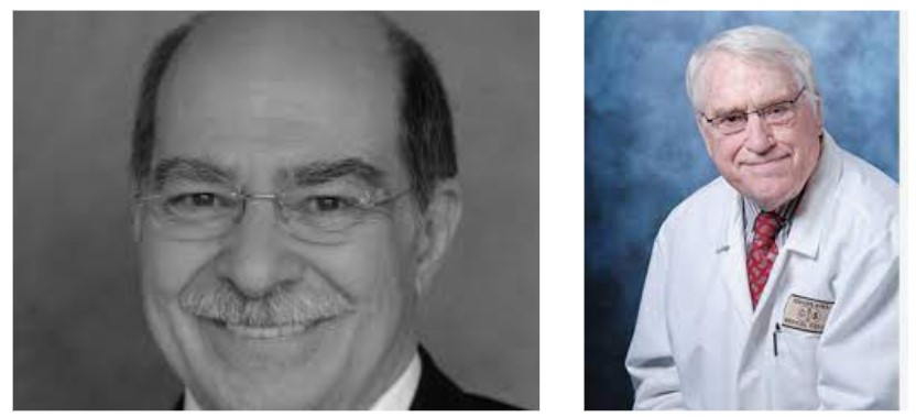
Dr. Diamond & Dr . Forrester
Posted in Uncategorized, tagged ACC AHA ESC guidelines for cad stable angina, cardiology journal club, COURAGE BARI 2D ORBITA ISCHEIMIA, ISCHEMIA trial, Limitations of ISCHEMIA trial on November 2, 2021|
There are a whole lot of scientists trying to jailbreak and expose the limitations of the hugely popular ISCHEMIA trial which put the emergency breaks in the way we used to practice cardiology. Not everyone is happy. While few are ready to apply the brake, many continue to love the accelerator.
This study (Ref 1) talks about an important issue. How much of the CAD populations in the real world will match the ISCHEMIA trial population? It concludes it is just 32%. It suggests caution to the cardiologists to understand this trial from a proper perspective. Don’t give too much importance, lest we may end up with Inappropriate non-intervention.
Sounds too good?
But is it real?
The authors of ISCHEMIA have countered this claim. (Ref 2)If we include all mild and moderate symptom cohort Ischemia study population is very much relevant in the true world and, actually constitutes about 68 % .
Final message
Clinical trials are the greatest gift of science and EBM. But why is that …it never fails to confuse us at each and every step, while we accumulate tons and tons of evidence.
I wish someone do a mega four-limbed study on what really our patients are getting in the overall CAD care.
I could easily guess the winning theme of this hypothetical trial. (That’s not good news though) However, response 4 If practiced in the right spirits would have the maximum impact on global cardiovascular health in terms of both healing and saving.
Reference
Posted in Cardiology Illistrations, Cardiology Illustrations, Cardiology teaching websites, Cardiology-Echocardiography, cardiomyopathy, hocm hcm, hypertrophic cardiomyopathy, Uncategorized, tagged Alcohol septal ablation, ASE posters on hocm hcm, best illistration for hyocm hcm hypertrophic cardiomyopathy, how to do genetic screening for hcm hocm, lvot vs mr gradient in hocm, myosin titin sarcomere mutations hocm, premyectomy echo work up on November 1, 2021|
Check out these two posters* for are a quick reference on HOCM with current updated evidence. The first one details about Echo evaluation. The second one illustrates the genetic screening flow chart of the HOCM families.
Some of the queries, you will find the answers from these posters are,
1. How to recognize Intrinsic mitral valve defect by MR jet morphology?
2. How to cross-check the true LVOT gradient from MR jet?
3. When to do a provocative test to document the LVOT gradient?
4. What are the standard pre-myectomy measurements by Echo?
5. How to screen a family member of HCM? Pros and cons of Phenotypic vs Genotyping screening
*Reference
Thanks to the American Society of Echocardiography.
Posted in Uncategorized, tagged cmc vellore cardiologist, Dr.A K.Abraham, great cardiologist of India, pioneering cardiologist india, tribute to dr k a abraham on October 24, 2021|
With deep regret, reporting the demise of Dr. KA Abraham, cardiologist, par excellence in our part of the world (Chennai, Tamil Nadu, India.) He was a man of great knowledge, wisdom, and integrity and was an inspiration to generations of cardiologists like us.

Dr.K.A Abraham 1942-2021
A life, that was fully dedicated to all those heart patients, many of them sick children in his den, the Railway hospital, Chennai for three decades. Though, never had the privilege to be associated with him either directly, nor does he knew me, one could feel instantly the greatness and simplicity in him. It’s 1994-95, vividly recall, the early morning classes, he took in the small auditorium in the Railway hospital,(For which, we used to rush to the far away Perambur from MMC in the peak Chennai traffic). His passion for teaching the basics of cardiac catheterization was phenomenal. Somehow, I used to think he was in the league of the Nobel trio of “Forssman, Richards & Cournand” who invented cardiac catheterization.
Those were ordinary days when the quest for knowledge and teaching was pure and Dr. Abraham was one of the great souls in pursuit of genuine and quality cardiac care. No surprise, his services were recognized and conferred a top award of India, Padmashree.
Yes, India truly lost a pioneering cardiologist.
Reference
https://en.wikipedia.org/wiki/K._A._Abraham
Posted in Uncategorized, tagged anti fibrinolytic system, thrombolysis on October 8, 2021|
Background
“Your husband is really lucky, his heart attack got spontaneously aborted. His ECG is near normal now. The angiogram is normal.No lysis, no stent is required. He secreted his own TPA and got rid of the clot. We will discharge him to tomorrow.”
Thank you very much, Doctor. How did this happen, doctor?
Don’t thank me. Definitely, I don’t have an answer. Spontaneous successful thrombolysis (Ref 4) happens up to 15 % of ACS. All I can say is he has a very disciplined fibrinolytic system backed up with an agile mast cell and basophil function.
Vascular events: Pathobiology
Vascular highway accidents that happen due to the sudden freezing of blood in any vital organs consume more human lives than any other disease. This can happen with or without a trigger from the vessel wall, the phenomenon which Virchow’s taught us a century ago.
By the way, who is keeping the 5 liters of blood in our body in a fluid status life long?
Blood coagulation and lysis is a fascinating balance of two pairs opposing hematological forces.
* Among the blood component cells platelet is the only cell that plays a critical role in the clotting process. Paradoxically the fiery red clot, that is loaded with RBCs has a little direct role in the clotting process while biochemical molecules like fibrinogen, thrombin plays a major role. The fact that blood is liquid forever, tells us a compelling fact, that innate anticoagulation and the fibrinolytic system are more critical than the extensively understood clotting mechanisms.
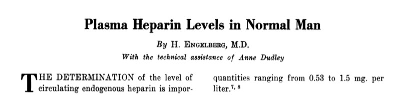
Are you aware we all have some amount of naturally secreted heparin in blood?
Circulation, Volume XXIII, April 1961
Which cells secrete heparin?
Basophils of blood and mast cells continuously secrete heparin that keep the blood fluid. Heparin is normally present in human plasma in values ranging from 10 to 24 units percent (1 to 2.4 mg. per liter). The range of average values is from 1.53 to 1.77 mg. The native heparin secretion by mast cell population is directly related to the anticoagulation activity (Bill Eksp Biol Med. 1984 Feb;97(2):131-4. Russian. PMID: 6230117.)
Where does TPA come from?
TPA normal levels not only confers the capacity to spontaneously abort a vascular event, but it is also a critical determinant of how streptokinase is going to act as this drug can’t act alone it simple accelerates the action of Tpa complex
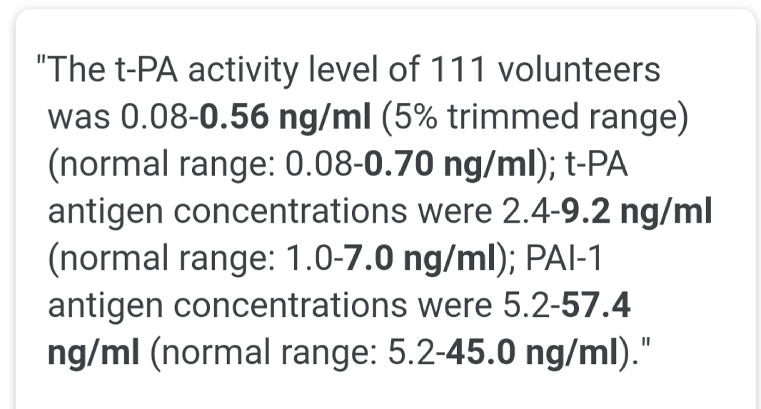
Mind you, the basal levels of TPA are injected instantly into the coronary thrombus with a zero-second time window. Now guess what is the potency of this enzyme cascade and who directs it?
Final message
If we think with a hematologic acumen, all vascular accidents are triggered by temporary aberrant behavior of blood or the interaction of its components with the immediate tissue interface. It is the inability to either prevent or fight the sudden occurrence of thrombus that is a primary problem than the tendency of the blood to clot spontaneously. The basal levels of natural Heparin, TPA, or anti thrombins and the way in which they get activated are much more important than the procoagulant forces, which we tend to blame often. (the famous vulnerable plaque vs vulnerable patient need to be reignited)
I think it’s time we dwell deeper into the native lytic mechanisms. This will throw an important vision on how we can replicate it in a pharmacological way. If only we have a drug that melts intravascular clots locally, vascular deaths of many vital organs can be prevented. The research in newer lytic agents has almost ceased & needs fresh Impetus.(TPA is a 25-year-old Invention)
Reference
Postamble
I think we have a problem with our basic science teaching methods. Tried a fact check. When I asked a few passing-out medical graduates, they could rattle everything about clotting factors but struggled to recall any anti-clotting mechanisms. We are still a long way to go, understanding the mysteries of how the blood keeps itself fluid, but always on the alert mode to clot whenever necessary.
DIC: The sine-qua-non of lytic dysfunction
DIC is one of the famous diagnoses in critical care medicine. It is a perfect example of defective fibrinolysis. (Also referred to as acute fibrinolytic shutdown*) classically occurs in septic shock and related conditions. We still lack good criteria to diagnose non-overt DIC-prone patients. A good review was written in 1999, still helps us understand the core concept.
*Both shutdown as well as Inappropriate activation
Levi M, Ten Cate H. Disseminated intravascular coagulation. N Engl J Med. 1999;341(8):586–92.
Posted in Diabetes and Heart, lipids lipid metabolism, Medical ethics, Uncategorized, tagged diabetes and statins, JUPITER study, risk of diabetes with statins, statin induced diabetes new onset and progression, statin side effects on October 5, 2021|
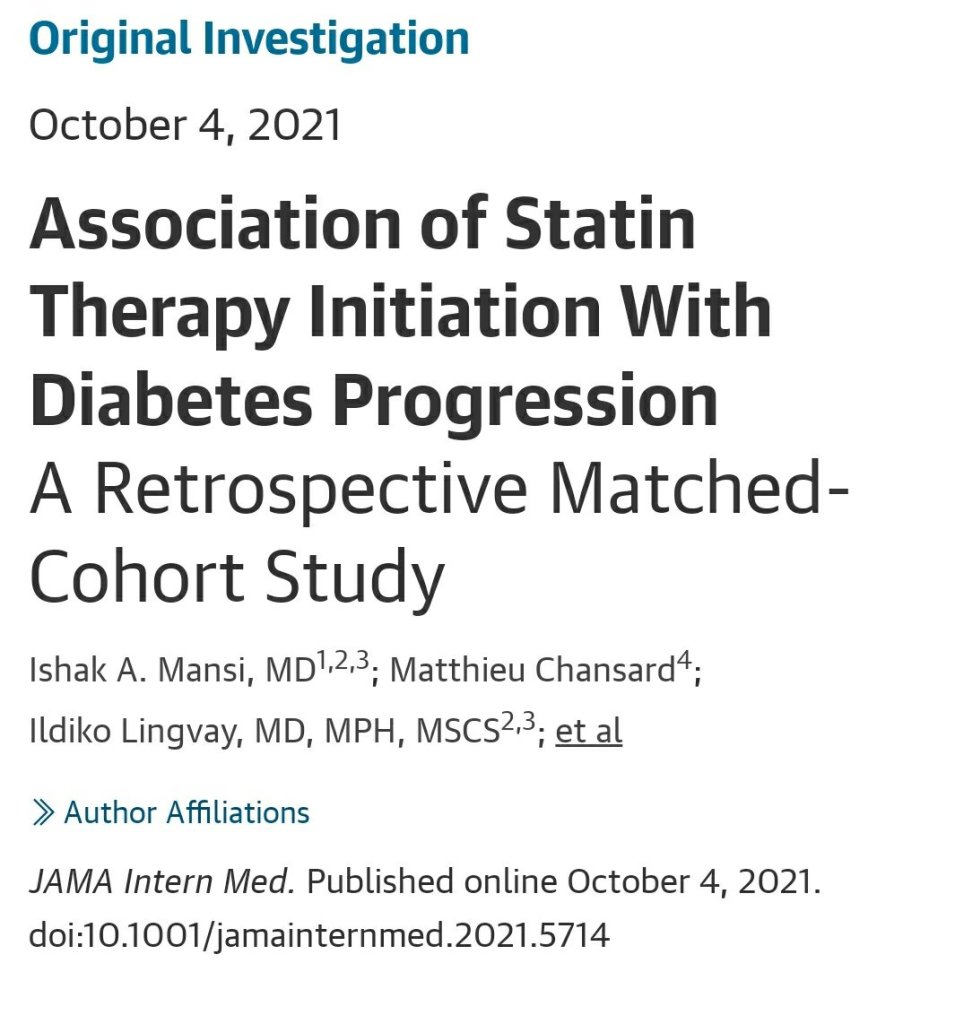
This is not a breaking news story. It’s the same old secret that was exposed in JUPITER trial with Rosuvostatin 14 years ago. Yes, I am talking about the relationship between the usage of statin and the occurrence of diabetes. Now, we have this huge study on possible diabetes progression with statins. It’s not from a small journal to ignore.
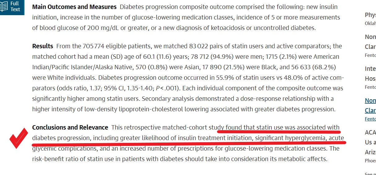
83 thousand patients data, the world’s largest series on link between statin therapy and diabetes.
This study has this to conclude
Diabetogenic statins
Something* happens as the statins antagonize the HMG COA enzyme that resides within the delicate membranes of the endoplasmic reticulum inside the most specialized cells in our human body, called hepatocytes.
*What is that something?
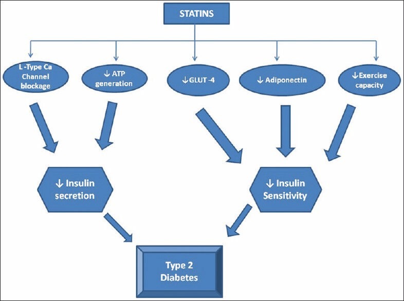
Image source Umme Aiman et al Journal of Pharmacology and Pharmacotherapeutics 5(3):181-5DOI: 10.4103/0976-500X.136097
How to go about this issue?
With-holding statin in as many as possible is the best thing for such diabetic (non-diabetic?) patients. But, the more pragmatic option is to ignore these negative studies, and instead intensify diabetes management if it worsens. After all, we can’t afford to lose the prodigious evidence-based cardio-vascular protective effects of statins and earn the wrath of our patients and peers you know!
Further Interest
Posted in Pregnancy and heart, pregnancy and heart disease, Uncategorized, tagged esc ropac zahara pregnancy, heart disease complicating pregnancy, indication for lscs in hcm hocm, lscs or normal delivery in hocm hcm, pregnancy in hocm, pregnancy in hypertrophic cardiomyoapthy, spinal anesthesis vs ga in pregnancy on August 27, 2021|
Background
Yesterday, my fellow informed me about a frantic call for cardiac fitness for an emergency cesarian section in 24-year-old woman with hypertrophic cardiomyopathy, who is asymptomatic and has a 20mmhg gradient across LVOT.
“Was she in labor”?
“No, she is 36 weeks term.
“Why LSCS? Why emergency”?
“I don’t know sir. Let me discuss and come again”.
HCM in pregnancy: An approach
Hypertrophic cardiomyopathy is a specific genetic disorder of myocyte (myosin and others) within the sarcomere. Though uncommon in pregnancy it raises considerable anxiety to the patient, family, and the obstetrician.
Hemodynamics
Though we tend to worry more about dynamic LVOT obstruction, it is actually the restrictive physiology of LV myocardium that might cause more concern. Three key variables operate in this entity namely preload, afterload, and contractility that determine the cardiac hemodynamics and possibly the symptoms. We know the classical consequence of pregnancy is a fall in systemic vascular resistance( SVR) ie afterload.
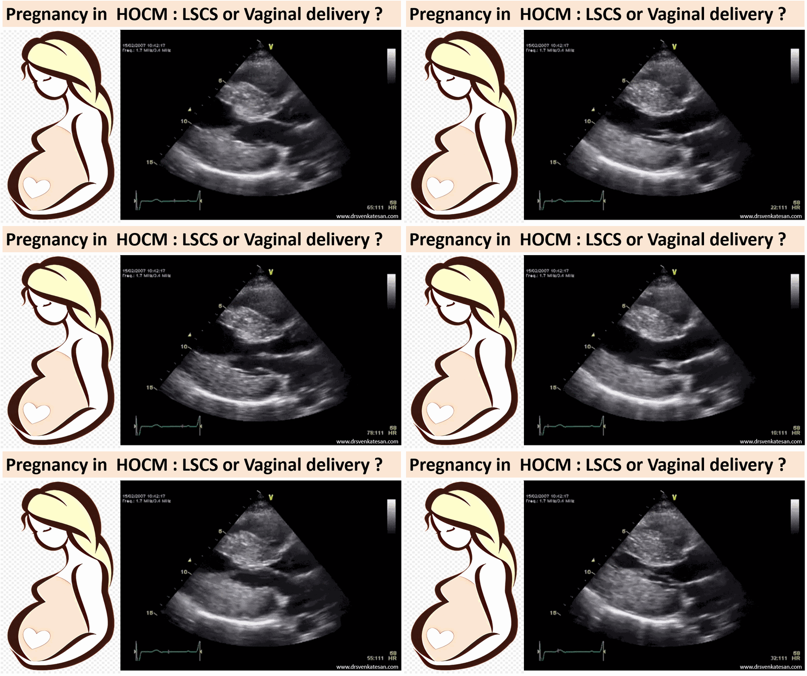
In pregnancy, there is a complex interaction between these three parameters along with heart rate. Fortunately, the net effect ends up favorable for LV performance. This is made possible because a major compensation occurs by a 50% increase in blood volume that effectively counters the deleterious effect of fall in SVR on LVOT gradient. (If mitral regurgitation is significant, the fall in SVR actually may help reduce regurgitant fraction especially if its intrinsic defect )
Maternal outcome
Is good (if not excellent). Maternal mortality reported in the literature, is gradually coming down (0 to .5% in various series) However, about 15 % of HCM patients with gross LVH or obstruction, may develop pulmonary congestion in the third trimester. In some patients, VPDs, nonsustained VT, even AF can lead to some tense cardiac consultations but are usually innocuous. I am not sure about the sudden death in pregnancy. I guess it should be negligible, unlike the non-pregnant HCM.
A mystery learning point
It is surprising both fetal and maternal outcome is little related to the severity of LVOT gradient (Ref 2)
Indication for cesarian
Fetal outcome
Premature birth, stillbirths, low weight are little more common than normal pregnancies. Fetal bradycardia due to beta-blockers has been noted but not troublesome.
What is the role of cardiologist?
The precise answer is “minuscule role”. I can vouch for this from a personal level. ( Consults are meant only for bringing some comfort to the obstetrical team). Active cardiac interventions are rarely required or rather desired. (Of course, patients who have significant symptoms, operated for HC, on OAC for AF, the rare ones with ICD needs expert care)
Final message
*It is worthwhile to note other forms of severe LVOT obstruction like valvular, supra valvular stenosis, and Aortic pathologies like Marfan, coarctation aorta are serious entities that deserve prompt cesarian sections.
Reference
2.
https://academic.oup.com/eurheartj/article/38/35/2683/3811991
Posted in Uncategorized, tagged ectatatic coronary rca, guidewire angioplasty, oac noac for coronary ectasia, plain balloon angioplasty, primary pci on August 22, 2021|
Inferior STEMI, and see the first shot in RCA below. The patient was pain-free and hemodynamically stable at the time of the angiogram. (Don’t wonder how this is possible, defying the fundamental rules learned from animal experiments after acute ligation of the coronary artery)
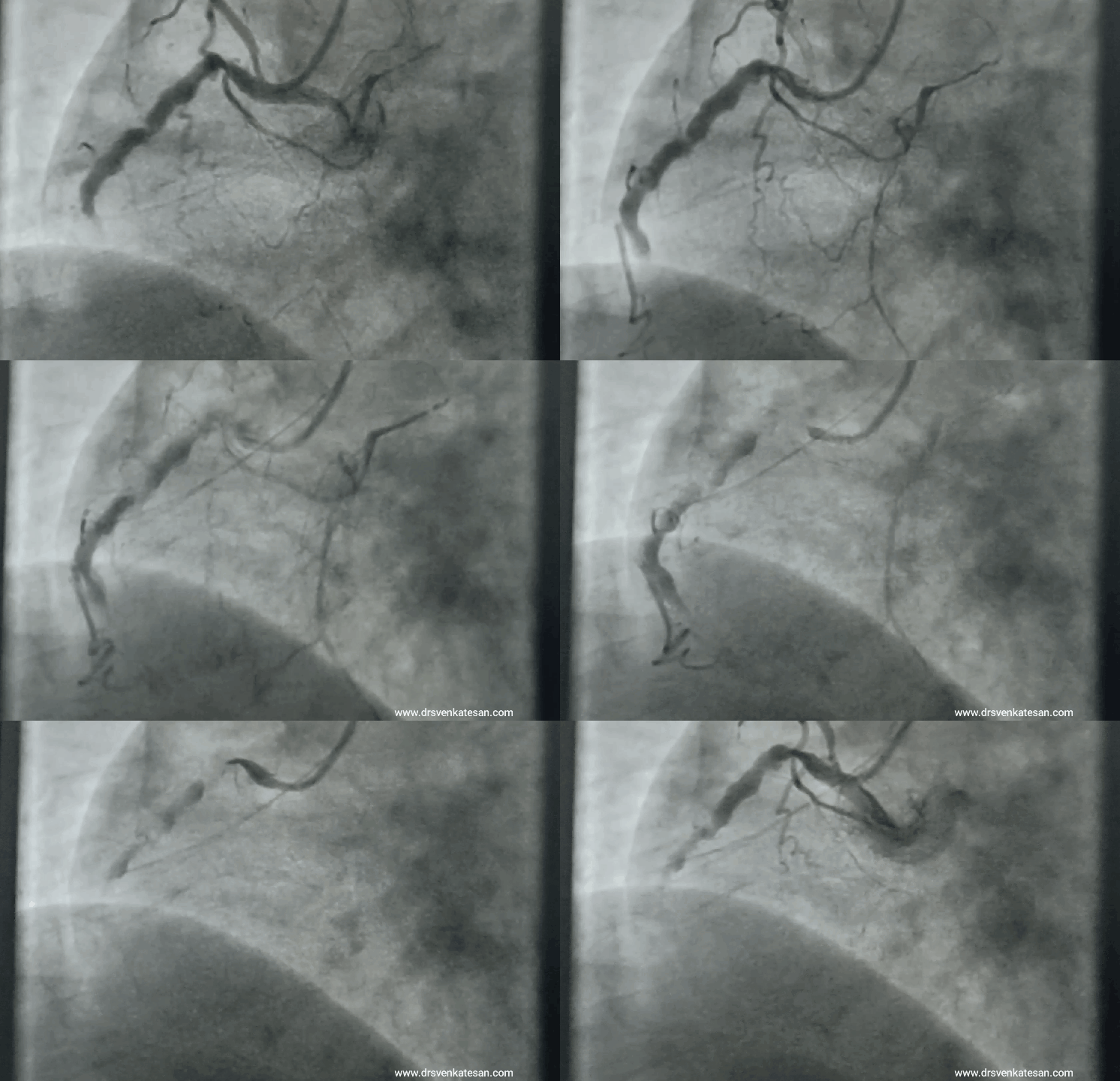
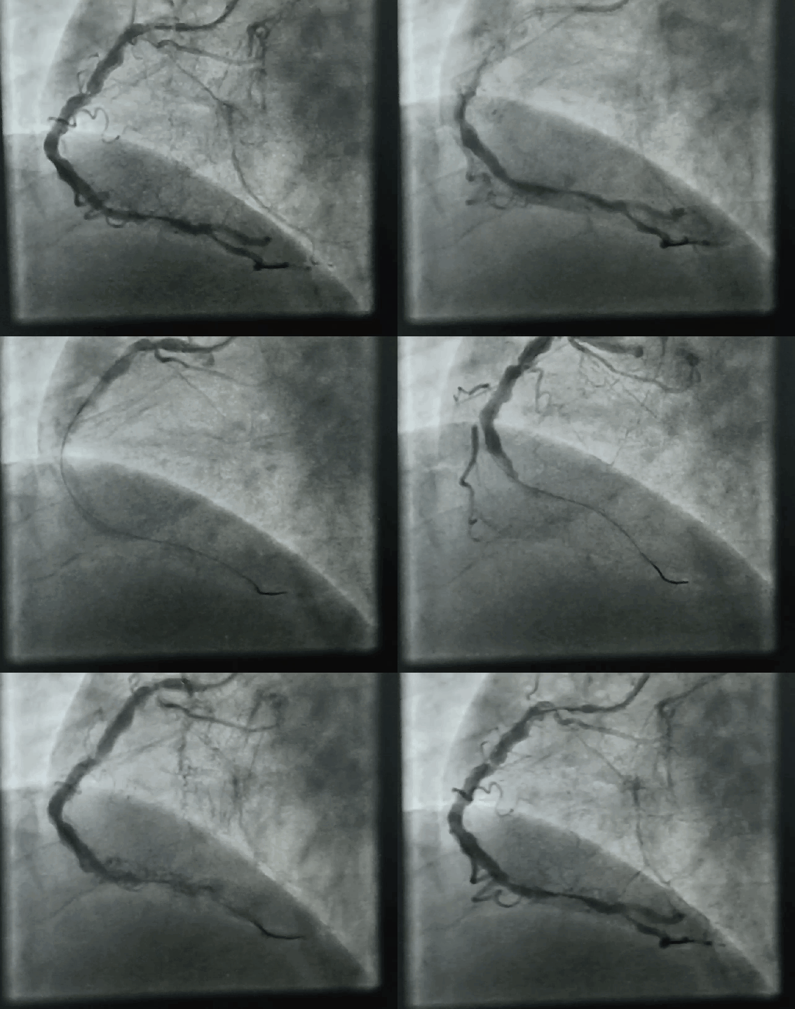
What was done? How is the patient?
Nothing was done & nothing happened to the patient as well. Just guidewire was crossed and few minutes of balloon touch-up work. Did the patient improve? Can’t say anything because he was fine even with this total occlusion.
Lessons to be learned
* By the way, who are all bothered to know LAD anatomy in this patient. Is it surprising the RCA is sending collaterals to the left side in its hour of crisis? Yes. LAD had chronic sub-total lesion as well.
Reference
2.
Posted in Cardiology - Clinical, Cardiology -Definitions, cardiology- coronary care, Ethics in Medicine, Uncategorized, tagged ACC AHA ESC Guidelines for multivessel cad, angina related artery ara vs ira, approach to tvd, cabg vs pci vs medical maangement ov cad, Chronic stable cad, courage ISCHEMIA ORBITA BARI 2D trials, IRA vs ARA coronary, triple vessel disease on August 20, 2021|
Yes, it is a triple vessel disease, with one tight lesion and at least two other significant lesions. One of them appears diffuse as well.

Representative Image: Source courtesy DOI: 10.14740/cr548w LicenseCC BY-NC 4.0
“What to do next?. Is he symptomatic? Yes. Definitely has significant angina” but LV function is normal.
“Ok then. If you are daring enough, ask this question”.
Which lesion is causing angina?
No easy answer at all. Try looking for some clues right from history, ECG, stress ECHO, meticulous assessment of individual lesions. Realize, even sophisticated imaging like SPECT, PET functional MR, may not help much either.
Oftentimes, we need to use the lean resources of collective common sense and clinical acumen.
Want to get more confused?
Finally some easy options.
Which lesion is causing angina? Never entertain that troubling question at all. (Need not squeeze your coronary intellect you know )
Consider every lesion as important
Final message
Which lesion is causing angina? is indeed an important query one should raise. This paves way for selective focussed PCI in deserving lesions alone. However, when dealing with complex lesions subsets. the most pragmatic way as of today is to educate the patient and include them in the decision-making process (Never forget to offer medical management as a permanent option, especially if there is no critical LAD disease, and say thanks to ISCHEMIA/COURAGE/ BARI 2D.)