Feeds:
Posts
Comments

Posts Tagged ‘hypoxic spells’

Catheter based interventions in TOF  has caught the imagination of  Interventional cardiologists.decades ago. (Quereshi reported first in 1988 Royal Liverpool hospital ) .Somehow it could not develop into a full-fledged modality. The key issue in TOF  is,  RVOT obstruction is infundibular with some degree of valvular involvement. While the valvular component is amenable for easy correction by balloon, the infundibular stenosis requires some form of cutting or splitting. Embryologically,  the malalignment of IVS is the primary mechanism of obstruction. The balloon catheter is will find it difficult to tackle the alignment defect. .Obviously, surgeons can do a comprehensive RVOT reconstruction.

Things are beginning to change. Cutting balloons are available. Various dedicated VSD devices are being developed. Closure of large sub-aortic VSD  followed by  RVOT dilatation appears challenging task but distinctly possible in the near future.

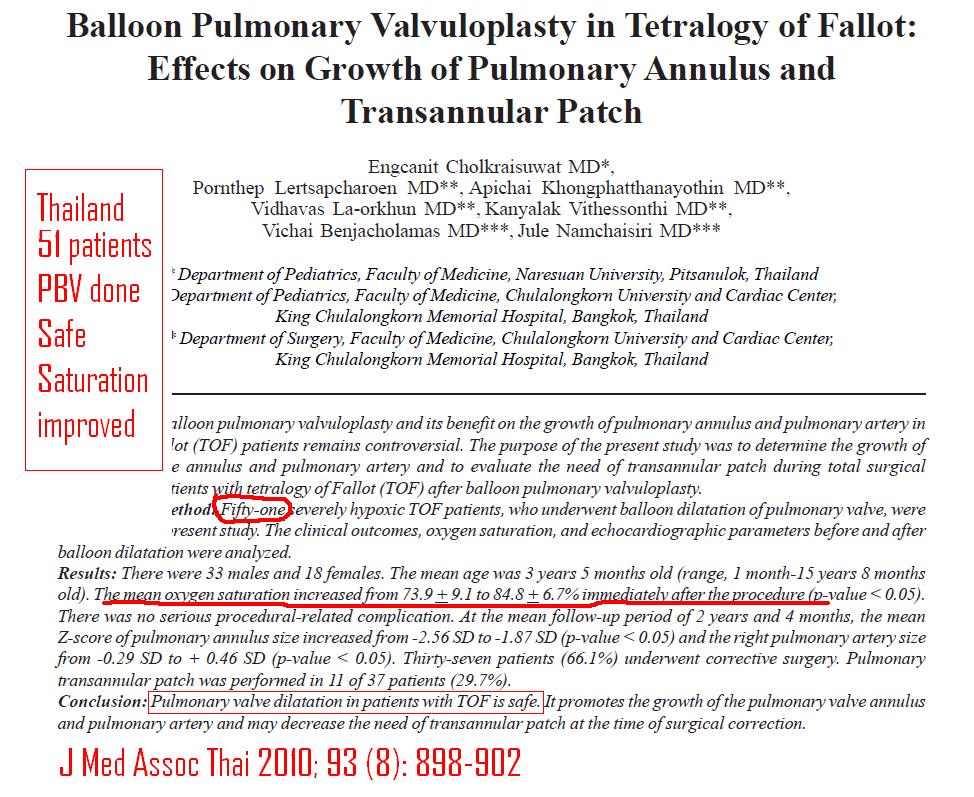
Few cases of palliative RVOT dilatation with a balloon  in critical TOF  is been attempted We hope, in the coming decades at least simple forms of TOF are conquered by the interventional cardiologists!

Hardware: A small profile  coronary  cutting balloon  from Boston scientific .

What is in store for the future ?

3D printing of live heart and designer device or deployable patches for the malaligned VSD is possible. Currently, intracardiac ultrasound would assist the procedure.

RVOT reconstruction with RVOT stenting and percutaneous valves (Melody or Right sided TAVR equivalents) is already been done in post-ICR residual obstructions or late RVOT failure

Coronary cutting balloon flextome tof pulmonary valvuloplasty coronary hard ware

Flextome -Coronary cutting balloon

Balloon pulmonary valvotomy for tof tetrology of fallot

balloon angioplasty for TOF cutting balloon

pulmonary valvotomy in tof tetrology

pulmonary valvotomy in tof tetrology 3

 Other References

1.Boucek MM, Webster HE, Orsmond GS, Ruttenberg HD. Balloon pulmonary valvotomy: palliation for cyanotic heart disease. Am Heart J. 1988;115:318-322.

2.Qureschi SA, Kirk CR, Lamb RK, Arnold R, Wilkinson JL. Balloon dilatation of the pulmonary valve in the first year of life in patients with tetralogy of Fallot: a preliminary study. Br Heart J. 1988; 60:232-235.

 3.Parsons JM, Ladusans EJ, Qureshi SA. Growth of the pulmonary artery after neonatal balloon dilatation of the right ventricular outflow tract in an infant with tetralogy of Fallot and atrioventricular septal defect. Br Heart J. 1989;62:65-68.

4.De Geeter P, Weisburd P, Dillenseger P, Willard D. Valvuloplastie pulmonaire percutanée palliative dans les formes néonatales de tétralogie de Fallot. Arch Fr Pediatr. 1989;46:117-119.

Read Full Post »

During  clinical examination of cyanotic congenital heart disease(CHD) ,  the major  task is to differentiate conditions with reduced  or increased pulmonary blood flow .

When a child with  CHD  is presented in clinical examinations , students are often asked to arrive at  the diagnosis  from history , physical examination before going in for ECG, X ray  or  echocardiography.

History,  surprisingly can  suggest  the  correct diagnosis in many (Most ?)

Reduced pulmonary blood flow is often associated with

  • Cyanosis   appearing with  /or worsening with   exertion*
  • Hypoxic spells.(Almost always occur in reduced pulmonary  blood flow )
  • History of squatting( Majority in reduced pulmonary flow)

Relief  of dyspnea   by assuming squatting position  convey   important hemodynamic information. It implies  there is significant reduction in pulmonary blood flow in standing posture , that  gets corrected  in the squat position.For squatting to improve pulmonary blood flow there must be a communication between right and left heart .This is most often due to a large VSD, rarely an  ASD .

Related article : How squatting relieves hypoxia in TOF ?

*Note :  Cyanosis  is  not  specific for reduced pulmonary  blood flow. In fact , simple reduction in pulmonary blood flow per se , cannot result in significant cyanosis .There need to be admixture /or right to left to shunt to produce cyanosis .Cyanosis in  pure admixutre states like TGV, Single ventricle , Common AV canal , Common atrium TAPVC,  are less Dependant on the reduction of  pulmonary  flow. In these situations RVOT obstruction if  present  will aggravate the baseline cyanosis.

Examination

Apart  from direct evidence for reduced pulmonary blood flow , lack of evidence for increased pulmonary  flow could  often mean ,  we are  actually  dealing  with  reduced pulmonary blood flow.

The following are the clinical clues to suspect  reduced pulmonary blood flow.

  • A quiet precardium*
  • A inconspicuous pulmonary component of S 2
  • Generally if  S 2 is well split  and both components are well heard it is highly likely the  pulmonary  blood flow is not reduced.
  • Lack of  pulmonary  arterial pulsations
  • Absence of mid diastolic  flow murmurs  in AV valves
  • Presence  of continuous murmur in a patient with cyanotic CHD almost always mean   reduced pulmonary flow and the lungs are perfused by alternate arterial collaterals (MAPCA)

* A silent  heart is the hall mark of Tetrology of Fallot which constitutes 80% of all CHD with reduced pulmonary blood flow.

Read Full Post »