Feeds:
Posts
Comments

Posts Tagged ‘ostium secundum asd’

We are taught embryology of the heart, right from the first year , when we enter the medical school.We learnt about various holes of the heart in a  sincere way .After 15 years or so , it is fascinating for a fellow to become full-fledged structural interventional cardiologist , and close an ASD with an Amplatzer device with absolute ease.

Can’t ignore the basics

Meanwhile, how many of us are aware, there is a big disconnect between basic science and the cath lab cardiology. When I ask my fellows,  what is the origin  ostium secundum ASD ? majority come  with wrong answer. Very few of us have time and interest to go deep into the different layers  of IAS development. Now, let us be aware,  there is a host of errors in the way we have understood the embryological basis of ASD.

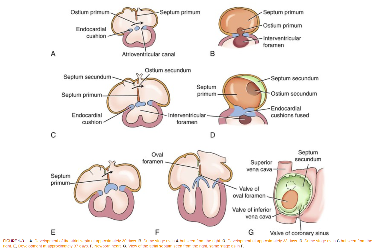
Image source : https://radiologykey.com/embryology-and-physiology-of-the-fetal-heart/

Misnomers galore

The first one  : The most common type of ASD, what we  call,  it as OS ASD , Is actually a defect in septum primum.

The second  is OP ASD is not due to defect in septum primum , but due to defect in AV cushion.

The third : The so called sinus venosus type of ASD is not an ASD at all, where IAS is totally intact.it is just an vascular Unroofing, between PV and SVC.

The fourth and an ultimate shocker  Embryologically, there may not be anything called true septum secundum, it is just interatrial fold, we named it as septum secundum and include it as apart of part of IAS .

The traditional NameWhat it Sounds LikeWhat it Actually Means Anatomically
Ostium Secundum ASDA defect in the septum secundum.A hole or deficiency in the septum primum (the fossa ovalis floor).
Ostium Primum ASDA defect in septum primum, that comes lower in IAS than OS ASDIt is an endocardial cushion defect at the AV junction , to precise they are defects of IAS rather Partial AVSD).
Sinus Venosus DefectA true hole within the interatrial septal wall.An unroofing/missing wall between the pulmonary vein and a vena cava (SVC or IVVC) outside the true septum.
Septum SecundumAn ingrowth of septal tissue left of
septum primum , that forms the upper part of IAS
It is not true component of IAS. It is a thick in-folding of the outer roof wall (interatrial groove) pushed downwards and incompletely fuse with septum primum, forming the fossa ovalis in the process.

Final message

A hole is a hole is a hole.If you have a device close it, can’t waste time to bother about the origin of it . May be you are right as a restless Interventional cardiologist. 150 years of congenital heart disease , thousands of literature and advanced imaging ,interventions, can’t be Ignored. Let us  be aware of the reality of  embryology and how we are still following the old cheat sheets .Fellows please make a note of it.

Read Full Post »

Spontaneous closure of VSD is  a well recognised  phenomenon, than ASD  though both happen in equal frequency.The simple reason being VSD is a noisy disease , ironically the smaller the size of VSD  more noisy it is . Hence  it is rarely  missed  while ASD is largely silent in children. For this reason  it is  possible ASD may be the most common congenital disease .

Natural history of ASD(OS) closure

  • ASDs of size 3-4 mm 100 % will close by 3  years
  • Bulk of the ASD < 8  mm close spontaneously by 5 years.
  • ASD> 10mm is unlikely to close

Factors that determine spontaneous closure

  • Apart from size and location
  • Closure  is accelerated by remnant of flap of foramen  ovale
  • Fenestrations and  Septal  aneurysms also  favor spontaneous closure.
  • Margins  of the defect if rough  triggers fibrotic reactions

spontaneous closure of asd

Why SVC and primum defect do not close easily ?

Plane of ASD  secundum is single and  bridging of tissue is possible .

Sinus venous and  primum defects exhibit  holes which run in multiple planes hence approximation not possible . They also do not  have a valve mechanism.

Un-natural history of ASD

In the current era, one more  force interferes  with spontaneous closure of ASD . It  comes from the  hyper trained aggressive Interventional cardiologists who compete with the nature and easily prevail over it !

Reference

asd spontaneous closure

Read Full Post »

This is a relatively common abnormality of IAS. It is  often observed  as  IAS bulging  into left  or right atrium  in routine echocardiogram.If this happens without  atrial hypertension it is termed as IAS aneurysm .

This is due to valve of foramen ovale bending into the RA/LA*  cavity for various distance. By definition , the radius of curvature of  the bulge should be more than 10 mm to label it as IAS aneurysm.

*Bulging into RA more common

Click on the Image to see the animation

General features

  • Mostly a benign entity.
  • More often observed  in  association with PFOs or ostium secundum ASD.
  • When occurs in isolation does not result in any shunting across it
  • The septal bulge can be static or  dynamic . It could swing  into LA, RA, and back to LA or vice versa.
  • Anatomically 5 types are proposed.
  • Multiple fenestration in the aneurysms have been noted.
  • Aneurysm  formation may aid in spontaneous closure of ASD.

Clinical  implications

  • IAS aneurysm tend to aggravate  stasis of LA  blood flow and predispose to minute LA clots and systemic thrombo embolism .
  • IAS aneurysm can act as an arrhythmic focus , generating focal atrial tachycardias.
  • A non ejection click  may be occasionally heard as  the IAS aneurysm  bulges and tenses within LA/RA cavity .

Reference


1 . Olivares -Reyes A, et al. Atrial Septal Aneurysm: A new classification in 205 adults. J Am Soc Echocardiogr
1997;10:644-56.

2. Longhini C, et al. Atrial septal aneurysm: echocardiographic study. Am J Cardiol 1985;56:653-67.

3. Gondi B, Nanda NC. Two-dimensional echocardiographic features of atrial septal aneurysm. Circulation 1981;63:452-57

4. http://www.fac.org.ar/revista/00v29n4/congreso/premio3.PDF

Read Full Post »