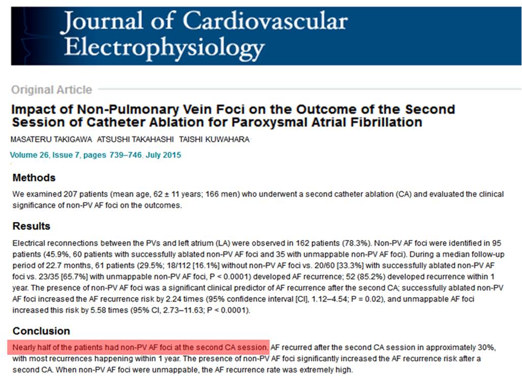
Feeds:
Posts
Comments

STEMI occurs due to acute occlusion of a coronary artery  (ATO) which needs emergency opening at the earliest, ideally within  3 hours , or up to 12h . The opening shall be either by pharmacological / catheter means  or both. After 24 hours opening  a ATO has questionable benefit unless the patient is hemodynamically unstable or symptomatic.

What is a CTO ?

Traditionally we believe 3 months is the period to call a coronary occlusion as chronic.(Previously it was 6 months) This time frame was considered appropriate based on our understanding of the infarct process , that may take up to 3 -6months for complete healing of infarct  .This 3 month period is arbitrary as the chronology of intra-coronary lesion organisation  is different from myocardial infarct healing. Its worthwhile to note the chronicity of  a coronary lesion and its morphology is nothing to do with the quantum of myocardial damage it inflicts .This is because in many patients with STEMI who present late , the ATO progresses to CTO silently without any clinical demarcation or progressive myocardial damage.

chronic total occlusion acute total coronary bridging collateral

If 24 h is the cut off point for opening the ATOs to accrue any meaningful benefit , can we call  all ATO’s beyond 24 h as  physiological  CTO equivalents ?It doesn’t make sense  isn’t ? But , consider this , how do you call an occluded coronary artery between 24 h t0 say  2 weeks or 2 weeks to  to 3 months ? Sub acute total occlusion (STO) ? .Some  experts have argued to remove CTO as an entity from acute coronary setting .This can’t be done as chronicity has to set in  for ACS lesion as well. Obviously , we have a nomenclature issue here. We require a  new terminology to differentiate CTO related to ACS and CTO related to chronic coronary syndromes.

Therapeutic implication

The moment we  diagnose  a true chronic total occlusion , not only the urgency of intervention but also the indication to open becomes  questionable in an otherwise asymptomatic population.

An ironical situation often arises , when we can’t technically open a ATO in a one week old STEMI .However , the same lesion one may  open after 3 months as it has acquired a new name by now as CTO , which is perceived  a lesser guilty act of violating the sacred PCI guidelines !

effect of inspiration on jvp and bp pulsus paradoxus bernhiem effect ventricular interdependence

Image  modified  from  http://www.anatomygallery.info

That’s  normal . . . what happens during pathological states ?

There are important diseases  that  restricts entry of blood into right heart chambers. They can occur either in an acute  (Tamponade) or in chronic  fashion like constrictive pericarditis  and restrictive cardiomyopathy.These entities  show distinctive impact on JVP and systemic pulse.

The two pathognomonic signs are Kussmaul sign and pulsus paradoxus* that go hand in hand in most  situations.Inappropriate elevation of JVP with inspiration is termed as Kussmaul sign , while exaggerated fall in systemic BP with inspiration is called Pulsus paradoxus.The later is the  arterial counter part of  Kussmaul sign in JVP .However, there can be dissociation between these two signs occasionally.

* Pulsus paradoxus is a term originally  used by Kussmaul when he noted heart sounds were  retained while pulse dissappeared  in  patients with cardiac  tamponade .Later we realised the loss of pulse was linked to inspiratory cycle  of respiration. To make  this sign objective  sphygmomanometery  criteria was formulated which measured the difference between inspiratory  and expiratory korotkoff’s  sounds .

Coming up next 

Why Kussmaul sign  is often absent in Tamponade while  its arterial counterpart pulsus  paradoxus may still be conspicuous ?

Right ventricle,being a venous chamber has distinct anatomical and physiological features to carry out this function.RV has a complex shape, its triangular in long axis and  crescent like in short axis , thin (<5mm)  more distendable  .Contraction of RV begins slightly early but ends later than LV  (30ms )

RV receives blood from RA and ejects in to PA in a sequential manner .The inflow, body and outflow contract somewhat like  intestinal peristalsis. This is facilitated  by the incremental  delay in the electrical depolarization of right ventricle.In physiological conditions, the later half of QRS  is  responsible for RV activity and RVOT is the last to contract. (This intrinsic electrical and mechanical  delay in RV contraction is a physiological inter ventricular  desynchrony . One  should be aware of this when planning cardiac resynchronisation therapy in cardiac failure.  )

Click over the image for an animation of RV contraction.

wpid-wp-1430586787404.gif

Image courtesey Oxford spcialist hand book in cardiology :Echocardiography Paul Leeson , Second edition ,.Oxford university press 2012 Multi media .

Note:LV is  a fairly   elliptical and strongly  muscular pump and contracts in a  single go with maximum force.(dp/dt).

Final message

Though both right and  left ventricle originate from same  straight heart tube , developmentally the right ventricle evolves for a different form and function . Now,we realise there are lots of sharing of parental muscle fibers that engulfs and bonds both chambers.(Mind you ,This is the fundamental mechanism of ventricular interdependence.Of course ,IVS is a common wall shared lifelong by both chambers  without any (sibling related?)  hemo-dynamic dispute !

3D echocardiography and MR imaging has helped us to understand the RV morphology better and exciting articles written by pioneers are available  free for those who are interested.

Reference

These two quotes  on practice of medicine are close to my heart , one from Voltaire , a non medical man (a French poet )  and  the other from ,one of the greatest medical professional of our times, William  Osler .

gretest medical quotes william osler voltaire

It is amazing ,how the thinking pattern of a  philosopher  and a true scientific professional living  centuries apart are almost in sync with a great medical reality !

J point is a critical point in the  ECG  when the ventricles hand over the baton in  the  electrical relay race from depolarization to repolarization .This the time the sodium  channels extinguish itself  and the potassium current begins its activity  from Phase 0 to 1 .

If the  potassium channels  activate little early and snatch the baton prematurely from sodium , we get early repolarization pattern .When this happens , the J point of ECG show a conspicuous wave  called J wave , originally  denoting  Junctional wave between QRS/ST segment  (Now  perceived as  Jitter waves ?) The other implication of premature K+ activity is , lifting up of  ST segment , making it the most common cause of non ischemic ST elevation.

* J wave in hypothermia is referred to as Osborne wave and  may not be  not related to ERS(Ref.4)

J wave and J point early repolarisation syndrome

Image source.www.cardiology.org

The Ito current is responsible for the phase  1 of action potential (AP), where a rapid outward k + ion flux take place and draws the dome of AP . The dynamics of Ito is complex .It depends  upon the density of epicardial K + channels , which are  clustered in a heterogeneous manner .There seems to be a concentration gradient   along the epicardium and endocardium , making the wave appear prominent in some. This is especially true in healthy, athletic  male population  where we have some evidence for androgen  to  play a role on how  these channels will behave.Here comes the overlap between Brugada  syndrome and ERS as well.

The subset of patients with J wave pattern were recently shown to have increased risk of primary VF due to phase 2 reentry ,  when they develop ACS. (Rather J wave pattern was more common in patients who had primary VF following STEMI(Ref 1).This resulted in a spate of worrying articles .Now we know , the  fear is  largely unfounded ,the risk is far less.

Current thinking is,  persons who have asymptomatic ERS pattern with prominent J waves should not be investigated electro-physiologically . (Please remember , every human  heart can be induced  to VF in EP lab  if appropriately  stimulated ! )

In fact , I used to tell the  young men  who  harbor  prominent J wave , as a marker of healthy heart  rather. Let us not  fear them with a remote risk  that could be as  negligible as risk of  intercontinental flight crashing into the ocean  !

References

1.Haissaguerre M, Derval N, Sacher F, et al. Sudden cardiac arrest associated with early repolarization. N Engl J Med. 2008;358:2016–2023.

2.Idiopathic Ventricular Fibrillation “Le Syndrome d’Haïssaguerre” and the Fear of J Waves , Sami Viskin, J Am Coll Cardiol. 2009;53(7):620-622. 11

Have you felt like this query any time in your office ?

If “Yes” is your answer, then you are not alone .There was a unique  conference  that  took place  in 2010 to answer the same query in Rome , Italy on behalf of Italian cardiology society  , where this entity is researched more than any other place.Its worth going through this.

Reference

PTMC involves a critical step , where one has to cross  the IAS to reach the LA.The septal puncture remains  somewhat a blind procedure in fluoroscopy .(Echo can still assist us. )

Stitch effect is a rare complication where the needle pierces the intrapericardial space from the right atrial side and re-enter the left atria .This wrong way entry into LA may not be recognised  untill the sheath is withdrawn and a cardiac tamponade ensues after removal.

Where exactly the stitch  occurs ? What are  the anatomical planes ?

This usually  happens in the superior aspects of   IAS , abutting the roof of RA and LA . The alignment of IAS with reference to RA and LA is key a determinant.We know in mitral stenosis LA can outgrow the RA , bringing   superior aspect of LA  in a different  plane with reference to IAS  .The IAS puncture site may overshoot , enter the pericardial  space and  stitches the non IAS aspect of RA and LA together , of course  still guiding us  into LA through a false pericardial track (Which is not recognized )

stitch effect ptmc stich phenemenon

Note : The intra-pericardial track can be more complex than we realise as a significant part of posterior LA is extra-pericardial and transverse sinus of pericardium can get involved as well.


Our understanding(mis ?)  suggests at least four different stitches are possible

  1. IAS-Pericardial space -LA roof
  2. RA-Pericardial space -LA roof

Other complex tracts (Based on theoretical assumptions . Please note , in  some of the fatal punctures the exact  route was not identified by surgeons even under direct  vision . )

3.RA-Pericardial  space -Extra cardiac-Reenter LA

4.RA-IAS -Pericardial space-Extracardiac -Reenter LA  ?

What are the possible bleeding sites in stitch effect ?

There can be two sites of active bleeding .One from RA exit point and other from LA entry point of needle.Extra-cardiac oozing can also occur if the needle has pierced the outer pericardium before entering LA.

Management

  • Recognition is the key. It requires extra anatomic acumen to diagnose the false track before we insert and withdraw the sheath.Echocardiography should be liberally  used if you suspect a false track .
  • Tamponade  is to be  drained promptly and emergency surgery is usually required if re-accumulation occurs.
  • Closing the puncture site with devices has been successfully  attempted in few patients .A small ASD device (or a Plug ? ) is expected to close  the site of  puncture . Since the anatomy  can be complex ,one may need to  close with two devices , one on LA side and other one RA side .The radial force that closes the tear and long term retention of these device are not known .

Related topic

Other mechanisms of cardiac  tamponade during  PTMC


Mconnell’s  sign is a distinct echocardiographic sign that occurs in Acute pulmonary embolism , where RA and RV dilates. RV shows a distinct regional wall motion abnormality in which RV free wall shows akinesia (or severe hypokinesia ) with well-preserved RV apical contraction.This is visible in apical 4 chamber view.

This sign is explained by  both anatomic  and hemo-dyanmic reasons.

  • RV when exposed to  sudden pressure overload  it not only dilates , it’s wall stress increases (Laplace law : Wall tension = P x Radius  )   and end up mechanically stunned . But , since the RV has a complex shape the distribution of this stress  is not uniform .As the RV assumes more spherical  shape the apical  part is not exposed to this stress as it tend to abut under LV.
  • RV apex is anatomically tethered with LV apex and share significant amount of circumferential fibres .In patients with acute pulmonary embolism ,  LV usually is hyperkinteic  due to tachycardia .This pulls the  RV apex  along with it for a proxy contraction .
  • Rarely , primary RV ischemia  due to RCA under perfusion* may be responsible for this unique  wall motion defect . Since RV apex  is mostly supplied by LAD it is free from ischemia . (*Acute elevation of RV intramural pressure due to PHT , compromising RCA perfusion pressure  )

Reference

1.McConnell MV, Solomon SD, Rayan ME, Come PC, Goldhaber SZ, Lee RT. Regional right ventricular dysfunction detected by echocardiography in acute pulmonary embolism. Am J Cardiol. 1996; 78: 469–473.

2. Rachel P. SoslandKamal Gupta,McConnell’s Sign circulation. 2008; 118: e517-e518

3. Link to the Echo clipping of McConnell sign in echocardiography


During exercise stress test , systolic pressures should raise at least by 20-40mmhg.(Max 60 mm from baseline )  Diastolic BP will remain at baseline or show a marginal elevation or even a  miniscule fall.This is primarily due to increased cardiac out-put , mediated by demand and dilatation of musculature  in exercising muscle.

If the systolic BP falls or does not raise during exercise , it implies either poor LV function , vascular insufficiency or autonomic dysfunction.

If there is a fall of > 20mm , even in the first stage,  accompanied by  chest discomfort or giddiness with ECG changes ,it could be an  ominous sign of serious left main or a tight proximal LAD disease and test should be terminated immediately.

Paradoxically , If BP raises more then 200 at any time we call it hypertensive response and may be a risk factor for future onset systemic HT or even a stroke. ( Stress test not only help us  detect ischemia of heart ,it also can reveal  how the vascular system would handle  and adopt to increased cardiac output at time of  hemodynamic demands)

Physical de-conditioning is an important cause for hypertensive response , as the entire vascular system lacks dynamism (Vascular tone ) ,  a  precursor for  hypertension, endothelial  dysfunction and cardio vascular events..

We generally believe ischemia and it’s clinical counterpart  angina would  go together .It is not true .Most patients with ischemic cardiomyopathy do not have any significant  angina in spite of  having one or more critically  narrowed coronary arteries.

The reasons could be many ,

  1. Little viable tissue to generate Ischemia.
  2. Less contractile elements and less MVO2 consumption.
  3. Severe LV dysfunction makes these patients adopt a very restrictive lifestyle.
  4. Loss of nerve fibers  along with myocyte necrosis and apoptosis.
  5. Post CABG patients often have no angina due to denervation..

The benefits of revascularisation in ischemic DCM is not clear. As the cardiomyopathy  progresses , intensity of angina regresses and dyspnea dominates .Presence of angina makes the decision to  revascularise easy .To consider dyspnea as an anginal equivalent in ischemic DCM and advising revascularisation can not be  justified .