Feeds:
Posts
Comments

Posts Tagged ‘journal of electrophysiology’

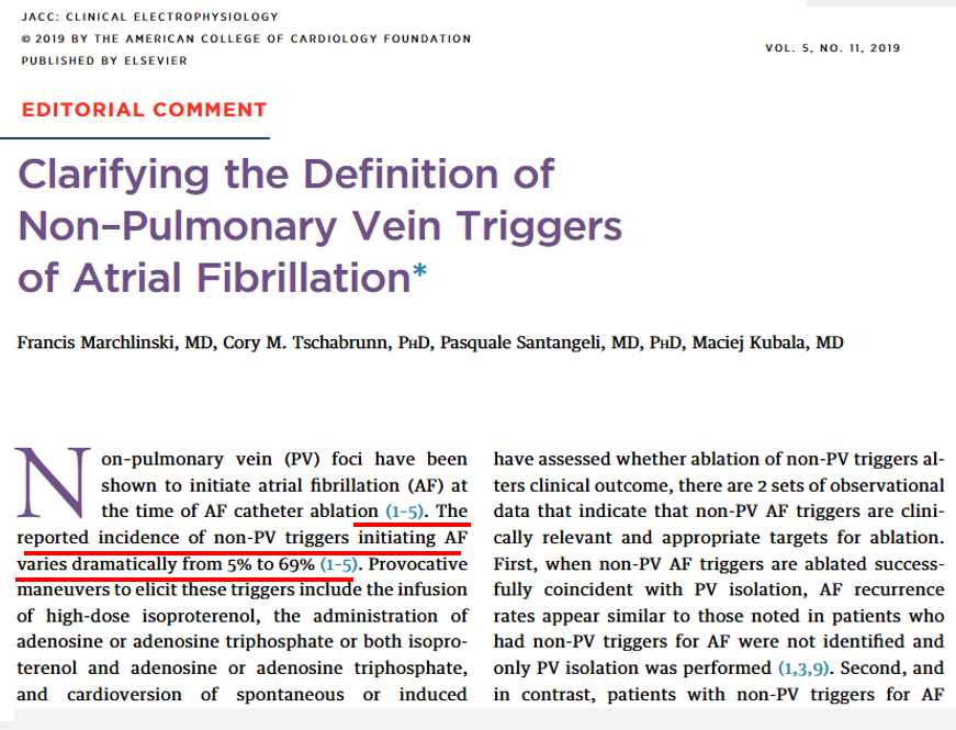
For right or wrong reasons, the world of electrophysiology has pushed us into a belief system that, if it is AF, the culprit must be pulmonary veins. In fact, non-pulmonary vein origins can be a staggering 70% in some series. (See below) It can be in the free walls of the left atrium, LA appendage, IAS, IV, SVC junctions, coronary sinus, ligament of Marshall, crista terminalis, etc. (Ref 2)

For example , where will be the initial focal trigger for AF in a pateint with COPD ?

Can you ever think of ablating PVs in a patient with AF and COPD, where the right atrium is under stress and strain? It doesn’t require any extraordinary intelligence to conclude any chronic focal atrial tachycardia can get degenerated to AF in the long run. In that case, the famous atrial tachycardia localizing map from Peter Kistler et al from Australia JACC 2006 holds good for location AF focus too.

If we look at the above map,RA prevails over LA convincigly in termes of focal atrial tachycardia. Only 20% of focal AT arise from pulmonary veins. I guess, the same should be true for AF.

Focus-less Atrial fibrillation

Right from the days of James Mckenzie, when AF was refered to as delirium cordis or ataxia of pulse, AF was always considered as a chaotic, focus-less arrhythmia. It is still true in many cases. The recent pulmonary vein triggers are just a small revelation and need not be a revolutionary paradigm shift , as we are taught. There are innumerable patients who develop de-novo AF without any focus. Hypoxic or acidotic milleu of a single atrial myocyte can iniitiate an AF, alosan episode of atrial ischemia, diffuse inflammation as in atrial epi-myocardiits can trigger AF from any spot on the atrium.

Reference

1.Francis Marchlinski Cory M. Tschabrunn Pasquale Santangeli , Maciej Kubala J Am Coll Cardiol EP. 2019 Nov, 5 (11) 1328–1330

2.Yang, S.Y., Cha, MJ., Oh, H.J. et al. Role of non-pulmonary vein triggers in persistent atrial fibrillation. Int J Arrhythm 24, 7 (2023). https://doi.org/10.1186/s42444-023-00088-0

3.Aronson JK. One hundred years of atrial fibrillation. Br J Clin Pharmacol. 2005 Oct;60(4):345-6. doi: 10.1111/j.1365-2125.2005.02501.x. PMID: 16187965; PMCID: PMC1884824.

Read Full Post »

The cell of origin of ventricular tachycardia is rarely discussed at bedside. It is still in research labs !

                                    Ventricles are not made up off entirely myocytes. Apart from myocytes it contains specialised  purkinje cells , fibrocytes, interstitial cells and  some times primitive mesenchymal cells. Ventricular tachycardia can arise either in purkinje cells, the myocytes  or even the fibrocytes. The myocyte  VT  classically occur during ACS or post infarct VTs.They are  more often hemodynamically unstable and quickly degenerate into ventricular fibrillation. Myocardial VT is likely to be pulseless and require DC cardiversion frequently. Purkinje VTs are relatively less unstable. If VT arise proximally in the septum near the distal his, or in bundle branches (BBR) the VT is more stable.They  are likely to respond to be medical management.

What is the therapeutic implication of knowing  myocardial VT ?

                               In fact  ,simply knowing the cell of origin of VT is not suffice .The ionic currents inside the cell that trigger and sustain the VT is more important. There are few ionic circuits responsible for VT. Sodium , Intra cellular calcium, potassium , beta receptor mediated calcium current.If we know the individual ionic culpirit we can block that specifically  . Now we have multi purpose ion blockers  like amiodarone which acts like a broad spectrum antibiotic and terminates a VT.

                              So as of now there is no real purpose of breaking our head  in locating the cell  of origin  and the ions responsible for VT  at  the bed side ,( Researchers will do that for us !).  We have only few  antiarrhythmic drugs available in our crash cart  .Our job is to choose the optimal  drug  which will fit in for our patient. In electro physiology labs, radio frequency ablation is done .This is  nothing but shooting down the abnormal electrical  focus (Cluster of cells or a samll segment of myocardium).  In future,  a single abnormal  cell could be selectively neutralised with cell based therapy assisted by  nanopore robots !

Read Full Post »