Feeds:
Posts
Comments

Posts Tagged ‘pulmonary embolism’

Pulmonary embolism continues to be  an intriguing entity,  both in the diagnosis and management. The spectrum is wide, from a clinically insignificant, or totally unrecognized episode to a massive cardiopulmonary event causing sudden death. Similarly, treatment can either be conservative (even sending the patient home ) or lead to  aggressive cath lab intervention.

The task of PE risk stratification

Obviously, risk stratifying is the mainstay in the management.The fact that we are grappling with multiple risk stratifying scores like PESI, simplified PESI or HESTIA expose our limitations. To segregate , low and high‑risk categories do not demand much expertise. It is the intermediate‑risk group that plays havoc in the ER. It is based on RV function and biomarkers like troponin and D‑dimer. But it was soon realised clinical parameters like blood pressure and hemodynamic stability are the more powerful markers of risk.

However,the reality is, many patients in the intermediate‑risk group are rushed to the cath lab, either due to overestimation of risk or fear of potential escalation to high risk. A third reason,is often non‑academic, it’s done  for showing our expertise.

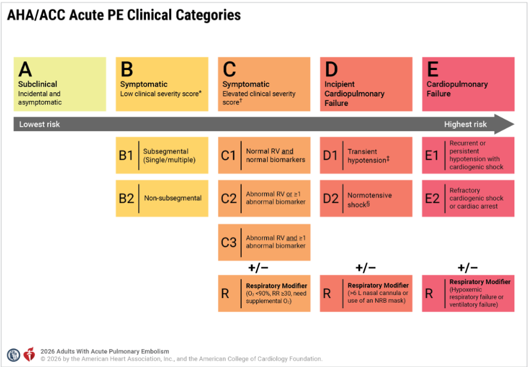
Now, to solve the puzzle of intermediate risk, we have created two new subcategories, C and D. ACC/AHA has been very careful in defining and labeling the entities. Still, there are a lot of gaps that need to be fixed.


In this post let us confabulate few aspects of the new grading of PE and in particular the curious case of D 2 shock .
series of iFAQ ( infrequently) asked questions will from the basis of this discussion


Does category C to D progress in a sequentially worsening hierarchy?

Though it looks that way, the clinical presentation rarely follows a sequential order.

Why is BP is not given adequate weightage in Category C?

A normal blood pressure is a mandatory baseline requirement to enter Category C. However, BP is an important factor used in the clinical severity assessment (PESI etc) .It is not clear why , the committee did not explicitly use the term Category C with reference to BP.

Why is the word “symptomatic” missing in Category D?

The word “symptomatic” is deliberately omitted from the title and subcategories of Category D in the 2026 AHA/ACC Pulmonary Embolism guidelines because symptoms alone are no longer a differentiating factor at these higher levels of severity. Alos, the patient needs to be alert, to tell the symptoms.

Does that mean symptomatic Class C is less risky than asymptomatic Class D?

Yes. A symptomatic patient in Category C is at a significantly lower risk of dying or collapsing than a patient in Category D.A patient in Category C might feel worse because they are aware of their chest pain. A patient in Category D may not complain of pain at all, perhaps because they are in altered sensorium.

What is D2 shock, and when and how do we diagnose it?

First, the patient must fulfill the baseline criteria of Category C, which includes right ventricular dysfunction and positive biochemical parameters of either Troponin or NT-Pro BNP. Then, they must have at least one parameter to confirm shock. Under Category D, the term normotensive shock defines the D2 category, where the blood pressure is normal.

The signs of shock include:

  1. Lactate greater than 2 mmol/L.
  2. Evidence of acute kidney injury, meaning urine output is less than 0.5 mL/kg/hr.
  3. Altered mental status changes.
  4. A cardiac index less than 2.2 L/min/m2.
  5. A mean arterial pressure less than 60 mmHg.(In D2 shock the MAP more than 60 mmhg)

Is elevated lactate mandatory to diagnose D2?

No. Any single criteria  from the above  list is enough. Oliguria alone or even altered mental status is sufficient.

Can D2 shock be diagnosed based only on altered sensorium?

Yes. it is possible to diagnose Category D2 based only on a vague, mild, or temporary change in mental status .This carries a huge risk of falsely classifying a stable Category C patient into the more critical Category D. To prevent this error, the 2026 AHA/ACC guidelines has strict definition of Category D2, ie the altered sensorium must be profound and persistent.

Why Category D1 patients can show a fall in BP, while Category D2 has normal BP?

In PE, the right heart is overwhelmed by the acute  rise in RV afterload, causing a short term  drop in the volume of blood until the RV recovers. This is the mechanism of fall in BP D1.In Category D2, the blood pressure stays completely normal (90 mmHg or higher), but the patient may be in ongoing organ shock spiral. In D2  the RV  is failing just like in D1, but the patient’s sympathetic response fires at it’s peak with maximum vasoconstriction to sustain the BP and life.

Why D2 shock  sounds a mystery ?

It may look implausible that a patient in D1 can have low blood pressure, but when they worsen, they seemingly gain blood pressure to reach D2 and get  the tag of normotensive shock. In the bed side this is not true. The hidden clinical reality is, D1 and D2 are not meant to be viewed as sequential, worsening hierarchical stages. A patient can move from Category C to D1 or D2 directly, and a direct jump from Category C to Category E is also possible without spending any time in Category D.

Dynamics of the C-D-E categories

It becomes problematic to have the term normotension in the title itself in D2 . This implies ,a patient with hypotension and underperfusion should exit from D2 shock. This could mean a worsening patient paradoxically enters Category D1 or Category C or E. To be precise, a fall in blood pressure represents is really a sign of worsening or not is debatable and decided on patient to patient basis. Also both category C will also feature a normal blood pressure. This can be mixed up with D2 if organ perfusion markers like oliguria occur transiently. This trichotomy between hemodynamic stability, organ perfusion , versus respiratory stability needs to be addressed. The PERT team need to be vigilant on this asseement.

Do we need to sub classify D2 shock ?

In realistic observations, the most common cause of normal BP in D2   shock is the intensive treatment these patients receive with RV inotropes and systemic catecholamines.   Perhaps we may   need to add another rider to Category D2, such as Normotensive with drug support versus normotensive without inotrope support. This would help us know the exact direction in which these patients will progress.

Can Category C3 be riskier than D2?

This is tricky and indeed seems possible . Category C3 can exceed the morbidity of Category D, especially when it features the respiratory modifier as C3R. It must be recalled that both the C and D categories present with right ventricular dysfunction and positive cardiac biomarkers, but their primary organ failure differs. A Category C3R patient experiences an acute oxygenation crisis. Conversely, a Category D2 patient, even with normal lactate, suffers from occult circulatory shock (an adequately functioning liver can clear the lactate efficiently.) Though C3R appears more dramatic at the bedside due to the need of respiratory support , Category D2 carries a higher mortality.

Final message

It looks like treating patients with PE is much easier task than risk stratifying them

Pulmonary embolism management is a fierce fight between the right ventricle,  the occluded pulmonary arteries and the lungs.The first 48 hrs is crucial. RV shock incidence rapidly reduce after this time window. We realised very late, the total thrombus burden actually has a poor correlation with the final outcome. Most episodes of PE can be managed medically.

Balancing pharmacological lysis versus catheter-based lysis requires a very delicate skill. While most patients are eligible for standard lysis, only a very few truly benefit from catheter-based therapies. The rate of RV failure and mortality drops dramatically once the right ventricle rises to the challenge. Therefore, there is no clinical point in doing a catheter-based lysis if the blood pressure is normal and the right ventricle is already recovering.

Finally and most importantly, the alarming entity D 2 (Normotensive shock) should not cause unnecessary panic. If the, saturation and RV function remains good or shows signs of recovery , catheter based modalities can be avoided.

Reference

1.Writing Committee Members; Creager MA, Barnes GD, Giri J, Mukherjee 2026 AHA/ACC/ACCP/ACEP/CHEST/SCAI/SHM/SIR/SVM/SVN Guideline for the Evaluation and Management of Acute Pulmonary Embolism in Adults: A Report of the American College of Cardiology/American Heart Association Joint Committee on Clinical Practice Guidelines. Circulation. 2026 Mar 24;153(12):e977-e1051.

2Yuriditsky E, Zhang RS, Zhang Right Ventricular-Pulmonary Arterial Uncoupling as a Predictor of Invasive Hemodynamics and Normotensive Shock in Acute Pulmonary Embolism. Am J Cardiol. 2025 Feb 1;236:1-7. doi: 10.1016/j.amjcard.2024.10.036. Epub 2024 Nov 4. PMID: 39505227.

3.Dominick Roto, Raquel Lamarche, Navigating normotensive shock Chest physician May 6, 2026
Read Full Post »

Role of d dimer in acute aortic syndromes

D -Dimer is a marker of  intravascular fibrinolysis .It is a degradation product of fibrinogen. A level more than 500ng/ml is significant.In acute aortic dissection this level is reported to be more than 2000ng/ml.

The beauty of this molecule is it is elevated in three important chest pain emergencies.

  • Acute myocardial infarction
  • Pulmonary embolism
  • And now aortic dissection.

The issue is not simple , as we know any intravascular coagulation and lysis can elevate this molecule.In patients with chronic CAD as like a chronic thrombotic lesions within the coronary arteries can also elevate d dimer.

Similarly , in portal, cortical, deep venous thrombosis all result in elevated D dimer.

So , such a non specific test  , how can be  useful in the diagnosis of aortic dissection ?

Yes, you are right ,

D Dimer helps us  not in diagnosing aortic dissection but  helps us in ruling out a possible dissection

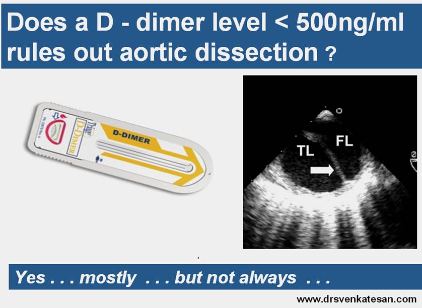
D-Dimer levels <500 has a negative predictive value of 98% .

What is the bio- chemical  dynamics of  D dimer in dissection ?

D dimer in aortic dissection is mainly secreted within the false lumen. For d dimer to secrete into  systemic circulation  the clotted area should be exposed to a adequately flushed systemic blood at a good perfusion pressure.The contact area between the clot and fresh blood  is of critical importance.

d dimer aortic dissection false lumen

So ,  even though it has been reported d dimer has near 100% negative predictive value . . . is there a chance a dissections might occur with normal  d dimer levels ?

Yes, very well possible with due credits to published data

  • A dissection without thrombus(Rare . . . but still possible !)
  • A clot confined to false lumen with entry or exit points sealed.
  • A dissection without a exit point.
  • Intramural hematoma with no communication with aorta

Infrequently asked questions

  1. Time window ? Dimers are mainly useful in  patients who report before 24h after the onset of chestpain.
  2. How long it takes for the dimers to  get excreted ?
  3. Can coronary dissections in STEMI elevate dimer ?

Final message

D dimer is  mainly useful in  “not making a diagnosing” aortic dissection.

If  dimer levels are strongly  positive and  clinically the patient  has  has no evidence for acute MI or acute pulmonary embolism  and continues to have chest/back/atypically located pain  suspect aortic dissection , and order for further imaging like TEE,MRI, MDCT etc.

* Do not forget the role of routine , simple bedside transthoracic and suprasternal echocardiogram.It can diagnose dissection correctly in good number of patients.

** Never oder for costly thoracic imaging whenever d dimer is elevated.

*** When you send the sample for dimer make sure to mention  the clinical likelyhood of dissection .If it is very high the lab has every reason to reject the sample and suggest you to go ahead with thoracic images.

This is because ,  it could be costly miss . . . if you depend on dimer to diagnose a dissection

Imagine this scenerio , while your patient has a absent left radial pulse due to dissection and you are waiting for the lab report to arrive !

Never use it for diagnosing aortic dissection.

Read Full Post »

The importance of venous system

Cardiovascular system  consists  not only of  heart  but also   the blood delivery and retrieval   system namely  the arterial and venous circulation .As  the heart pumps , 6  liters   of blood  every minute  , it  has to   traverse the  entire venous circulation promptly ,  to  complete  the hemodynamic circuit.

ei_2401

Source : From web. Thanks to whoever created this Image

While  physicians are preoccupied with disease  of heart there is an important  groups of disorder of venous system .The  deep vein thrombosis  (Also called venous thrombo embolism . VTE ) is the  most important  entity .This disorder even though is a cardiovascular  disease  ,  it   rarely presents to a cardiologist .

As  we tended  to ignore the  veins   for decades  now, “sudden venous deaths ” other wise called massive pulmonary embolism  is calling for our attention

What are the deep veins ?

Popleteal , femoral , iliac (External , Internal , common ) . Upper limb axillary and subclavian veins.

Clinical classification of DVT  :  Femoro popleteal  , Pelvic  ( Ilio femoral) , Mesentric DVT.

What are the high risk population for development  DVT ?

  • Genetic predisposition  constitute  the  strongest risk(Factor V lieden mutation )
  • Major orthopedic surgery
  • Pregnancy /Oral contraception
  • Disseminated  malignancy

And now  ,  the more fashionable risk factor “DVT after long distance flights”

What is key diagnostic issues in DVT ?

Key to diagnosis is clinical alertness .Local swelling  , edema legs and inflammation should alert the physician.

  • Homan’s sign(Pain on  dorsiflextion of  ankle) .
  • Louvel’s sign (Leg pain on coughing ),
  • Lowerberg’s sign  (BP cuff induced pain on affected leg at low level inflation  )

( Well’s score  is based on pre test propabilityLancet 1997 )

Many times DVT is diagnosed only after it embolises into lung.So remember shortness of breath and acute dyspnea  could be the first manifestation of DVT.

Once diagnosed  DVT  it should be  risk stratified either as low risk or high risk .

Biochemical diagnosis of DVT : DVT is a form of intra vascular coagulation and it activates fibrinolysis.D Dimer estimation has strong negative predictive value .If D dimer is negative it excludes DVT by 99% .Positive D dimer does not confirm it .

Is  it  necessary  t0 image  the venous clots ?

No . It is rarely required.  Instead we need to know the site of occlusion .Doppler and ultrasound scan can help locate the site of obstruction .

Other modalities *  may help evaluate the thrombus

Venous angiography (Filling defect, collateral )

MR angiogram

Fibrinogen tagged nuclear scan

Management

  • Acute management
  • Long term management .

Acute management

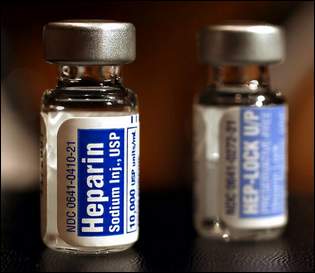
Immediate Heparin ,  bolus followed by infusion ( 5000U, 1000U/h) followed by oral anticoagulation forms the corner stone of  management of DVT .

Once a DVT is documented should we attempt  to  improve the venous circulation or try to slow down  the venous circulation ?

There is a paradox here.The therapeutic strategy is to improve the venous circulation . A sluggish venous circulation predisposes fresh thrombus. So even though ,  it is  logical to expect some  migration of  thrombus  proximally  with the standard  therapeutic methods of DVT  ,  it is the ultimate principle of management of DVT.

How heparin infusion achieves  it’s  therapeutic goal  of clearing  thrombus burden in the venous circulation is not clear .It is believed sub clinical PE  occurs in every case with large DVT and these thrombi get   microlysed either  within focus of  DVT or in transit circulation  or within the pulmonary vascular  bed.

What is effect of intensive anti coagulation on DVT ?

  • Lyses the thrombus
  • Dissolves the thrombus
  • Dislodges thrombus
  • It can prevent only fresh thrombus

Answer : All of the above can occur

Can we track the movement of deep vein thrombus ?

It is not an easy thing to track the movement. Doppler will give an idea. Invasive investigation to track the thrombus is neither practical nor necessary.

What is aggressive management * for DVT ?

* Aggression is rarely required in DVT management.

Interventional

  • Thrombolysis : Systemic/local catheter based
  • Venous angioplasty/Stenting
  • IVC filters

Surgical

Indication for thrombolytic therapy

Surprisingly,   thrombolytic therapy has limited role in the  management of  DVT. There  is absolutely no role for routine thrombolysis in DVT (Heparin does the same job , more consistently with less risk )

It is used only when there is limb threatening or lung threatening situation .

Pulmonary embolism already occured

Massive iliofemoral thrombosis .

When will you call a vascular  surgeon ?

Thromboembolectomy as a treatment for DVT is rarely advocated .

The 2004 American College of Chest Physicians consensus statement on the treatment of thromboembolic disease recommended against the routine use of venous thrombectomy in acute DVT except in cases of phlegmasia cerulea dolens . ( Severe necrotising venous edema )

The issues against surgery are

  • Generally these patients are more sick and co morbid conditions
  • Complex nature of surgery in deep iliac veins
  • Blood loss  from deep friable veins
  • The surgery further traumatizes the vein, recurrence  of DVT is  very much possible
  • Primary cause is not addressed by surgery

What are the indications for IVC filter ? What  are the types of filter available ?

The indication for IVC filter in the acute management of DVT * has been ( Rather continues to be  . . .) controversial .The major reason for the controversy is the risk  ( The wasted effort  too !)   to benefit ratio and  is not clear.

gunther-tulip-ivc-filter

There are three types of IVC filter available

  1. Temporary venous filters
  2. Temporary retrieval IV filters ( Gunther tulip ) Nitinol recovery filter Bard
  3. Permanent IVC filters

*In long term prevention of PE the indications are fairly established.

Differential diagnosis

Is there an entity  called  superficial venous Thrombosis (SVT)

Superficial venous thrombosis and thrombophlebitis are more common than DVT and should not be confused with DVT. ( Easier said, some confusion is bound to occur !especially ,  when it occurs  over  thighs ) .This is common  following IV line  and  varicose veins in lower limb Present with pain, tenderness, or an indurated cord along a palpable  superficial vein  with erythema. It is less likely to propagate into pulmonary circulation.

How often a superficial venous thrombosis convert into deep vein thrombosis ?

Patients with superficial phlebitis above the knee have an increased risk of deep venous thrombosis and should probably have ultrasonography.They may require  warm compression , NSAIDS and  local thrombectomy.

What is the post-thrombotic syndrome? How to differentiate it from recurrent  DVT ?

Post-thrombotic syndrome is  due to the damage  to the  valves in the veins  that leads to chronic venous edema of extremities. It may mimic like an DVT . usually occur within 2 years of DVT.

Unanswered questions

1. What will happen to  the thrombus following filter insertion ?

Large thrombus gets trapped in IVC .The problem gets shifted form the legs to the vena cava .This makes it mandatory  for these  IVC  clots  to be cleared either manually or  pharmacologically. Small thrombus and embolic showers continue to cross the filter without  difficulty.

2. IVC filters are recommended in DVT , if a patient has an absolute contraindication to heparin but , is it not a fact ,  filters also demand  anticoagualtion ?

It is true , filters demand anticoagulation. So ,  oral anticoagualtion should be given whenever possible in all  even after IVC filter. This is  , not only to make sure filter does not get clogged but also prevent further  clot formation in the legs and also   distal to the filter in   (Procoagualnt )  individuals .Further , anticoagulation forms the mainstay treatment in patients with chronic thromboembolic PAH ,  which the filter does not address to.

3. Is there safe venous clots that the pulmonary circulation can effectively tackle ?

Typically PE occurs  as

  • Massive acute PE
  • Sub acute PE
  • Chronic pulmonary thrombo embolism (Showers of microemboli lead to PAH )

Consider the following pulmonary  vascular anatomy : MPA 2.5cm ,  RPA,  LPA 1cm segmental pulmonary artery 5 mm ,  pulmonary arteriole 3  mm ,  pulmonary capillaries 200 microns . The  deep venous thrombus typically has  a diameter of  up to 1-1.5 cm . It needs a at least  2. 5 cm   diameter  clot to occlude the main pulmonary artery.Micro thrombus may get cleared by pulmonary vascular bed.There can be safe venous clots.

Final message

  • DVT and PE are the common venous emergencies.
  • Prevention of PE is the major aim of acute managment.
  • Identifying the underlying cause and prevention of DVT per se is the long term aim.
  • Aggressive local approach is largely  unnecessary except in leg / lung  /life  saving situations

Intensive Heparin protocol  followed by long term oral anticoagualnats (1 year or more)  is  an excellent approach in most patients.

heparin

The most important point to remember is the treatment for high risk DVT and suspected  or established PE is exactly same

*Only 10% of PE  are candidates for thrombolytic or surgical  therapy so at times of real dilemma , there is nothing wrong in administering  heparin in all patients with suspected  high risk  DVT/PE  even without confirmation.

All those hi fi stuff of V/Q scan , pulmonary angiography may be a misadventure .Remember empirical (Some call it as unscientiifc ! )  therapy  too , can save many lifes

Further reading

Best Link for  IVC filters   http://www.tigc.org/eguidelines/VenaCava.htm

References

  1. Decousus H, Leizorovicz A, Parent F, et al. A clinical trial of vena caval filters in the prevention of pulmonary embolism in patients with proximal deep-vein thrombosis. N Engl J Med 1998;338:409-415.

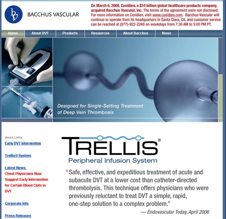
What is new in catheter thrombolysis in DVT ?

dvt thrombolysis

http://www.bacchus-vascular.com

A interventional catheter based clot lysis for  DVT

For the comprehensive  ACCP 2008 guidelines of managing DVT  reach the following site

Read Full Post »