Feeds:
Posts
Comments

Posts Tagged ‘wpw syndrome’

None exposes the expertise and stretches the electrophysiological acumen of a cardiologist, more than a strip of wide QRS tachycardia. Here is a patient who comes with palpitations, with mild hypotension , still comfortably entering the OPD with a non-emergency appointment.

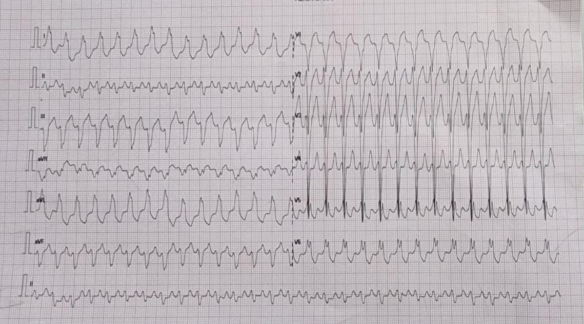
How many diagnosis are running through your mind when you see this ECG . How much it got narrowed after applying criteria like Brugada, Vereckei  etc ( VT, FT, OTVT, Mahaim Tachycardia, AVRT /AVNRT with aberrancy etc)

Now let me reveal the age of the patient .He is a 13 year old boy. Does this help you to narrow down the list of possibilities ? Logically, It should , but for some it can widen the list as well .( Brugada, Anderson, Long QT, Naxos all runs through the mind)

What happed next ?

As the cardiologist was wondering how to tackle this arrhythmia . Adenosine? Amiodarone or DC shock, thankfully, the boy got to spontaneous sinus rhythm .

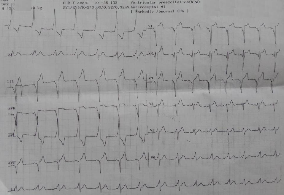
Click here to see the ECG after restored sinus rhythm & the diagnosis is Instant .

Yes it is a WPW substrate. Localization of pathway is a must and a headache for the fellows. .For me, It looks like a posterior pathway in the para-spetal area. 90% of AVRT are narrow QRS . Here it is wide. Why ?

It’s simple. The tachycardia is anti-dromic.

Is it ? Look at the strip again and confirm is this antidromic ?

I am not sure , I still think it is still antidromic .

I am saying No. This is Ortho-dromic wide QRS tachycardia .

How do you say so ?

Antidromic will still more wide and rapid and won’t terminate spontaneously .Of course one problem here aberrancy usually take an RBBB morphology.

How are you so sure . Let us send this to an EP guy.

*What did the EP say? Well, he didn’t commit to anything as I expected. He said until he measures the refractory period of the accessory path, he won’t comment anything about ortho or antidromic query of AVRT. He asked us to send the case for ablation immediately,


Ok. Let us analyse the reason for orthodromic wide QRS AVRT

1.Rate dependent aberrancy

2.Functional abberancy.

3.Preexisting BBB

4.If patients are on drugs that can prolong his Purkinje conduction


Final message

Two lessons may be learnt.

Lesson 1: Trying to decode a wide QRS tachycardia, without knowing clinical background, should be forbidden. This topic is intentionally made complicated by a flawed teaching methodology of wide QRS tachycardia for over 4 decades. Maybe the single lead AVR algorithm promises to be a quick remedy. If AVR is positive, it is VT; cannot be SVT.

(For the curious readers , please go through Dr. Masood Akhtar’s article from Wisconsin on how to differentiate VT & SVT without even looking at the ECG by law of statistics that beat the invasive EP studies).

Lesson 2 : Wide QRS AVRT is not always antidromic . In fact, orthodromic wide QRS AVRT is 2 to 3 times more common. Many of us don’t realise this bias in our learning.

Related topic

How common is orthodromic wide QRS atrial fibrillation ?

Reference

1.Vereckei A. Current algorithms for the diagnosis of wide QRS complex tachycardias. Curr Cardiol Rev. 2014 Aug;10(3):262-76. doi: 10.2174/1573403×10666140514103309. PMID: 24827795; PMCID:

2.Vereckei A, Duray G, Szénási G, Altemose GT, Miller JM. New algorithm using only lead aVR for differential diagnosis of wide QRS complex tachycardia. Heart Rhythm. 2008 Jan;5(1):89-98. doi: 10.1016/j.hrthm.2007.09.020. Epub 2007 Sep 20. PMID: 18180024.

3.Akhtar M, Shenasa M, Jazayeri M, Caceres J, Tchou PJ. Wide QRS complex tachycardia. Reappraisal of a common clinical problem. Ann Intern Med. 1988 Dec 1;109(11):905-12. doi: 10.7326/0003-4819-109-11-905. PMID: 3190044.

4.Tchou P, Young P, Mahmud R, Denker S, Jazayeri M, Akhtar M. Useful clinical criteria for the diagnosis of ventricular tachycardia. Am J Med. 1988 Jan;84(1):53-6. doi: 10.1016/0002-9343(88)90008-3. PMID: 3337132.

Acknowledgemt

The ECG is posted with courtesy of my colleague Dr. C.Moorthy, Cardiologist, Chennai.

Read Full Post »

If some body is struggling with same problem, say for over three decades , there is something seriously wrong with the way we deal with the problem. How do you localise accessory pathway in WPW syndrome from 12 lead ECG ? is one such entity, This question is asked exclusively in cardiology board exams. Now a 2023 paper from EUROPACE claims, it has come out with a simple algorithm bettering all the previous ones. Please check it for yourself.

One thing we can say with conviction is most of these embryological remnant pathways are posteriorly or laterally dragged in peri AV annular region or the para septal region. Very few appear anteriorly, if anterior it is more often placed on the right side.

Why should we take this question easy ?

Try asking any experienced EP specialist* to localise a pathway in given 12 lead ECG . Don’t get surprised by a long silence before they commit, because they know the truth, and how delicate this question might sound on quiet a few occasion, because of various anatomical and physiological reasons.

*Never fail to appreciate their hard long hours in cath lab to spot, analyse and shoot these tracts.(EP stuff is not like angioplasties, which, many can do even in half sleep!)

Final message

Yes, localising WPW can be either a fascinating or frustrating exercise depending on our understanding about the attitudinal cardiac anatomy, variable autonomic tone dependent morphological behaviour of delta waves, PR intervals, QRS axis ,the transition zones etc. Shrewd fellows may go through this 12 lead stress test. ,

For others just try to localise right from left , & then posterior or lateral Forget the anterior ones. This is more than suffice. Unlike drug trials, where statistics are often battered , here the Incident numbers are the key measure of truth. (Even without seeing a ECG you are likely to be correct in 80 % times, if you localise the pathway to posterior, para-septal or left lateral zones. )

Reference

Read Full Post »

As I expected ,  my earlier algorithm “An Idiot’s approach to tachy-arrhythmias” has  elicited  mixed reactions  .Some  EPs calling it a dud while few  physicians termed it awesome . Here is  a follow up .

Heart rate of a tachycardia is the most neglected parameter by physicians .  They are often seen spending  hours together for decoding  arrhythmia , splitting the brains   for P wave  location , VA conduction, Fusion beats etc .Finally they end up  either administering  Amiodarone a broad spectrum anti arrhythmic agent or DC shock.

Here is an unusual algorithm  for arriving at a diagnosis in all tachy-arrhythmias  based only on heart rate and the width of he qrs complex.

(Click over the table for high resolution image )

approach to cardiac arrhythmias narrow qrs vs wide qrs brugada wellens criteria

General principles in diagnosis of tachycardia

Narrow  qrs tachycardias.

90 % rule : If regular It is sinus tachy if irregular it is A-FIB . Take some efforts to r/o sinus  tachycardia . (In children and young adult it can be extremely difficult at times )* Please note : Sinus tachycardia can show some irregularity due to sinus arrhythmia and  frequent  APDs and JPDS . Further at  fast rates P may fuse with T it should not be confused with  A-fib .

Wide qrs tachycardia

Common things  are common , if  you sight a large animal with a huge trunk  in a Kenyan safari ,  it is most likely to be an  elephant and not a Dinosaur !  Please diagnose VT  when you encounter wide qrs tachycardia by default especially when the BP drops  !

  Management issues

It  would be  foolish to split our heads for decoding an arrhythmia when a patient is unstable .Any hemo-dyanmic unstable tachycardia needs DC shock . (Synchronized will be better unless it is dire emergency )There are very few arrhythmia where DC shock is contraindicated   ( MAT/Dig toxicity/Underlying sinus node dysfunction )

Only if the patient is hemo-dynamically  very much stable   the  physicians  have enough time to  confuse themselves  and the real  ordeal begins .Please remember  the 5 arrhythmias  constitute 98  % of all known tachy-arrhytmia . So where ever  you practice ,  whether  in remote Nigerian village  or  sophisticated  Cleveland  university hospital , when you are  confronted with a tachycardia  the diagnosis  should be one among the  following  five  !)

  1. Sinus tachycardia .
  2. AF/A-fib
  3. Atrial tachycardia  with  or without blocks
  4. ventricular tachycardia /VF
  5. AVNR/AVRT with or without aberrancy

All  other tachy-arrhythmiaa  are  largely  academic !

Regarding  drugs

Knowing the mechanism of  arrhythmia genesis  is less important  at bed side . They are  triggered , sustained, and maintained by either functional or structural component .Ionic basis operates in every arrhythmia  , but it is the anatomical  substrate that maintains it .This happens in only diseased heart.The only point worth remembering regarding mechanism of arrhythmia  genesis  is ,  automatic and focal tachycardias  will not respond to DC shock . All other can be termed some form of re-entry . Micro reentry  for all practical purposes behave like  triggered  activity. Ischemic and electrolytic VTs are primarily ionic based and often polymorphic.Structural VT are commonly mono-morphic. Any VT just prior to degeneration to VF become polymorphic

Every patient with cardiac arrhythmia should be checked for hypoxia,acidois , electrolyte defect or exposure to any  pro arrhythmic drugs. (The commonest  cause of tachycardia in any  IMCU , is inotropic induced (dopamine /doubtamine ) tachycardia .

We  have  5  pharmacological options

  1. Blocking  adrenergic  receptors(IV Esmolol, Metoprolol)
  2. Blocking calcium channel (Dilitazem,Verapamil)
  3. Blocking potassium channel  (Amiodarone  ,Sotolol Adenosine  to a cetian extent )
  4. Blocking sodium channel . ( Procainamide , Lignocaine (Wonder drug almost forgotten now ! ) Flecanide Mexilitene etc)
  5. Digoxin ,Adenosine  magnesium are special  anti-arrhythmic  agent which  has very useful role in certain specific situations (Magnesium -Torsades/Polymorphic VT / Adenosine in LVOT/RVOT VT etc)

General principle is ventricular arrhythmias  are blocked successfully  by sodium or potassium blockade  Atrial and functional tachycardia are blocked by calcium or adrenegic blockade  .Of course,  there would be  some degree of overlap  when the arrhythmia  origin  hovers  around the junction  on either side of the AV  ring . This is basis of verapamil sensitive VT .Clusters of  calcium  channels are scattered  in the junctional  region

Refractory tachycardia

  1. Consider ablation  in AVNRT/AVRT
  2. ICD +Drugs  in VT
  3. Ablate and  Pace(Some A-fibs)
  4. Ablate and ICD (Some  incessant VTs)
  5. Surgery in minority

In AVNRT/AVRT 90 % success can be achieved  in most EP centers .VT ablation  is still a complex process  with  success rate around 60 % ICDs  are indicated in all recurrent VTs except incessant forms .(Where the battery will deplete within a month !) Surgical cure (Maze etc  ) is possible in selected few while undergoing mitral valve surgery.Contrary  to the modern scientific  mood ,  I can ay with conviction most A-fibs can be managed medically except a fraction will require pulmonary vein ablation / isolation .

Final message

Mastering the field of of  cardiac  arrhythmias ,  though  appear a daunting task ,  it does not  require   immense  sense  to understand real world problems are  only a  few and can be tackled in a simplistic manner !

Read Full Post »

Heart rate of a tachycardia is the simplest of all  . . . but   neglected parameter by physicians.  They are often seen spending  hours together for decoding  arrhythmia , splitting their brain for locating P waves ,  VA conduction, Fusion beats etc Finally , most end up  either administering  Amiodarone a broad spectrum anti arrhythmic agent or a DC shock  without arriving at a correct diagnosis.

Here is an unusual algorithm  for arriving at a diagnosis in all tachy-arrhythmais  based only on heart rate and the width of  the qrs complex with acceptable accuracy.

(Click over the table for high resolution image )

approach to cardiac arrhythmias narrow qrs vs wide qrs brugada wellens criteria

Caution :

The above table is  an extremely simplified approach for tachy arrhythmias. Not applicable for scientifically inclined . But in my personal opinion ,  in an emergency room  pure science matters less !

Read Full Post »

When I posed the above question  to few  cardiologists including electro physiologists , the answer I got was surprising .  In the process ,  I could understand why cardiology is such fascinating subject !   Each one gave a different answer and all the 5 responses were forth coming .

The following post in my blog which  I wrote years ago tries to decode the reason for such wide variation in our understanding of AVRT of WPW.

By the way ,  is there a  real risk   for an  ortho-dromic AVRT into anti-dromic AVRT by a definite block in AV node  ?

Read Full Post »

AV nodal tissue is a not compact structure as we would be believe  . But it is a fact ,  AV  node  do attempt  to compact after birth.

It is never complete.

Note the islands of his bundle entrapped .   Image source : M. Paz Sua´rez-Mier J Am Coll Cardiol 1998;32:1885–90 From the Section of Histopathology, Institute of Toxicology and †Department of Pathology, La Paz Hospital, Madrid, Spain.

All specialised cells should coalesce  to form the compact zone .This fails to happen in many . Failure of AV nodal compaction  results  in islands of slow conducting cells in around AV node . Some of them can mutate  , and   acquire fast conducting properties as well . (Accessory pathway )

This failure of AV nodal compaction is termed as  persistent fetal dispersion of AV node .

In the his bundle  same phenomenon is called as his bundle  de-fragmentation .These abnormalities are noted in pathological  specimens  of  Pokkuri sudden death in Asians .

Unexplained sudden deaths and instant bradycardias and complete heart blocks are related to these dispersion of  AV nodal cells downstream . This also explains some of patients with infra hisian escape show junctional characteristics.

Many cardiac pathologists have observed this . Still  there is a  missing  link  .

References

M. Paz Sua´rez-Mier,Carlos Gamallo  J Am Coll Cardiol 1998;32:1885–90) Atrioventricular Node Fetal Dispersion and His Bundle Fragmentation  of the Cardiac Conduction System in Sudden Cardiac Death

Kirschner RH, Eckner FAO, Baron RC. The cardiac pathology of sudden,unexplained nocturnal death in Southeast Asian refugees. JAMA 1986;256:2700–5.

Hudson REB. The conducting system: anatomy, histology and pathology in acquired heart disease. In: Silver MD, editor. Cardiovascular Pathology. New York: Churchill Livingstone, 1991:1367– 427.

James TN. Normal variations and pathologic changes in structure of the cardiac conduction system and their functional significance. J Am Coll Cardiol 1985;5:71B– 8B.

James TN, Marshall TK. De Subitaneis Mortibus XVIII. Persistent fetal dispersion of the atrioventricular node and His bundle within the central  fibrous body. Circulation 1976;53:1026 –34.

Persistent Fetal Dispersion of the Atrioventricular Node  Association With the Wolff-Parkinson-White Syndrome Claude Brechenmacher, MD; Jean-Paul Fauchier, MD; Thomas N. James, MD Arch Intern Med. 1980;140(3):377-382.

Read Full Post »

AVRT is  a second commonest cause of  narrow qrs tachycardia.  While , all narrow qrs tachycardia in AVRT must be  ortho-dromic. wide qrs tachycardia in WPW  can either be ortho-dromic or anti-dromic ,

The classical one is the much popular and fancied Antidromic  AVRT . Please be reminded  AVRT can conduct  orthodromically  through AV nodal tissue  but still  become  aberrant , as it travel downwards thorough the bundles   and result in a wide qrs tachycardia .

Among the two which  is more common ?

My observation is  ortho-dromic  wide qrs  AVRT  is  more  prevalent . Do you agree ?

Final message

Not all wide qrs tachycardia  in WPW  is anti-dromic !

Read Full Post »

The fundamental difference between  accessory pathways (APs) and AV nodal tissue is the former lacks decremental properties . That is  , APs continue  to conduct whatever the  impulse   it receives. (Unlike  the AV node which has a filtering  mechanism , A heart rate sinker / Dampener) . This is what we were taught and we believe in that .

If it is true  , every episode of   atrial fibrillation should conduct with 400-600 ventricular responses . In reality it does not happen .  The usual ventricular rate in AF with WPW is  250-300 /mt .

What happens to the rest of atrial impulses ?

I am sure it must  get   blocked in APs . Of course it is possible the block need not be in a fixed ratio  .It  changes in a  dynamic   manner with  reference to the   refractory period . (Please note , blocks and increased refractory  periods  can be  used inter changeably in most  physiological situations .

Final message

All APs are not dangerous .They do have a   restrictive mechanism in place .This is evident in every patient with AF and WPW syndrome with a fairly controlled ventricular  response  . Hence  one can conclude   APs in WPW syndrome do have a physiological block in most episodes of  Antidromic AF . The cut off  for safe  refractory period is defined empirically as > 250 ms.

Coming to the title  question , Is  there a physiological  2 : 1  block  in accessory pathway  during AF and WPW syndrome  ?

Yes . It seems so !  A WPW  patient who has  just recovered from a  well tolerated AF ,  is  sort of a natural screening test which effectively rules out a future SCD .(Unless of course he has multiple APs with varying RPs  , one for AF other for VF !)

Is that a correct way of reasoning ?  Experts may provide further  input .

Read Full Post »

Identifying the P wave is the key to decode  any  narrow QRS  tachycardia . Though the  the relationship to  p and  qrs is vita ,  many times it is  not  easy to  relate them.More easily one  may  get  a  clue to the mechanism by analysing   P wave timing .This is the basis of calling narrow qrs tachycardia as short RP and long RP.

Wonder   . . .  why  the  relation “P to R” became  “R to P” here !

Since  in the   common narrow qrs tachycardias  AVNRT/AVRT  ,  atria  activates  the atria  in a  retrograde manner , we look  for the relationship of qrs complex on subsequent P wave . Hence the interval between R to P become the focus.

In other words RP interval indicates retrograde  conduction property of AV tissue .

If it is slow the P wave will be well separated from QRS .

If it is fast it will be close to QRS complex .

If it is ultra fast as in some AVNRT ,it can fall within the qrs complex and completely invisible .

(The so called  r’ prime in classical AVNRT is nothing but a distorted p wave on the terminal qrs complex.)

Based on  RP interval  the following classification is used (List is incomplete)

Short RP Tachycardia

  • AVNRT (Slow-Fast )
  • AVRT

Long  RP tachycardia

  • Atypical AVNRT(Fast -slow)
  • Atrial tachycardia*
  • Sinus tachycardia*
  • SA nodal re-entry*
  • Some forms of AVRT

* Please note ,  here the P wave is not determined by the preceding qrs unlike other tachycardia in the list.

What is the  cut off point to call it is Short RP /Long RP ?

It is arbitrary . Following may help

If RP interval > PR interval it is long RP.

If the absolute RP interval is >  100  ms  with the heart rate of > 160 it would  generally  Indicate a long RP tachycardia .

The timing  of  retrograde P can be very complex than we believe  as the following factors heavily influence it.

  • The autonomic tone
  • Site of retrograde atrial  breakthrough point .
  • Atrial size ,
  • Atrial  refractionaries
  • Effect of drugs
  • Intact-ness of inter atrial conduction
  • Chances of the retrograde atrial activation capturing Internodal pathway

Final message

The P wave location in narrow qrs tachycardia is primarily determined by the retrograde VA  conduction and less  on the antegrade AV conduction  . Looking at the interval between R and P is a  quick way of getting the VA conduction in the bed side.

Once we get an  idea how the VA  circuit  conducts , we can narrow down the possibilities  in  Narrow qrs tachycardias !

Comming  soon

What determines the morphology of retrograde P waves in AVNRT/AVRT ?

Read Full Post »

Localising  WPW syndrome is a favorite  time pass  for cardiologists in spite of  serious  limitations of surface ECG .Still , it is vital to generate a rough idea about the location of  these pathways ,  so that we can focus  our efforts  on  some sort of ablation procedure .

Arruda algorithm is probably a simple and fairly useful technique to remember. It asks us to climb 4 steps   and pause at each  step and look sideways   for the accessory  pathways !

Step 1 (Left free wall step )

Initially one need to look only two leads .

Look at lead 1  and  V1 for   delta wave and R/S ratio .After Identifying delta wave look for the polarity of delta wave (This can sometimes be really difficult ) .If there is iso-electric or negative delta it immediately  fixes the pathway  in left free wall . Similarly if V1 R >  S it also fixes in left free wall. To locate more precisely in left free wall  look  for  delta  wave polarity in  AVF  and proceed down*

If none of these finding are present then  Go to step 2 .

Step 2 (Coronary sinus step )

It is the most simple step . If negative delta  located in lead 2 (often mimic inferior MI)

Here the pathway is often located in coronary sinus /middle cardiac vein often as diverticulum.

After excluding left free wall and coronary sinus origin one has to look at possible septal  pathway  .

For this  go to step 3

Step 3  (Septal step ) And  again v1 lead  becomes important if v1 shows negative or iso-electric  go down  to septal  pathway decoding

After ruling out septal origin the scheme takes us to right free wall by default.

Step 4  (Right free wall step)  If the delta wave does not fit in  any   of the above three steps (Including  positive  delta in V 1 )  it  fixes  the right free wall  pathway

Arruda scheme summary

Arruda scheme  guides  us  to scan  systematically  for pathway from left free wall  to  septum and lastly  the right free  wall  (The key  to  locate  the APs is  to look at  delta waves in lead  1, 2  AVF and R/S ratio In V1 )

Here is a  simplified version for basic localization

Reference

  1. Arruda MS, McClelland JH and Wang X , et al. Development and validation of an ECG algorithm for identifying accessory pathway ablation site in Wolff-Parkinson-White syndrome. J Cardiovasc Electrophysiol 1998;9:2–12.

Read Full Post »

Older Posts »