Feeds:
Posts
Comments

Archive for the ‘Cardiology – Electrophysiology -Pacemaker’ Category

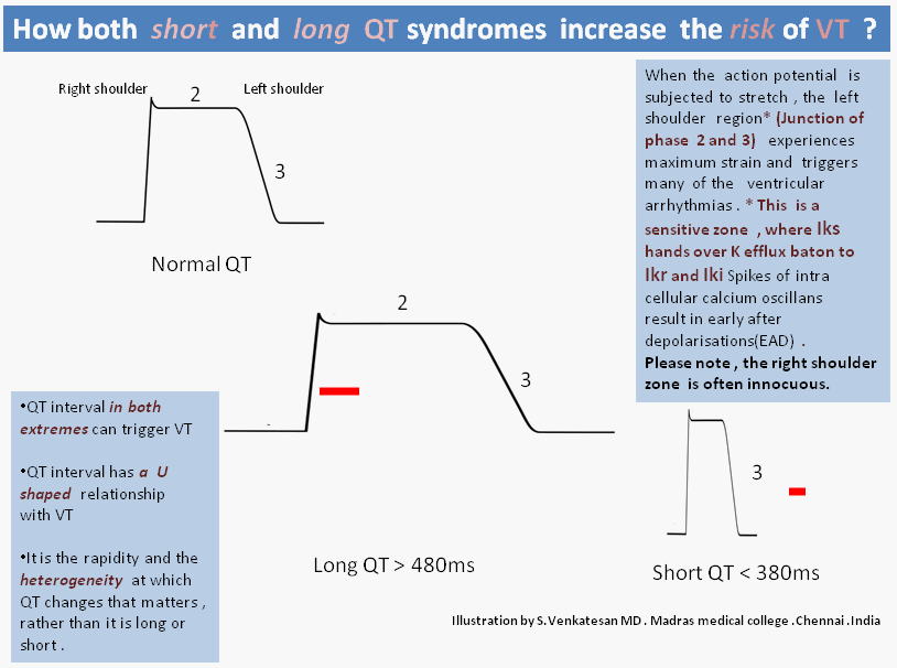
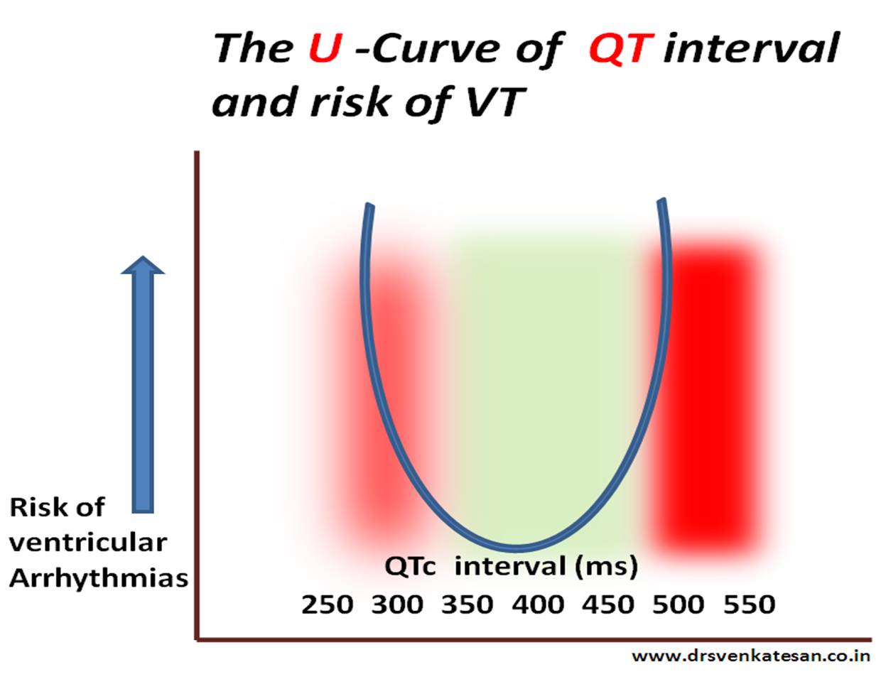
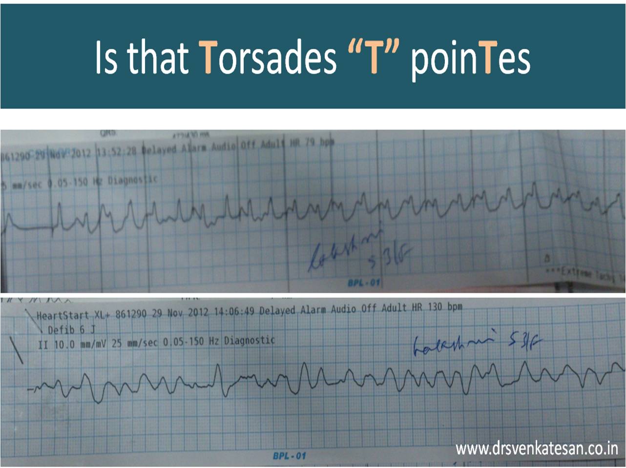
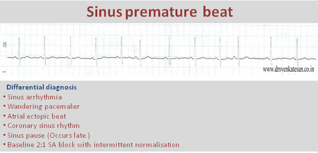
Syncope in CHB is due to unsafe escape rhythm, changing focus of VPDs,  extreme bradycardia, (<20 /minute),  pause induced VT, (Usually polymorphic and torsades is quite common .)  ultimately may end with convulsions,  ventricular fibrillation, and death.

Syncope in SND is due to extreme slowing of SA node . Sinus pauses or even arrest can happen resulting in ventricular standstill. Fortunately, a stable escape rhythm ensues more often than in CHB. (It may just be around 20 or 30/mt. still, ventricular arrhythmias are uncommon. ) This implies an important fact that stability is more important than slowness.Fatality is rare in SND.However, the mechanism of syncope in  SND is influenced by the integrity of AV conduction also. If it is severely impaired it can trigger ventricular arrhythmias as well as the escape focus becomes unstable infra hisian location.

Paradoxically, in patients with SND, an episode of palpitation due to AF  or sinus tachycardia precedes the episode of syncope. An intelligent patient may recognize this as a warning and can take lying posture after runs of palpitation.This is because of tachycardia-induced suppression of  SA node prolong the sinus node recovery time still further.

How to differentiate cardiac syncope from simple vasovagal syncope?

Cardiac syncope  is differentiated by common vaso-vagal syncope (VVS) as the latter occurs during erect posture . It may be entirely due to vascular component and hence it may simply represent hypotension without a true cardiac limb .(Vasodepressor syncope)

Hence the pulse rate and volume may take some time to recover in VVS, while Stokes  Admas of CHB  usually have a well-formed bounding pulse in the recovery phase, as the rate is low and systemic hypoxia is a consistent feature.

How is the respiration during Stokes – Adams syndrome ?

Intact. Oxygenation in the lungs goes on for time being. The pooled pulmonary blood gushes after the termination of syncope and causes  the classical flushing. Since the hypoxia causes systemic vasodilatation the flushing is more obvious.(Unlike vasovagal syncope where they are often pale)

History of stokes Adam’s syndrome Morgagni is the  one who gave credit to their  discovery

Though Morgagni first described the clinical picture of this syndrome in 1761,  It was published much later by Two Irish Physicians  Stokes, Adams. Wish this entity is referred to as Morgagni-Stokes-Adam’s syndrome

Reference

1.R. Adams. Cases of Diseases of the Heart, Accompanied with Pathological Observations. Dublin Hospital Reports, 1827, 4: 353–453.

2.W. Stokes. Observations on some cases of  permanently slow pulse. Dublin Quarterly Journal of Medical Science, 1846, 2: 73–85

Read Full Post »

qt interval animation short and long002

The stretch and strain experienced by the action potential’s left shoulder region is almost similar* in both long and short QT syndromes that trigger a VT.(* Hope this explanation makes some electrical sense !)

*Click  over the image for  high resolution

What is short QT interval ?
It is a range . Any thing less than 380 ms can be considered  short.Generally It becomes important only at < 320ms.
Relationship between QTc and risk of VT .
U curve of long and short qt syndrome

Read Full Post »

Scientific studies can be fun .In our spare time we often Indulge in rapid  fire sessions. We tested 30  wide qrs ECGs from our archives  (All proven VTs)  and  asked  our  cardiology fellows to apply Brugada criteria . They could   correctly  diagnose  VT  in 18* patients.The same ECGs were shown to the staff nurses of coronary care unit . 24  VTs were correctly identified  it.They did it by  their clinical sense and Instinct. (*12 vs 6  VTs missed)

wide qrs tachycardia svt with aberrancy and vt brugada verecki  griffith002

And now , four  clinical data was  provided. (Age , sex , Blood pressure , and  past H/o  MI were given )   The Nurses were able to predict it  28/30  VTs correctly.(97 % accuracy ) and the cardiologists  were able to equal the score now. So obviously clinical sense  was far superior .

Cardiology fellows were more likely to  mistake VT as SVT. This is far more common than SVT mistaken as VT. It is a strange academic  irony ,even the junior most nurses never missed a VT !

Summary

Simple sequence of history and clinical presentation is still far more powerful than ECG data in predicting wide qrs  arrhythmias . Nurses guess work is far superior than cardiologists  in predicting a wide QRS tachycardia as VT.

In fact , the  cardiology fellows are  preconditioned to   get confused   whenever they get a wide qrs tachycardia . Why  not aberrancy ?  In my  experience I have seen this question keeps  erupting inappropriately .Even  shrewd fellows suffer  from an  oscillatory  mind between VT and SVT .This is primarily because , every wide qrs ECG  is likely to  have at least two  criteria that fulfill both VT and SVT.

The implications are  genuine  and far reaching . While nurses  show a patient centric thinking  cardiology fellows  thought process revolves around ECG . Many modern-day cardiac physicians  are disconnected from clinical reality  and are obsessed with  complex EP concepts  and end up with a miserable face in the bed side !

This is not a new  revelation in 2013 . Masood Akthar told this  three decades ago.

Caution
Never try to glorify  guess-work . EP is a great science .The  pioneering concepts have made us understand how a VT emanates, travels , and exit from myocardium . We are able to localise it and ablate it .All credit goes to science . But , when it comes to bedside recognition of VT ,  clinical  sense  is a clear winner .With a  consistently > 90 % predictive value   it  can no longer be called as  a  guesswork   and becomes a hard scientific fact. Especially so , when the  intellectual  analysis of surface ECG   could predict  it  with paltry 70 %  accuracy (Read Reference 1)
Reference
This  analysis startlingly reveal  a fact .The over all accuracy  rate of predicting the wide qrs criteria  by  popular algorithms  is   between 66-77% ,  just 16 numerals   more than  gross   guess work  of 50 : 50 ( This  . . . or  . . . that )
Link to  Masood Akthar article

Read Full Post »

Pacemaker current is   strangely  referred  by physiologists  as  funny current (I f ) . I am yet to find the exact reason .  This is the current  that  sustain  our life right from the day 22  of  embryonic life when the  cardiac jelly beats for the first time.   SA node  solemnly  follow our  entire life  before  making  a  bid-adieu !

pacemaker  potential sa node 5


pacemaker current if funny current poential 002

pacemaker current if funny current poential 003

What is contribution of  If  current in the overall Pace-making  activity ?
This  has not been quantified . The fact that ,  Ivabradine induced  If  current  blockade does not result in serious bradycardia indicate  , SA node has alternate reserve currents as well . ( SA node  is a such a mystery  structure , it would never be a  surprise , if we  find many more  “not so funny”  currents !)

Read Full Post »

It is believed  T wave alternans  is a marker of impending ventricular fibrillation. Though it is not applicable in every clinical setting it is indeed true if we observe T wave alternans in an acute ischemic setting .Here is a patient with  ACS and inferior MI who developed T wave alternans after temporary pacing.

T waves alternans torades de pointes 3

t wave alternans

T waves alternans torades de pointes

went in  for a chaotic  T wave rhtythm  and ended up in VF that  required s shock.T wave  alternans is other wise known as repolarisation alternans .

Twist dance of Heart

Torsedes is twist around it’s axis.   Any   ECG wave  can twist in it’s axis .If T wave alternans  becomes gross it will twist 180 degrees   .Once this happens the heart can go for  fibrillation any moment !

Final message

Extreme form of T wave alternans would result in  complete twisting of repolarization vector which is a  harbinger of ventricular fibrillation

I wish  this can be referred to as Torsades  “T” pointes instead of  Torseades “de” pointes


Read Full Post »

Ventricular tachycardia is a common cardiac arrhythmia. The significance of which can be very dangerous to relatively benign  depending upon the etiology and underlying heart disease . The ECG during VT is rarely useful to identify the etiology .Often times  ECG after reversal will  throw more light .

What are the ECG clues one should look for once VT is  reverted ?

  • Any evidence for old MI
  • Low voltage QRS/ LBBB/RBBB may indicate DCM
  • LVH -HOCM features
  • VPDs – Multiple , LBBB morphology / suggest  RVOT  VT
  • QRS slur or notching  indicating scars
  • Epsilon waves indicate  ARVD
  • RBBB pattern would  suggest  Brugada
  • Prolonged QT interval
  • Tall  T waves/ U waves /  Inverted  T -and other electrolytic abnormality.
  • Delta waves would indicate anti-dromic tachycardia.

Read Full Post »

We are taught in  medical schools  early in our career ,   ventricular premature  depolarization (VPD ) takes  LBBB morphology if it arise from right ventricle , and  RBBB morphology if it arise from left ventricle .This is a rough rule of thumb.

Why this rule is  unreliable ?

VPDs have a focus of origin—–a short circuit——and an epicardial  breakthrough . All these together influence the morphology. Within  the left ventricle , a deep endocardial focus  can  behave  vastly different  from superficial epicardial focus  . The  course of VPD is influenced by the myocardial status ( scars etc ) . Further,  the electrical  properties of  interventricular septum is shared  by both ventricles .

  • Generally – LBBB morphology  has  more localizing value .
  • Most RV focus have LBBB morphology (but not vice versa!)
  • LV focus can either have LBBB or RBBB

What happens to  a VPD  arising from  interventricular septum ?

IVS is  not only shared by both ventricles , it does  not have  true  epicardial  surface  (Both side  bordered by endocardium ) In most septal VPDs , breakthrough occur on either side of the ventricle  . However , It  keeps trying  to break through  epicardial surface  !  .  Hence , septal VPD  is like cat on wall situation .So the morphology varies quiet frequently.Further , the VPD can capture  the specialised conduction tissue occurs  more commonly with septal VPDs. This can alter both the width and morphology of QRS.

Read Full Post »

Modern human life is driven by technology  . We are gradually taken over by gadgets .Heart is not an exception . Implantable cardiovertor defibrillator (ICD )  is major innovation  where in , an  electrically  wayward heart  is brought under control  by series of automatic shocks . One of my patients with old MI and significant LV  dysfunction for whom ICD was an option  taught me a lesson in physics  . During  counseling ,  he was asking me about  the quality of life issues etc .
ICD  shocks  during sex 2
First,   he wanted to know  how  stressful  the  sexual acts  are ?  . . .Then he  surprised me with this  question .  He wanted to know ,  the  risk of  developing a VT/VF  during the act and  will the ICD  shock  his spouse as  well ?  That was a real  cracker of  a  question  I thought .   I had to do  a mini  research   and found that  these are  low energy shocks (30 Joules)  and transmitted electrical injury is a non issue  .
I realised   then  . . .  we live in a era  where   physicians often get educated from their patients !
Here is the  FAQs on ICD from the patient pages of circulation . With  due courtesy I  reproduce it here.
ICD  shocks  during sex
Final message
ICD shocks are  low energy shocks delivered  Intra cardiac ,  and  rarely  reach the surface  ,  hence has little risk for electrical injury  for those in contact.
References
1. Vazquez LD, Sears SF, Shea JB, Vazquez PM. Sexual health for patients with an implantable cardioverter defibrillator.

2.Steinke EE. Sexual concerns of patients and partners after an implantable cardioverter defibrillator.
Dimens Crit Care Nurs. 2003;22:89–96
3. A  review article on the  broad issue.
sexual activity in cardiovascular disease  circulation 2012

Read Full Post »

This is an  ECG which  I reported  yesterday in my clinic . I thought it was a  near perfect example for sinus node premature beat .

sinus premature beat spb 2

(Of course I need to explain  why the  P morphology  slightly  differs )

A  sudden unexpected  QRS  complex is often called as  ectopic beat . If it occurs prematurely (ie earlier than anticipated )  it is called as premature beat. If it occurs late it is refereed  to as escape beat .Please note the difference is not absolute .

Sinus node is a dramatic bundle of energy with divine powers that  drives rhythm of life !

The pacemaker cells are arranged in a compact fashion with  differential properties from cranial cells firing fast and caudal cells little slower. The neural control is under constant Neuro/electro/humoral  servo control mechanism.It is well known the pacemaker shifts it’s firing location within the SA node in fairly regular fashion .The entire SA node has rich adrenergic and  cholinergic  innervation , with  a dominant control by the later . (This is  why the intrinsic heart rate is  in the tachycardia  range (around 116 )  when SA node is denerved  pharmacologically )

wandering-pacemaker

sinus premature systole spd sinus node ectopics002

SA node ,  being  a complex structure ,  it is not surprising to note  few beats to fire  slightly late  or  prematurely.If it occurs late it is called sinus pause ,  if it occurs early it is sinus premature beat , if  both occurs  interchangeably  we refer it as  sinus arhhytmia. (Read  about sinus pause here)

What is the clinical significance  of   SPD ? (Sinus premature depolarisation )

It is a  very benign entity that it is  merely an  academic fascination . By  stretching my  imagination  I  can  correlate  it  with few possible  clinical issues.

  • May be it has potenital to trigger a  SA nodal reentry tachycardia  or In appropriate sinus tachycardia/bradycardia.
  • It may be imporatnt in sinus node modification process.
  • However ,the main issue is  thee  cardiac physicians  in their enthusiasm should not mistake it for some serious  cardiac arrhythmia !

Related article

https://drsvenkatesan.wordpress.com/2009/04/14/can-premature-ectopic-beats-occur-in-sa-node/

Read Full Post »

Q waves are  neither  sacred  nor sinister waves . It represents   either of the  following .

  1. Electrical activity that goes away from the recording electrode.
  2. Or whenever there is a  electrical insulation or hurdle that interrupts the flow  of current  towards the electrode ( and if it is sustained )  it  can result in q waves (Minor interruption produces  a notch or  slurs . Please note a major slur becomes a q wave equivalent  )

Here is young women of 42 years with  a diagnosis of  old  anterior MI for   over 5 years ( Getting a dedicated care from a cardiologist!  The prescription included Imdur/Betaloc/ Statin/Clopidogrel and Aspirin )

This was the ECG . It was very convincing for  old ASMI.

epicardial fat and poor r wave in v 1 v 2 v3 q  waves

It  happened ,  I did an echo for her .

epicardial fat and q waves in ecg pesudo infarct non infarct 2  q

She lacked wall motion defects even after a meticulous search .  Instead   she had a   good layer of epicardial fat measuring 9 mm .That was more localised in  anterior wall extending little to LV apex.Her EF 65 % .

*She was a  fairly obese (not gross )  individual with a BMI of 34 .The fat pad thickness was not that huge  , I thought , still it was producing the q waves . I  have seen much thicker fat pads with good R waves in ECG . I  wonder ,  is it the type of fat that adds up to electrical insulation ?

This patient was sent back to me  again  for ruling out ASMI .  Echo was  done  two weeks  later . No evidence for  ASMI  could be detected.

epicardial fat and q waves in ecg pesudo infarct non infarct fat 2  q

What is the normal thickness of epicardial fat pad ?

It is less than few mm . Exact normality is not known .(Empirically < 5mm ) it is very rare for fat deposition  in infero posterior aspect , except in morbid obesity.

What is the function of epicardial fat ?

  • Long considered inert . Now , found to be a metabolically  active lipid pool.
  • We also know  heart  consumes more fatty acid than an other organs for moment to moment energy consumption .
  • Inflammatory mediator in atherosclerosis ?
  • It may also act as a mechanical cushion effect along with pericardium
  • Rarely fat infiltration can compress the heart and may result in restrictive  AV filling defects in doppler  .(May explain the unexpanded dyspnea  in many obese patients )
epicardial fat a dynamic depot athreosclerosis

Role of epicardial fat depot in the genesis of atherosclerosis

Subcutaneous vs  Epicardial fat.

We know thick chest wall can also interfere with ECG. Epicardial fat is more likely to record  q waves than  sub cutaneous fat ,  as the insulation is more closer to heart in epicardial fat . In thick chest wall current leaks from heart and  well scattered  hence  poor R wave is more common in such situations rather than q waves !

Following things can generate  q waves (Other than Infarct  )

  1. Fibrosis-Myocardial /Interstitial
  2. LVH
  3. Thickened pericardium
  4. Thick chest wall/ Epicardial fat
  5. Air/Fluid in pericardial space

Final message

In obese men and women  anterior Q wave can be  misleading .Such  medical errors can be so convincing .

After thought

If epicardial fat can  extinguish   R waves  and  replace it with  q waves  , these  innocuous  looking fat pads has every reason to  influence the ST segment shifts during  an episode  of ACS  as well !  .  Isn’t ?  . If so  . . . how reliable is  our ECG criterias  to diagnose  acute coronary syndromes  in grossly obese men and women ?

Read Full Post »

« Newer Posts - Older Posts »