Feeds:
Posts
Comments

Pacemaker implantation is one of the few high impact interventions done by cardiologists.Traditionally , RV apex was paced for many decades with tined or screwing leads. Serious concern was raised in recent years about the ideal site of pacemaker lead position. .Today, some argue  pacing RV from RV apex is a electro-physiological crime (Of-course it appears  more of hyped up fear ! )

What is the concern with RV apical pacing  ?

RV apical pacing is  unphysiological .To mimic physiology we have to move the lead as  cranial as possible within RV. (*Obviously  pacing anywhere in RV is non-physiological .Best would be atrial pacing . This is possible only  in sinus node dysfunction with Intact AV conduction.) Hence RVOT and mid septal pacing was proposed and practiced as a relatively  more physiological site as the spike could capture the natural conduction system early and ventricle is activated from above down.

How to localise the RV lead in mid septum ?

Inter ventricular septum , hardly occupies an area of 5/5 sq cm  with a  complex structure , shape and alignment with RV and LV cavity .In pathological states it can further get distorted. To catch it’s profile in fluoroscopy and imagine the plane requires a mind of “cardiac architect”.So, to locate the  IVS various views are suggested. Currently LAO 40 is typically  used for fixing  mid septal position. In midseptal position ECG is expected to show small q  in lead 1 and AVL. QRS axis will be more left .We seemed to agree with this.

rv pacing mid septal postion rvot rv apical
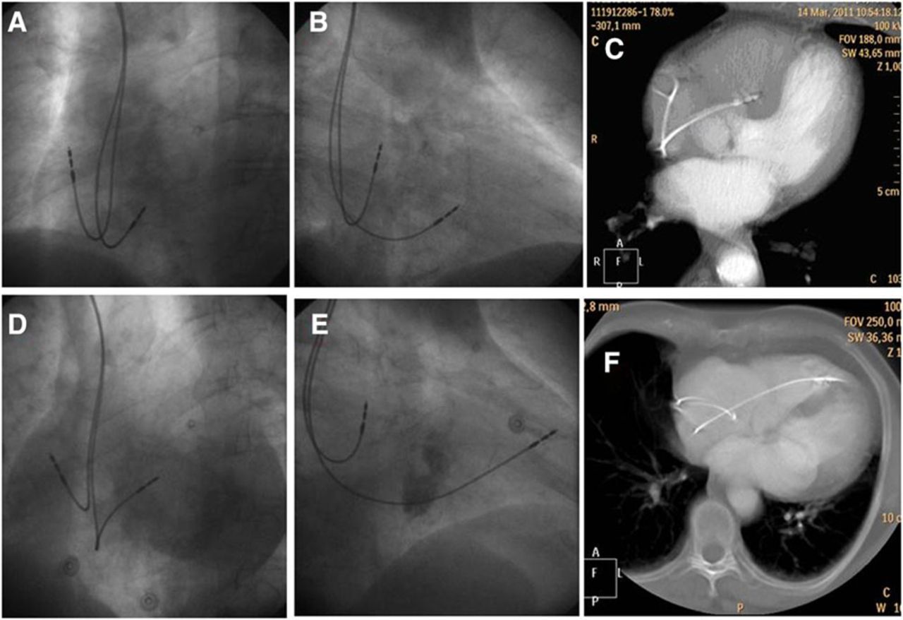
Image courtesy: Pavel Osmancik Circulation: Arrhythmia and Electrophysiology. 2013; 6: 719-725 Fluoroscopic examples of 2 patients. One with correct lead placement in the septum (A–C) and the other with the lead placed in the anterior wall (D–F). See the similarity of the left anterior oblique 40 view, the difference of the right anterior oblique 30 and the location of the lead on computed tomography image.

And suddenly  this paper  from  the picturesque  city of  Prague , Czechoslovakia throws a stunner.

What we have been thinking as  mid septal pacing with LAO protocol turns out to be   anterior RV pacing in 60% of times.The study showed LA0 40 view  failed to differentiate anterior RV pacing from mid septal pacing .CT scan correlates have documented this . Further ,we have never given a thought to the wayward trabecuale coming across mid septal zone. It has happened time and again (Atleast i have witnessed) the lead would  just fall short of IVS and get screwed in the trabeculae  with high risk for dislodgement . RAO 30 view  consistently separates the mid septal lead from anterior  RV position.It is suggested a carefully done per-procedure echo would also be useful in locating the lead tip .

What hemodynamic implication of mid vs RV anterior pacing ?

Could be significant . No reliable data available. It s tempting to think the hemodynamic inadequacies of an improperly placed mid septal RV pacing may not be different from a conventional RV apex pacing .

Final message

The complex shape of RV and IVS in  fluoroscopy can fool us. If you really want to pace the mid septum, please ensure with multiple views and confirm with RAO 30  . Also remember echocardiography is not a forbidden tool for localising pacemaker lead.

Reference

1.The Insufficiency of Left Anterior Oblique and the Usefulness of Right Anterior Oblique Projection for Correct Localization of a Computed Tomography–Verified Right Ventricular Lead Into the Midseptum Pavel Osmancik, Petr Stros, Dalibor Heart 2013; 6: 719-725

A land mark concept , that changed our understanding about the mechansim of genesis of Atrial fibrillation happened  in the year 1998 .( Haïssaguerre ,Spontaneous Initiation of Atrial Fibrillation by Ectopic Beats Originating in the Pulmonary Veins N Engl J Med 1998; 339:659-666). He proved AF originates in specific focal points from the pulmonary vein ostia at its draining point in LA.Even though there only few selected focal points it was difficult identify those and hence empirical RF ablation of all 4 pulmonary vein became a standard practice. 100s of thousand of these invasive procedure were carried out in patients with chronic AF.

Now, in a span of 10 years , we realise many of these patients require second or third siting of ablation.The irony is , there are many non pulmonary connections  that require repeat ablation.

Common mechanism for recurrences are

  1. Inadequate first ablation
  2. Reconnections
  3. Inflammation and fresh scars
  4. Additional venous focal  sites (coronary sinus  ,SVC, Vien of marshall)
  5. Multiple mechanisms /*Non- focal , systemi AF mediated by neurohumoral triggers ?

A study from the prestigious JCE in May issue of 2015, reveals a starling fact , that about 50% of AF patients  have additional connections  other than pulmonary vein that require ablations at a future date.

If proven right ,  just wonder how much of knowledge and its dissemination  , efforts from  EP industry , technology transfer  over the years is threatening to become redundant .Let us hope,we will somehow conquer the AF either electrically or pharmacologically.

pulmonary vien ablatioan atrial fibrillation carto non pulmonary vien connection

A strong message comes out from this. In modern science, one need not be unduly excited about a new breakthrough.Proof of concept will have to overcome the ultimate test , ie time .

We essentially live in our blood vessels and age in our arteries.CAD is the principal  cardio vascular disease, which  God has created in Homo-sapiens to ensure they  do not stay more than “allotted life span” in this planet. Of course , the current generation cardiologists equipped with scientific weapons , have since decided to take the fight directly in the Almighty’s domain .Contrary to the popular medical doctrine, treating an established CAD seems to be easier task than preventing a new onset CAD.

While , arteriosclerosis is a normal aging process, atherosclerosis could be an  aging as well as a distinctive pathological process. However , athero-thrombosis  is a definite pathology of vessel wall .We know at any time atherosclerosis  can transform into  athero-thrombosis  and result in  clinical event depending upon the  triggers and other associated conditions, which we refer to as major or minor  risk factors.

In scientific terms ,

  • Primary prevention  of CAD is preventing first episode of  Coronary  event*(Typically , CSA, STEMI/UA,NSTEMI/SCD,)
  • Secondary prevention  is  prevention  of  second  or subsequent episodes following the first clinical event.(*What if , if  the first event is silent and never known ? )

For all practical  purposes CAD and  coronary atherosclerosis is synonymous. Can we  prevent atherosclerosis in human biological system ?  What are we  supposed to refer to  such a preventive measure , if any ?

We are biased towards obstructive CAD as we often  think it is the  the only form of CAD .Then , how do we diagnose , treat and prevent a minimal non flow limiting plaque , coronary endothelial dysfunction , or  acute coronary erosion that can occur in very early stages of atherosclerosis, in other wise healthy persons.(Routine IVUS ,OCT ? Futile isn’t )

  • Preventing  sub-clinical  CAD  from manifesting  as clinical CAD , is technically   primary prevention”  but still  patho-biologically  secondary prevention.
  • Preventing a CAD in a patient  with peripheral vascular  disease  or preventing CAD in a patient with TIA or stroke is secondary prevention for cerebrovascular disease but  falls within the definition  of primary prevention of  CAD.
  • Then  comes the new semantics :Primordial prevention .This could be same as primary (or another  version of primary prevention ).Primordial prevention is preventing development  the risk factor  itself (Like DM,HT, Dyslipidemia )

So ,whenever , we talk about primary prevention of CAD by Aspirin or Statins ,realize the complexities involved .Before i finish, let me make you  dizzy further with this quixotic one . In a multivessel CAD, as the  atherosclerotic  plaques are scattered across the coronary arteries in various stages of  maturity  , long term Aspirin  following  anterior STEMI has to secondary prevent an event  in LAD territory  . . . but   primary prevent a plaque disruption in RCA territory !

Reference

1.USPSTF Guidelines : Aspirin for the Prevention of Cardiovascular Disease: Preventive Medication

TAVR is the new state of the art Aortic valve replacement procedure done by cardiologists .Nearly 200 thousand implants have been done , and it is
backed up by major trials TAVI TAVR valve dislodgement embolism partent a b What keeps the Aortic prosthesis in situ in the aortic root/Annulus  ?

The valve is not actively fixed but passively positioned in aortic root by either self expanding or balloon expanded valve  system .It retains the position by two different forces acting on the valve in two difffernt directions , but work coherently to keep the valve static .The radial force of the hardware is centrifugal and the elastic force exerted by annulus is centripetal .It may appear mysterious how these oppose each other in a balanced way and arrest the valve in the desired site. Fortunately, there is little  supero- inferior force operating and hence the chances of dislodgement is low .It should also be mentioned we are not yet clear about the best site for TAVR. Annular , supra annular ,or is it at lower virtual annulus , all has some advantages and disadvantages.

Is progressive aortic annular dilatation possible in these degenerative aortic valve ?
Aortic stenosis is chronic degenerative disease. Generally we expect the annulus is narrow and fixed. However for some reason if the aortic annulus loses it constricting force or the root dilates or fresh calcium deposits, there is definite risk  (Not theoretical :See Reference ) of valve destabilisation  , dislodgement and embolisation . *It is vital to understand the para-valvular leak could be a  remote precursor of such potential dislodgement as it represents  micro gaps  in the prosthetic / tissue interface.

How many such  embolisation of valves  are reported following TAVR ?

While the incidence of para valvular leak is common ,(up to 20%) fortunately valve embolisation is reported between .3 to 7.5% (Ref 2). Stastically , subclinical   destabilisation, malpostion  and dislodgement should be more common. The timing of displacement is not clearly reported in literature .It can occur  at any time between few hours after implantation to a much delayed (months after ) complication .

Final message

TAVR is a major break through in Interventional cardiology .It gives us hope for possible TAMR (mitral) and other valve repair modalities .Though dislodgement of prosthesis appear a rare event it is tempting  to ask ,  whether we should work towards a actively fixing valve in aortic root ? That remains a open question !

Reference

1. A survivor of late prosthesis migration and rotation following percutaneous transcatheter aortic valve implantation. Pang PY, Chiam PT, Chua YL, et alEur J Cardiothorac Surg 2012;41:1195-6.

2.Thirty-day results of the SAPIEN aortic Bioprosthesis European Outcome (SOURCE) Registry: A European registry of transcatheter aortic valve implantation using the Edwards SAPIEN valve.Thomas M, Schymik G, Walther T, et al. Circulation 2010;122:62-9.

3.Migration of the transcatheter valve into the left ventricle Christopher Cao , Su C. Ang , Michael P. Vallely Mart Annals of Cardiothoracic Surgery Vol 1, No 2 (July 2012 4.Delayed Transcatheter Heart Valve Migration and Failure Vuyisile T. Nkomo, Rakesh M. Suri, ,J Am Coll Cardiol Img. 2014;7(9):960-962.

TGV is the most common cyanotic heart disease to present at birth.The outcome is dismal unless it is intervened at the earliest .It becomes a real  emergency if the dTGA is associated with intact IVS and IAS. (Though foramen ovale /ductus  may help for a while prolonging the survival . Often times , it is restrictive and demands immediate septostomy if primary arterial switch correction is not attempted )

Presence of VSD provides vital time interval to plan  surgery in a less emergent fashion. Otherwise , arterial switch if  contemplated one has to do it before 2-3 weeks (Rarely up to a month ) .The principle is to reverse the circulation before the  systemic left ventricle regress its myocyte function as it is exposed to low pressure pulmonary circuit .

Hemodynamics

We know , dTGA  has two parallel circuits in a bizzare hemodynamic disconnect .The right sided  pulmonary circuit sustains  the systemic blood flow with deoxygenated blood.The left sided systemic circuit recirculates oxygenated blood into pulmonary circulation again and again.

Survival depends upon , the anatomical communication between two circuits and the effective quantum (Deoxygenated)  of blood flow to the lungs .

Which communication is best for survival d TGA ?

The key is to understand   what we mean by “mixing  vs  shunting”  and the effective pulmonary blood flow (ie quantum of truly deoxygenated blood reaching lungs  vs oxygenated blood recirculating  the lungs in a hemodyamically wasted loop

It appears both atrial  ventricular level  mixing is good for maintaining adequate O2 saturation than PDA .

What does PDA do ?

Ductus is expected to persist in  dTGA  when  IVS and IAS are intact  . It is not clear  what determines the persistence of duct  in dTGA with intact IVS. Principles of natural  hemodynamic adoption would argue , all patients with dTGA should never close their ductus and foramen ovale. While , In reality it may be true for PFO to persist, ductus often gets closed on schedule* , aggravating  the hypoxia .(Both flow mediated myogenic resposne, O2 content of ductal blood and size are the determinants of ductal closure)

The ductal circulation in dTGA can be complex . It initially  acts as a channel to mix both arterial and venous blood flow .It can become a liablity  if its large , as the pulmonary vascular resistance falls,  blood is shunted from aorta to pulmonary circulation and potentially precipitate failure .Please note,  this shunting  also can  improve  the saturation as it diverts aortic blood into lungs ,still the ultimate usefulness will depend upon intermixing  at some other level  other than ducuts , ie either atria  or ventricle .One should  realise  ductus can never bring  good admixture as it happens outside the heart .It is obvious intra cardiac  mixing  with ASD and VSD  is always better and devoid of providing differentially saturated blood  to the extremities  , which is an inherent feature  PDA mediated  mixing .If ductus  is the only means of mixing and shunting  the lower half of the body has  an advantage as it is perfused continuously by oxygenated blood .This  may manifest as  reversed differential cyanosis in few combinations of TGA physiology. (Relatively pink  lower limbs and cyanosed upper extremity )

Summary

Ductus can be a double edged friend or single edged foe  in dTGV . It depends upon the size , quantum of shunt and associated  channels of mixing like ASD and VSD . If it occurs with  intact IAS and IVS it plays a role of  life sustainer.

Presence of ductus is definitely useful  initially.It can either help by mixing, intermittent bi-drectional shunting  or committed left to right shunting .This is why we attempt to  preserve its patency or even recannalise it by stenting  in such situations .The later can be used to buy time and train the regressing LV .However ,large ductus can be counter productive  if additional shunts are also present where one should even contemplate closing it .

Reference

1.

pda in dtga ductus ductal flow in tga2.Transposition of the great arteries with intact ventricular septum and patent ductus arteriosus. Waldman JD, Paul MH, Newfeld EA, Muster AJ, Idriss FS.Am J Cardiol. 1977 Feb;39(2):232-8.

Beta blockers(BBs)  have become  key drugs in  management of CHF .It helps by blocking  toxic effects of inappropriately  elevated   catecholamine  , which is actually a  compensatory response(A fight and survival reaction )  from the sympathetic system  to a failing  heart . This process becomes a liability in the long run  as the  adrenergic receptors either down regulate or even promote apoptosis and cell death .Along with  RASS-ACE  it affects every cell in the body promoting neuro- humoral catabolic state.

By trial and error  methodology we have found blocking the sympathetic system by BBs confer  consistent benefits in CHF .This is in contrary to the days we were ignorantly stimulating the beta receptors with positive inotropic  agents and  wary to give BBs in cardiac failure .This is one the most dramatic 180 degree turn around in the annals of clinical cardiac therapeutics last century.

Is all BBs  same ? Is it the class effect ?

It is tempting to think  all  BBs are  equal and to conclude they simply represent a class effect.But carvidilol  seems to be the flag bearer , for whatever  reason . (Apart from the outcome of  landmark studies , there is a pharmacological basis for it’s superiority COMET/COPERNICUS)

What is the secret of carvidilol’s superiority in CHF than other BBs ?

We know CHF is a systemic syndrome as do the  sympathetic activation .Hence , In CHF , it would require a non selective , systemically acting beta blocker to reverse and reset the adverse  effects of  catechlamine.surge.(Does that mean Propronolol (Inderal ) could be the best ?)

Carvidilol being a non selective BB  fits  perfectly  for the job . Of course , additionally  it has alpha blocking action that reduce the  after-load  reliving he LV wall stress during systole enabling further lowering of MVO2 and   promoting  regression of LV size as well.

Having said  that  prototype cardioselctive agents  like  Metoprolol , Bisoprolol are also  backed by robust evidence  for survival benefit in CHF . How to explain this paradox ? (CIBIS/MERIT )

“Thinking wildly(Evidence  would come later ) it is possible the benefits  from cardio selective agents are  accrued much  later as the dosage is titrated upwards . I would believe the  “inflection point”  of benefit could be same time they lose the cardioselectivity”

Final message

Cardioselectivity is  boasted as a gifted property of BBs .It may be true  in HT, arrhythmia and angina , but  in cardiac  failure it plays a different  ball game .The simple logic is the target  receptors need to be blocked  in systemic fashion.

 Reference

2. http://www.jfponline.com/fileadmin/qhi/jfp/pdfs/6402/JFP_06402_ClinInq1.pdf

Syncope is a classical feature of LVOT obstruction especially with valvular aortic stenosis.The mechanism of exertional syncope in Aortic stenosis is traditionally attributable to the fixed obstruction .This fixed obstruction is not able to cope up with increased cardiac output as demanded by the exercising muscles . But exercise  induced reflex as well as local vaso-dilatation mechanism is intact . The consequence is predictable. A critical fall in SVR amidst a obstructed LVOT precipitating a syncope.

However , If the above mechanism is the sole reason for syncope in Aortic stenosis , we have a problem to explain why syncope is  rare even in critical mitral stenosis which is also fixed LV inflow obstruction ?

Is there some thing unique in LVOT obstruction that causes syncope ?

No, it is nothing to do with LVOT .To generate a true pathological syncope, reduction in cardiac output per-se may not be enough . It appears there should be an inappropriate systemic vasodilatation as well to precipitate a syncope.This can happen only if the parasympathetic system gets activated by some means . The trigger is located in the mechano- receptors of left ventricle . Hypertophied left ventricle with high Intra cavitory pressure (Often above 200mmhg) generated due to LVOT obstruction activates the syncope circuit.The same rule may apply for RVOT as well .One could get syncope with critical valvular PS or severe pulmonary hypertension when RV mechanical receptors get a triggered.

What happens in mitral stenosis ?

In mitral stenosis , LV is under- filled ,  wall thickness is normal .There is little likely-hood of LV mechno-receptors to get stimulated as the LV wall stress is normal. This is the reason syncope is less common in mitral stenosis. However , this is not  absolute rule , syncope can still occur in severely narrowed orifice of mitral valve due to low flow state alone or a ball valve thrombus and paroxysms of arrhythmia .

Heart transplantation  as a treatment modality was conceptualized  by Christian Barnard in 1967 . Still considered as an  “Act of God” this  surgery is regularly performed worldwide by dedicated  transplant team consisting of cardiac surgeon , physician , Anesthetist , pathologist and others .Unlike other organs , heart transplant cannot have a “live donor” .Though  started  half a century ago, the real  pace has  picked up only in last 2 decades .Currently it is  “globally accepted  standard” intervention in terminal cardiac failure (Including pediatric heart conditions)

How are the survival rates ?

HEART TRANSPLANT SURVIVAL

  • Now, many centers  are able to reach the  bench mark Stanford- statistical rates with a consistent  five  year survival rate crossing   75% .
  • The median survival rates is 10.5 years
  • One of the estimate indicate , If they cross first year, median survival reach 13.5 years
  • There has been many living survivors who have  crossed 30  years.

Looking at these numbers  , there is dramatic impact  in terms of global disease burden and the life gained.Statistically  speaking  successful treatment by heart transplant is equivalent to overwhelming  many  cancers in a human body !

Can these  results reproducible in all centers ?

These excellent outcome is  the reward to highly dedicated teams with  pioneering work culture . One should be cautious to start new transplant center without proper facility and expertise.Unregulated heart transplant  centers is vested with risk of pulling down the excellent  statistics of this unique form of human organ exchange.

Newer developments

Patient  selection criteria and  strategies to prevent rejection is being streamlined .The major  issue is availability of donor heart and how to optimize organ procurement and increase transport survival time .Transmedics has deviced a state of the art organ transport system . The other exciting thing expected to happen is potential (Ironical though !) heart donors from  even cardiac  /circulatory deaths . (Dhital KK, Iyer A, Connellan M Lancet. Adult heart transplantation with distant procurement and ex-vivo preservation of donor hearts after circulatory death: a case series.2015 Jun 27;385(9987)

What about artificial hearts ?

As of  now biological heart has definite edge over  artificial heart.Meanwhile rapid development of LV  assist  device and near total artificial  hearts may  end up with destination therapy rather than bridge to transplant. The “INTERMACS ” data are very promising and let us wait for the day artificial heart  can  score over the biological  ones.

Reference

1. The Registry of the International Society for Heart and Lung Transplantation: Twenty-eighth Adult Heart Transplant Report–2011.Stehlik J1, Edwards LB, Kucheryavaya  J Heart Lung Transplant. 2011 Oct;30(10):1078-94.


2.
heart transplantation
longest survival after heart transplanation
longest survivor of heart transplant

Aortic stenosis is diagnosed by 2D valve morphology, area ,and pressure gradient across the aortic valve.Though anatomical 2D images and indices are good enough to diagnose severe AS , we are obsessed *  with pressures  which are subjected moment to moment hemodynamic and contractile variables. To record a good gradient we need a normally contracting ventricle and good flow across the narrowed LVOT. If any one of the is critically compromised  gradients can’t be picked up by Doppler.(A new entity of AS was recently included , which fails to generate the gradient in spite of good LV function and the AS being significant.)

So ,whenever one records a “Low gradient AS” there are 4 distinct possibilities.

  1. Truly mild AS
  2. Technical inadequate Doppler alignment , with possible true moderate /severe anatomical  AS .
  3. Low gradient AS due to LV dysfunction, with true moderate /severe anatomical AS
  4. Low gradient AS with Low flow but normal LV function, with true anatomically moderate/severe AS

Echocardiographer should rule out 1 and 2 before going to the complex world of low gradient severe AS.In my personal opinion , the entity of Low flow , Low gradient with Normal LV function appear  redundant ( or is it beyond my understanding ) .One should look at the valve morphology and decide in such situations.

Then , one will shortly bump into this query  is it 2 or 4 ?

How to differentiate a  technically defective  recording  of low gradient AS  from  true low flow due to narrowed LVOT.(Low gradient for me , high gradient for my professor !)

Now, basic readers  may please leave ,

Few inquisitive may ask   ( naturally though)

Does the ” low flow -low gradient AS”  is an exclusive phenomenon  that can occur only with normal LV function  or can it  occur in  dysfunctional left ventricle as well ,  who also have small cavity size and narrow LVOT  ?  (Within the low gradient AS due to LV dysfunction subset ,  How much is attributable  due to anatomial low flow  and how much is related to depressed LV contractile force ?)

Another googly . . .

Why can’t  Doubutamine* stress test  routinely  undertaken in the subset of patients with  with subjects with Low gradient /normal LV function to augment the anatomical low flow and find whether it is true  low flow or not ? *This would mean , a most impractical situation wherein every patient  with even mild AS should need to undergo dobutamine testing to rule out significant AS.

Final message

As of now ,this new concept   “Low flow , Low gradient, with Normal LV function” appears an  intellectual excess with little impact on patient outcome.The proposed new entity ultimately increase the likelyhood of over diagnosing  severe AS.Iam still expecting  more clarity  on the issue. ( or else for the moment forget the pressures and  simply fall back on  a meticulous assessment of 2D valve morphology and take a call , you will be surprised how often we get into man made scientific traps. )

Reference

1.Low-flow, low-gradient aortic stenosis with normal and depressed left ventricular ejection fraction.Pibarot P1, Dumesnil JG.J Am Coll Cardiol. 2012 Nov 6;60(19):1845-53

Degenerative Aortic stenosis occur with either normal  or congenitally malformed/ bicuspid valve.This contributes to the major chunk of  aortic valve surgeries and interventions (TAVR) in elderly population  . The optimal  timing of aortic valve replacement in patients with AS is debatable inspite of  well formed guidelines.

Three factors determine it .Symptoms , severity of aortic valve narrowing  and the tactness of LV function .The last parameter is a tricky one .We used to think in the past , severe LV dysfunction is a contraindication to aortic valve surgery. Now we realise ,however severe the LV dysfunction may be , relieving the obstruction will benefit  the patient and  the LV function is  also  likely to improve.

Cardiac physicians  face a dilemma when confronted with a patient  with low gradient and severe LV dysfunction .In this situation they are advised to do doubtamine stress Echo and watch for the gradient .If the gradient  increases that would  imply true fixed stenosis . (In pseudo aortic stenosis increased contractility opens the aortic valve and gradient will fall )

While this concept appears simple .There  are few  important issues that goes unaddressed  as we have not yet fully understood the  mechanism of LV dysfunction in aortic stenosis .(Link to mechansim of LV dysfunction in Aortic stenosis.)

At what degree of aortic stenosis LV goes down fighting and fail to generate the required  gradient ?

Myocardial function  and behaviour at times of hemodynamic stress can be highly  variable and most of us believe it is determined primarily by the genetic switches of myosin and other contractile elements .This is naturally proven at times of hypertensive left ventricular failure (Only in a fraction of the hypertensive population  LV is set to fail  when BP acutely raises.)

Proposed concept

Considering the complexities in cardiac mechanics , hemodynamics (and not to forget the vast control exhibited by genetic imprints over the hemodynamic behavior of LV) , it seems highly plausible even mild degrees of Aortic stenosis can inflict significant myocardial dysfunction in certain patients . Hence the phenomenon of pseudo aortic stenosis needs further critical analysis If this is proven to be true there could be a realistic indication for aortic valve intervention even in patient with low gradient / true Mild AS with LV dysfunction.
A word of caution is required .Relying too much (Which we often do ) on gradients in the assessment of aortic stenosis has skewed our common sense. Its wiser to have a  meaningful look at the valve morphology . A normal appearing  valve in 2D can never cause significant stenosis. Pressure recovery phenomenon also is to be given due respect as it over estimates gradient .This will effectively avoid surprises and guilt on table when we find a relatively good looking valve posted for AVR /TAVI