Feeds:
Posts
Comments

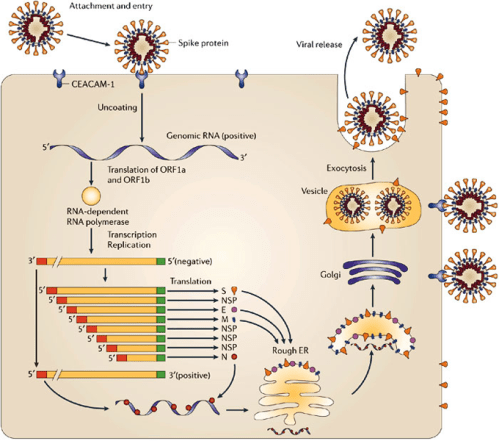
Viruses are essentially lifeless molecules (A nucleic acid RNA/DNA) .In the case of CoRoNA, it is the inbuilt RNA that acts as a commander in mischief. Ironically, it gets to life only when it attaches to the host cell. How a small bit of nucleic acid with a lipid cap infects a cell and becomes a deadly factory of new viruses and spread through the body remains a deep biological mystery.

Someone asked me , “Can we kill the Corona en masse”? The answer is frighteningly simple, we can’t kill them really, because they don’t have life in the first place. At best, we can deactivate or make it dormant and reduce its spread.

I just got contaminated with coronavirus . . .Is it a death sentence?

Even if you forcibly feed COvid 19 at random to 100 persons 80 % will be near normal or with a mild respiratory infection. You may wonder how can so many people are positive for Covid 19, and comfortable. We are still far away from understanding the complexity of how this virus will behave in a given human body.

This is because we are not clear what is the exact port of entry and how the first cell reacts to it.The way the body deals with it is entirely different if it attacks the respiratory tract through aerosol or it enters gastric tract (Imminent death with acidic PH ? we don’t know )

The mysterious interaction of genetic susceptibility, response to initial entry, epigenetic memory and subsequent immunological activation, will determine whether one is going to get simply infected and completely decimated.

Healthy humans enriched with good protein diet are expected to have good immunity. However, it can never be foolproof. It is obvious, there is something more than a host stress response readiness.(The fighting power and the fitness of your Immune cells T,B ,K,NK cells, infinite number of Interstitial scavenger cells and molecules)

When you are stressed the Immune system is activated or deactivated?

As expected the answer can be both. Then, how does the body will fight it over? Cells start synthesizing defense molecules. Unfortunately and paradoxically, cortisol is a major hormone released at times of emotional stress, that can severely compromise the immunity. Steroids are firefighters but cause collateral damage.

So its easy to conclude, positive emotions have positive immunity and negative emotions like fear, anger, distress can pull our immunity down.

Fear is a thought virus

Technically and biochemically, every human thought is a neurotransmitter. A neurotransmitter is nothing but a chain of amino acids synthesized in response to DNA/RNA codecs. So, straight away there is an obvious link between thoughts we harbor and the fighting power of the body. Why depression and anxiety affect the infection rate ?
The effect of various emotions on the Immune system is a big emerging topic. Fear-mongering about Corona and the manic digital dissemination of the virus of fear could turn out to be a great Immune system dampener.

One of my wonderfully healthy friends wanted to estimate the fighting power of his body’s immune system in case he is affected by Corona. He asked, whether his blood can be mapped and give a reassuring report?

I said no, it is not possible. Just take all the precautions. Reassure yourself that you have all the Immunity to fight. That’s it.

This following article elegantly explores the link between emotions and Immune response.

Final message

Can viruses befriend “fear” that reduces host Immunity and help self propagate? No one can be sure. But, I wish “unrestricted courage” acts as a vaccine to Corona, which can ooze from the brain free of cost.

Postample: What will happen to this pandemic?

It will (and should) settle down taking its toll. Preventive measures are gratifying. We need to learn from China, how they blunted the steep ascending curve of propagation.(Of course it started from there)

Who is responsible for such global pandemics?

I am sure, this is the toughest question, probably with no answer. Is it man-made or God made ? If you strongly believe, God will never punish human beings without a reason, then the answer is simple. Now, the world is under freeze. Its one way of arresting the mad growth of artificial,materialistic, biased economy. Corona could be a whipping force on mankind and let us use it to heal and unite fellow human beings.

Now, some positive news from CoVid 19 positive population

The false positive results are too high with currently done active screening tests.

This study from China says positive predictive value of a positive test is just 19% .It would mean 80% error rate. So, don’t really get unnerved with a positive test.

https://www.ncbi.nlm.nih.gov/m/pubmed/32133832/

Aortic dissection is a unique cardiac emergency that tests our collective understanding of vascular anatomy and pathology .It poses the ultimate challenge to the expertise and wisdom of both cardiologists, and surgeons.

It’s all about freezing the Time 

The philosophy of management swings between near-total Inaction* in some (As in most Type B & few Type A as well ) to “No holds barred” approach in others. (In most Type A and few Type B).

*Read it (also) as medical management that includes powerful Aortic pulse attenuation therapy with beta-blockers ( Unfortunately medical management is considered as Inaction by many current generation cardiologists)

Advanced Aortic Imaging is the key

A rapidly focussed TTE usually confirms the diagnosis.TEE might be used but carries a small risk of directly aggravating dissection when performed in an unstable patient. Conventional CT provides good (but limited) information, spiral MDCT delineates the vascular anatomy in a more clear way. MRI probably scores over and adds flow dynamics.

(4D Phase-contrast MRI showing slow helical flow in the false lumen and high-velocity flow in the true lumen. Computational flow dynamics will help assess entry point, plane of dissection, calculate false lumen Index, pressure and wall stress in true lumen and Aortic branch compromise etc . Image courtesy. The Lancet Volume 385, Issue 9970, 28 February–6 March 2015, Pages 800-811)

What is Non-A Non-B dissection?

This is a newly recognized subgroup. It’s not a surprise as the imaging modality improved we found the existence of this subset. In Non-A -Non-B Dissection initial tear involves the Arch and threaten to go retrograde A or antegrade B. We also realized there could be an apparently illogical transformation of type B becoming Type A, later on, due to late retrograde dissection.

The incidence of Non A , Non B dissection is about 10% (Ref 1). Here the outcome between surgery and medical is confusingly sitting at equipoise.

The traditional Debaky and Stanford classification didn’t address this subset.Though some other classification Like (DISSECT (Ref 2) /PENN (Ref 3) tried to clarify .

A new classification based on Type ,entry and malperfusion appear perfect.

TEM classification of Aortic dissection

This is a practical classification that uses Stanford as a base model but adds entry point and branch vessel compromise. This is analogous to TNM classification of tumours.(Hans Hinrich Sievers et al)

Where does EVAR stand ?

The management strategy of dissection of Aorta got an important makeover in the last decade (for good mostly ) as interventional cardiologists and radiologists landed in the hitherto to surgical domain with endovascular reconstruction (EVAR) .It is handy mainly in the complicated type B and complicated Non A and Non B . One more viable possibility is the hybrid approach of combining EVAR and surgery in delayed presentation of Type-A.

Single point Principle in Aortic dissection management

Rapid sealing of the entry point and arresting the false lumen progression wherever it is and by whatever means (especially in Type A dissection ) reduces mortality significantly.

Though exit points are important for comprehensive management, one need not waste time to locate and search for exit points. In a specific group of patients, it may not be visible or even absent altogether.

Another critical determinant

Detection and tackling the retrograde dissection and involvement of coronary ostium distortion and damage to Aortic valve , and pericardial invasion is the key to reducing early mortality in Type A dissection

The final outcome in God’s domain

The IRAD and other global registries in Aortic dissection has taught us important lessons. We are continuously learning and the patient outcome is improving.

Still, one unresolved statistical ( metaphysical ) mystery is how to identify? that small subset of patients who are lost because of inappropriately early aggressive Intervention who would have otherwise been pushed into natural survivors of Inaction or less action.

Reference

1.Sievers H-H, Rylski B, Czerny M, Baier ALM, Kreibich M, Siepe M et al. Aortic dissection reconsidered: type, entry site, malperfusion classification adding clarity and enabling outcome prediction. Interact CardioVasc Thorac Surg 2020;30:451–7.

2.Dake MD, Thompson M, van Sambeek M, Vermassen F, Morales JP;DEFINE Investigators. DISSECT: a new mnemonic-based approach to the
categorization of aortic dissection. Eur J Vasc Endovasc Surg 2013;46:175–90

3.Augoustides JG, Szeto WY, Desai ND, Pochettino A, Cheung AT, Savino JS et al. Classification of acute type A dissection: focus on clinical presentation and extent. Eur J Cardiothorac Surg 2011;39:519–22

Metanalysisfor Non A Non B dissection

Professional medical practice demands to put always the patient’s interest first. Unfortunately, current practicing methods threaten doctors to yield to patient’s whims & wishes which are influenced by significant non-academic forces. The principle of Informed consent is gradually losing its true meaning. Who is informing what ? and to whom?  is becoming a hazy conundrum in complex two-way confabulation based on severely battered evidence-based medicine.

pateint empowerment

Some of the conversations not heard in silent corridors of big hospitals

Why did you stent his LAD ? , He had triple vessel disease Is’int CABG Ideal?

“Yes , I agree. What  can I do , the patient  chose to get stented”

Why did you replace his keen joint, it was not that bad isn’t ?

“Yes, I agree but the patient chose it.”

Why did you do the cesarian madam? the pelvis was fine, she was contracting well  Isn’t?

“Yes, I agree, what to do. The patient decided it”

Why did you do the endoscopy, you are sure it was simple dyspepsia right? 

“I agree. What to do? The patient wanted it”

Final message 

We all agree patient empowerment is a critical component of health care delivery and management. I am afraid it can very easily go wrong and take a bizarre direction. Many times I felt it has seriously Interfered with professional decision making.

Still, I am not able to come to terms with this awkward situation. “How can  patients  (or their health care provider) enforce me to do a procedure on them , which I feel is Inappropriate or Injurious to them !

Missing you Dr.Hippocrates

Bernoulli equation is the most critical equation on which the foundation of clinical Doppler echocardiography is built. Bernoulli equation tells about fluid mechanics. Bernoulli’s principle states that the sum of potential and kinetic energy of fluid per unit volume flowing through a tube is constant.

A more detailed explanation regarding Bernoulli equation is linked in this video

Applying Bernoulli in Echocardiography

So, if we can somehow measure the velocity gained across a point inside the heart we can deduce the pressure gradient. Here comes the Doppler principle that helps us calculate the velocity. Doppler is based on the reflection of sound and the Doppler shift. With the Doppler shift, we can arrive difference in velocity across a valve, or conduit. When fluid flows across a narrowed orifice (Valve /Outflow) it accelerates and builds up velocity. This gain in velocity is equal to the pressure lost ie as given by the Bernoulli equation. Since potential energy is related to height and gravity same intracardiac zones it cancels out on either side. Hence, essentially the Bernoulli pressure gradient is equal to the difference between the kinetic energy on either side.

Let us see how this 1/2 of mass becomes 4. We have to convert density of blood which is 1.060 to mass.

mass to density

Note : Mass = ρV . Density is mass per unit volume. So the “m” in the equation is some times referred to synonymously with the density of blood.

modified simplified bernouli equation doppler pressure gradient mass density velocity drsvenkatesan madras medical college echocardiography 2 tr jet lvot gradient

Application in clinical echocardiography

There has been pioneering work from Holen, Hatle and Angleson who proved the value of this equation in the clinical situation in the late 1970s. Of course, Gorlin and Gorlin worked on this similar concept in the cath lab derived pressure gradients

Reference

1.Gorlin R, Gorlin SJ. The hydraulic formula for calculation of the area of the stenotic mitral valve, other cardiac valves, and circulatory shunts. I. Am Heart J 1951:41:1-29.

2.Holen J, Aaslfd R, Landmark K, Sknonsen S, Ostrem 1. Determination of effective orifice area in mitral stenosis from noninvasive ultrasound Doppler data and mitral flow rate. Acta Med Stand 1977;201:83-88.

3.Hatle L. Noninvasive assessment and differentiation of left ventricular outflow obstruction with Doppler ultrasound. Circulation 1981;84:381-
4.Hatle L. Brubakk A. Tromsdal A. Angelsen B. Noninvasive assessment of pressure drop p in. mitral stenosis by Doppler ultrasound. Br Heart J 1978:40:131-140.

Here is a 3-minute algorithm for the management of acute pulmonary embolism. Just need to ask 3 questions.

Caution: User discretion is advised. Tainted with reasonably acceptable levels of non-scientific content.

Click over the image for a high-resolution slide

Some more critical  questions need to be answered.

What is hemodynamic stability?

It is purely based on clinical signs and judgment.(One need to be doubly sure to rule out hypovolemia and sepsis-related hypotension)

Is RV dysfunction equivalent to hemodynamic stability?

No, it is not. Clinical instability must be associated.( The dogma is,  if the patient is stable even if there is significant RV dysfunction by echo , that RV dysfunction is not attributable to the current episode of PE)

Can we diagnose and proceed with lysis without CT pulmonary angiogram confirmation?

Yes, you can, provided your suspicion is too strong or you have the extraordinary talent to argue/defend even a fatal bleed ( with your boss or in medical audit ) in a patient who was subsequently proven not to suffer from PE .

How to switch over to Lysis from Heparin alone protocol?

Occasionally one may require to do it. There is an added risk of bleeding here. It can’t be avoided in some situations as Initially, it appear as low-risk PE later on becoming more Intense. Generally, high-risk unstable patients should receive lysis straightaway.

Is 60/60 sign is really useful in deciding lysis?

60 /60 sign tell us if Pulmonary artery acceleration time (PAT) and the TR jet both are less than 60 the likely hood of PE is high in a patient with suspected PE.

  • This sign recently got popular not because of its utility, rather because of its simplicity and attractive caption.
  • It may be very specific but least sensitive (<20%) So it can never be used as a screening test.
  • It also fails to differentiate chronic RV dysfunction from acute RV dysfunction.
  • The PAT is strongly influenced by RV dysfunction (It pulls it down below 60 as PAT is dependent on RV Dp/Dt and falsely diagnosing PE
  • 60/60 sign adds up to the value of  Mconllels sign and can confirm PE with almost 100% specificity.

Pulmonary atresia with VSD is one of the complex CHD subsets that requires a meticulous understanding of anatomy, physiology of pulmonary circulation. It can be termed as TAPAC -Total anomalous pulmonary arterial connection in extreme cases. Should we attempt to reverse this total chaotic pulmonary blood supply is the question?

It demands a highly focused cath study(hands & brain) and CT Imaging which might actually throw more light. Post-study Interaction with surgeon and team of cardiologist are vital. The decision to take up the challenge of surgery or abandoning poses equal intellectual stress. Continuous and critical decisions need to be taken. Repeat surgeries and cath based Interventions are often a rule.  Very few centers have mastered this surgery.

A single slide presentation


pulmonary atresia 4

In spite of all technological developments in pediatric cardiac surgery, there is considerable variations and expectation of the surgical outcome. The major surprise is the original Melbourne group(Ref 1 )  that advocated the uni-focalization as a  concept has almost abandoned this. Stanford and other groups still continue to use this technique more often as a single-stage procedure to improve the outcome.

Let us hope these children get the best of the right mix of technology and natural survival power and more importantly we must ensure the former do not interfere with the later

Reference 

1

Post ample

Surgery has definitely  revolutionized the outcome in neonates and children in less severe forms of PA with good central pulmonary arteries ( Most of the Barbero Marcial Type A and many type B) The perceived negativity in this post regarding the outcome of surgery is primarily belong to some of the  Barbero Type B and many of  C.

gr1

Barbero-Marcial M , Jatene A Semin Thorac Cardiovasc Surg. Surgical management of the anomalies of the pulmonary arteries in the tetralogy of Fallot with pulmonary atresia.1990 Jan;2(1):93-107. 


Pericardial effusion is often detected in patients with Infective endocarditis. Incidence can be as high as 25% . Most often it is mild, can be moderate in few.

Mechanism

  1. Sympathetic effusion in response to endocardial infection. It’s never more than minimal. (Evidence ? it’s only an assumption)
  2. IE related cardiac failure (Raised systemic venous pressure to which pericardial veins drain)
  3. Local sepsis, Abcess formation tracks to pericardial space through transmural lymphatics
  4. Fungal , granulomatous , Tuberculous IE (Rare) Here IE and PE  share the same pathology
  5. Part of systemic sepsis activated Immune mechanism (Polyseroists)
  6. Renal Involvement of IE-Renal failure
  7. Postoperative pericardial effusion in Prosthetic valve IE (Common, often loculated)

Clinical Implication

  • If the pericardial effusion is more than mild, it often denotes worse outcome. This implies more extensive infection or a marker of extracardiac causes of effusion like renal dysfunction.
  • Effusion may predispose to local dissemination of infection and ends up as peri-annular abscess is whether it is a cause or effect of effusion remains to be understood.It is often exudate as one would expect, but transudative  effusions also occur and would indicate more benign course.
  • The sterility of pericardial fluid has not been proven. Culture studies are rarely done from effusions associated with IE.
  • Pericardial effusions appear more often seen in IE of right heart valves. They turn out to be  IV drug abusers.
  • Contained rupture of an abscess needs to be differentiated from effusion

Can we give steroids for PE associated with IE?

Steroids can rapidly plug the inflammatory pores in the from the pericardial surface.It may also prevent future constriction. Currently, routine steroid therapy is not advised in infective pathology . If the infection is confirmed and is being taken care of by antimicrobial therapy there could be a role for steroids with user discretion.

Final message

During the echocardiographic evaluation of IE, the presence of pericardial effusion should be specifically looked for. These patients should be flagged and will require monitoring as the prognosis of PE complicating IE is a concern unless proved benign.

Reference

Two studies one from Spain and other from Egypt looked into this issue specifically.


Science is a journey in pursuit of truth. Hence, we search for it again and again.  (Thus, recurrent search becomes Re-Search)

As we try to progress in our knowledge towards absolute truth,  we need to admit our errors first. I think, one such error is blinking right in front of us in the vibrant corridors of coronary care and cath labs every day!  It is about the definition with which we deal the success of primary PCI. (A supposedly revolutionary acute coronary therapeutics  this century)

Waiting for the day . . . when all those fancy primary PCIs that leave the myocardium hurt (& retire ) with significant LV dysfunction to be reclassified as clear cases of primary PCI failures.

Severe aortic stenosis will cut off the systolic BP and hence classical pulsus parvus et tardus occurs. This is what , we have been taught all along.

How for it is true?  

One thing is clear from clinical observation. Systolic BP need not be low, often its normal even in severe Aortic stenosis. The issue becomes curious when  high BP is associated with severe Aortic stenosis. This can happen by a variety of mechanisms.(Aging/Loss of Aortic elasticity /Pressure recovery/Hypertension)  I think, there have been little correlative studies of pulsus parvus with central aortic pressure.

Can Aortic stenosis be a cause for systolic Hypertension?   (This academically murky question rose after I stumbled upon this paper )

This paper from the journal of Human Hypertension which was published many decades ago.It sincerely documented high BP in spite of severe AS . The most crucial aspect of this study , however, was the fact that hypertension was completely corrected after Aortic valve replacement. The authors attributed to this high systolic BP as the transmission of LV chamber pressure. This is a frontal attack on the traditional concept of pulsus parvus and systolic decapitation in LVOT obstruction.

I am not sure, whether knowledge always breeds knowledge. Medical science is equally affected by new-onset Ignorance or not recognizing past knowledge.( Like this paper of 1996.) I think this study is done with a good scientific basis and unable to find any serious flaws. Hats of to the authors. This could lead to a further breakthrough in our understanding of transvalvular gradients in Aortic stenosis and the poorly understood vascular- valvular Interactions. With, catheter-based TAVRs become so common, we can exactly measure the pressure dynamics in the Aortic root pre and post valve replacement. (* My take is , systolic BP in severe Aortic stenosis  is preserved until the onset of LV dysfunction)

Reference