Feeds:
Posts
Comments

We learn from basic physiology  lessons that human body is made up of 60 % water. What about heart ? There is no reason for the heart should behave differently from rest of the body . If my  assumptions are correct when the normal heart weighs 300g  , 180g of which should be  be water. The same thing could be applicable for LV mass( * Reference requested)

Is there myocardial congestion in cardiac failure ?

Genesis of edema in any tissue depends on local hydrostatic pressures, tissue resistive forces, osmotic balance, and cell membrane permeability. In the myocardium individual contribution of above factors are not known. Apart from total myocardial water content  , myocardial water logging depends upon the trans myocardial venous gradient and the coronary sinus exit pressure in right atrium.Technically ,any severe right heart failure should lead to myocardial congestion at least to some degree.Unlike the lungs , the myocardial edema fluid doesn’t produce crackle , (May cause S -3 gallop instead) . However ,we have modern technology to image water inside the myocardium. Yes, it is called proton / hydrogen imaging or simply called MRI .

This is especially evident In chronic kidney disease , where in the fractional water content within the myocardium is expected to increase further as the whole body is water logged.


myocardial edema by mri in ckd 2

We have seen time and again patients with CKD  improve in LV function immediately  after dialysis . It happens like a magic . The mechanism  is simple .The over-hydrated cardiac Interstitium  threatening to drown the myocytes  is promptly dehydrated by dialysis. This was my wild guess  until I came across this paper which proved the exact points.

myocardial edema in ckd chronic renal failure recovery of lv function after dialysis


Other situations  where myocardial edema may play a significant clinical Impact  (*Includes increased permeability of myocardial capillaries)

  1. Myocarditis
  2. Transplant rejection
  3. Stress cardiomyopathy (Takotsubo)
  4. Congestive heart failure
  5. Acute ischemic injury
  6. No Reflow situation after PCI

Final message

Myocardial interstitial edema in cardiac failure is a grossly under diagnosed  entity.  A water-logged myocardium is classical at least in CKD. We know it can severely compromise the LV function especially, the diastolic function that explains the all too common flash pulmonary edema in CKD.

The number of studies in this  topic (Myocardial Hydrology !)  is minuscule compared to other areas of research in cardiology literature.There is a need to involve both  Nephrologist and cardiologists to explore this curious concept of  dialysable  left ventricular mass in CKD/Cardio renal syndromes !

medical quotes new idea

One more area of research 

It is reasonable to believe,  cardiomegaly in cardiac failure  is primarily related to the increased end diastolic volumes .Still , we are not clear whether there is net increase in cardiac mass as the surface area of the heart increases with dilatation. (Even in DCM ? ) Whenever myocardial mass increases relative increase water is likely. Does the beneficial effect of diuretics in cardiac failure , and the restoration of  LV dimension is due to myocardial interstitial diuresis as well ?

Reference

myocardail edema water content congestion mri n myocardial diuresis interstitial

2.Andrés-Villarreal, M., Barba, I., Poncelas, M., Inserte, J., Rodriguez-Palomares, J., Pineda, V., & Garcia-Dorado, D. (2016). Measuring Water Distribution in the Heart: Preventing Edema Reduces Ischemia-Reperfusion Injury. Journal of the American Heart Association5(12), e003843. doi:10.1161/JAHA.116.003843

3.Cardiovascular magnetic resonance of myocardial edema using a short inversion time inversion recovery (STIR) black-blood technique: Diagnostic accuracy of visual and semi-quantitative assessment Darach O h-Ici, John P Ridgway, Titus Kuehne , Journal of Cardiovascular Magnetic Resonance 2012, 14:22  


Evening cardiac clinic is generally a relaxing place until an occasional patient  with vague chest discomfort present with this sort of an ECG .

He was a 68 y old hypertensive and was on Tablet Amlodipine .After a cursory look at his ECG , eyes wandered to look for some specifics. Suddenly ,my  ECG reading skill was stretched . Is it really LVH ? or Ischemia ? I asked for any old ECG which he couldn’t provide. I had to blink  more than a moment , before asking him to describe his chest pain in detail one more time . I got almost convinced it was not ACS  since he was having only localized pain over left side of chest. Still , I didn’t have the courage to send him home. An Echo was done.

Curious to know what the Echo showed ?


Yes , you also probably guessed right . It is LVH and there is no wall motion defect either.

How to differentiate between Ischemia and LVH ?

This is a common question asked in the board exams.There are number of ECG clues to differentiate the two .Mostly it will help  fellows  to pass exams. Academics rarely comes to your rescue when patients land with  chest pain  especially at odd hours.

Find the answer in this link

How to differentiate LV strain pattern from primary LV ischemia ?


Now comes the real twister , Does presence of LVH  exclude Ischemia in any way ?

Why can’t be ACS in a patient with LVH ?

When I posed these questions , some one suggested global longitudinal strain  with speckle  tracking to rule out ischemic wall motion defect, my resident suggested  high sensitivity Troponin and Ischemia modified albumin.

Whatever is the technological assistance , one thing  is certain,  we need to finally fall back  on patient’s symptoms . Unstable angina is neither an ECG diagnosis nor biochemical  or Echo diagnosis . (Its all about patient description about his angina , that clinches the  diagnosis !)

It remains a fact normal spot Troponin can never rule out  ACS on time ,( Even patients with unstable angina who  harbor  tight  LAD lesion can be both ECG /Enzyme negative )

When we are not sure ,the traditional coronary care dictum  shall operate .It demands admit, observe, with serial ECGs and enzymes. This protocol cant’t be followed strictly for a variety of reasons . We may have to rely entirely on our clinical Intuition accrued over the years.

A modern-day cardiologist might have a different dictum .The simplest solution in such situations is a diagnostic radial snap shot coronary angiogram .It is an easy way out  . . . , and avoids the trouble of spending wasteful minutes of personal conversation with the patient .The ethics of outsourcing history taking , patient record review or even clinical examination has pushed the definition of professional competence in coronary care into murky grey zone.

One more reason we should hesitate to rush these patient to cath lab is the detection of  incidental insignificant CAD (Which will loom larger than life when they are wheeled in from  ER)  that will  lead on to  further inappropriate chain of events.

How relevant is clinical acumen in modern era ?

Clinical acumen in medicine can never be taught in class rooms or read in text books. It is the innate ability to  combine  knowledge, experience ,skill  and  lastly (and most importantly) courage to ignore conflicting and pervasive data from new generation Investigations. Instead of helping us ,they often directly affect our increasingly vulnerable native medical cognition.

Even if some one is blessed with a good clinical acumen it seems to have little value many times as power of Investigations and fear of missing a event will prevail over it ! I have been victim of this phenomenon many times and not able to follow what my mind preach me !

What happened to this patient ?

I had to admit him against my  wish (Of course I was safe!)  He too got admitted reluctantly and was observed till morning , spent Rs 5000 for pack of investigations and stay , was discharged without any issues with a diagnosis of simple Hypertension and LVH.

The non academic bug  didn’t stop there  . . . again  contrary to my conscience  I had to suggest  optional coronary angiogram to rule out true CAD as a precautionary statutory advisory !


Counterpoint 

How do you know this is really not CAD  ? I won’t believe unless and until I see the CAG and its normal.

Ok, Let me post his angiogram if he decides to  undergo it.

We know pleural effusion (hydrothorax) is disproportionately more common on right side in cardiac failure.Though its a well observed phenomenon, the mechanism of which  has not been clear to us. It could be due to multiple  anatomical , physiological factors.


*The are  right and left lymphatic (Thoracic) ducts that drain the corresponding lungs and pleural space . There can be overlap and contribute to the differential occurrence of pleural effusion


Reference 

A meticulous paper written some 75 years ago (1946) from Harvard medical school teach us some important points in this phenomenon.

There is still lot, to be understood about pleural effusion in cardiac failure. We need to know why some pleural effusions tend to occur independent of hydrostatic forces.  It is also noted long-standing transudative effusions can become true exudates. Role of local pleural capillary hypoxia resulting increasing permeability is underestimated.Hepatic congestion and trans-abdominal seepage of fluid is a distinct possibility.

One more area we are not clear is  the relationship  between the  genesis of  pericardial effusion in cardiac failure and concomitant pleural effusion. Post operatively , after univentricular repair (as in Fontan ), pleural effusions can be much problematic with high venous pressure interfering with  pleural drainage.

Impact on symptoms

Finally, even mild pleural effusion can increase the work of breathing and result in dyspnea which is out of proportion to cardiac dysfunction.While we expect the diurteics to clear the effusion of cardiac failure, it doesn’t happen always arguing for a non transudative mechanism in at least some of them.

Further reading

Discerned readers are advised to study the pleural space dynamics in detail.

Link to the original Article of Edgar Mcpeak and Levine 1946


Though heart is known primarily as a pump, it is the four cardiac valves that ensures the critical , non-stop unidirectional flow that sustain the circulation . It is to be recalled these valves originate in the very early days of cardiac development when the primitive heart tube loops , even as the chambers expand from primitive ventricle. Mesenchymal differentiation controlled by various genes that cleaves the valve from myocardial tissue.

While heart by itself is the supreme vascular organ ,  its surprising  few structures inside the heart , like the valves are quiet avascular ( or is it really so ?)

The valves that beat average of 30 billion time in life time ,seem  to get its nourishment from Nowhere ?

Layers of heart valve 


Image source : Huk D., Lincoln J. (2017) Oxidative Stress in Cardiac Valve Development. In: Rodriguez-Porcel M., Chade A., Miller J. (eds) Studies on Atherosclerosis. Oxidative Stress in Applied Basic Research and Clinical Practice. Humana Press, Boston, MA

It’s primarily made up of  complex stratified connective tissue covered by endothelial cells with intervening interstitial cells. The extracellular matrix (ECM) layers are rich in elastin proteoglycan (spongiosa) and collagen (fibrosa). It has been found myocytes and dormant fibroblasts are scattered in the valve leaflets. This forms the basis of focal origins of  Cuspal Ventricular tachycardia J Cardiovasc Electrophysiol. 2002 Jun;13(6):551-6.  from electricaly activated myocytes.

Is there a dynamic cell turnover within the valve tissue ? Where does it get the nourishment ? What is the effect of  aging and CAD on valve tissue ? 

There is growing evidence for valvular extra cellular  matrix are indeed dynamic. This explains valvular lesions in connective tissue disorders like Rheumatoid arthritis. Progressive degenerative changes of aging  involves  remodelling , tissue engineering . Calcification of cardiac valves in endocrine disorders like  hyperparathyroid states implies  cardiac valvular interstitium is in continuum of systemic metabolic pool. There has been very little published evidence  correlating  CAD and Ischemic degeneration of valve.

Evidence for vascularity of valves

It is surprising , this simple question of  vascular supply to cardiac valves has  confronted the scientists for too long.

1.Histopathological and  autopsy studies have revealed vascular channels.(Harper 1938 BMJ)

2.X-ray projection microscopy was used to image the valves after injecting radio opaque microparticles in the ascending aorta. This is probably the one of the few original studies done in UK in 1968 (Clarke et al ) it revealed significant  vascularity of the valve .It was found 16 % the tricuspid valve, in 10% of the mitral valve, and was maximum in Aortic valve (24%).

3.The fact that statins reach the Aortic valve tissue in lipomatous Aortic stenosis indicate significant vascular port of entry to valves.

Direct evidence : Why don’t we ask the surgeon ?

When I asked my surgical colleagues  Does the valve  bleed  as they cut it  ? Most of them were amused with my query , still Iam not sure I got the answer right. At least one of them said since the heart is on pump ,  it won’t bleed any way !


Conclusive evidence : From a 1968 study 

It is academically humiliating to note we have to go back 50 years in time to get a proof for vascularity in cardiac valve.(Or anything new I am missing ? readers may share !) I think , this is strongest proof  for valve cusps are supplied by end coronary arteries. However the penetrance is not deep into all layers of the valve and distally

Is hematogenous spread of Infective endocarditis(IE) occur ?

How does systemic infection reach the valves ? We still believe the IE is due to direct colonisation from circulating blood .Hematogenous spread of IE do occur but difficult to prove. It seems infection of valves primarily happen from outside initiated by  endothelial injury over the valves .

*There seems to be a contribution from  systemic bacteremia  reaching  from within through the pre-existing capillary twigs as well.(Evidence elusive !)


Final message

Is cardiac valves vascular ?  It is curious even in this era , we struggle to answer this query with certainity.

Yes, they are vascular structures , but at best it has partial vascular capillary network. It is also possible  the valves can live a durable life even without these vascular Innervation.(I haven’t heard of a condition called mitral valve infarct or necrosis in RCA/LCX STEMI which supply the base of the heart)

Still, this partial  vasculaity can become Important at times of pathology like infection or degeneration. Expecting more research in this unique area of valvular heart disease

Reference

1.JOHN A. CLARKE AN X-RAY MICROSCOPIC STUDY OF THE BLOOD SUPPLY TO
THE VALVES OF THE HUMAN HEART BY   From the Department of Anatomy, University of Glasgow, Glasgow Brit. Heart J., 1965, 27, 420

2.https://europepmc.org/backend/ptpmcrender.fcgi?accid=PMC1252531&blobtype=pdf

3.Dow, D.R. and Harper, W.F. The vascularity of the valves of the human heart. J Anat193266610–617

4.Ritter, S.A., Gross, L., and Kugel, M.A. Blood vessels in the valves of normal human hearts: from a study of 700 human hearts. Am Heart J19283433–446

5. Saini N, Saikia UN, Sahni D, Singh RS.  Vascularity of human atrioventricular valves: a myth or fact? J Thorac Cardiovasc Surg. 2014 Jan;147(1):517-21. 

Cardiologist are always worried about the supply side of coronary blood flow. It’s fair enough, we can condone our brain for this one way thinking , afterall arterial supply remain the life-line for the heart. Some of us could (should) realise the importance of these  humble coronary veins which are anatomically and physiologically tied together.Its existence is as unique as their arterial counterpart.Coronary blood flow of about 250 ml traverses both the arms every minute.Imagine the scenario if the veins refuse to clear the blood from previous cardiac cycle . . . total hemodynamic chaos right ? Luckily such situations are rare !

See how the the two coronary arteries and its branches interwine with the 4 major coronary veins.

J. M. Bourgery from Atlas of Human Anatomy and Surgery / Atlas d’antomie Humaine et de Chirurgie by Jean Marc Bourgery (1797-1849) Los Angeles: Taschen, 2005. Atlas Case QM 25 .B67 2005

Is the LAD flow coupled with Great cardiac venous flow ?

It is curious to see the LAD  hugging its spouse great cardiac vein within the anterior Inter-ventricular groove , but directing the flow exactly in the opposite direction . One should  wonder is it the same stream of blood from LAD ?(Near 100% So2) goes out into myocardial tissue comes back with 30 % *saturation in GCV ? If this is true , one can measure the “LAD micro-circulatory bed” integrity by computing the arrival time of levo phase blood in GCV.

J. M. Bourgery from Atlas of Human Anatomy and Surgery / Atlas d’antomie Humaine et de Chirurgie by Jean Marc Bourgery (1797-1849) Los Angeles: Taschen, 2005. Atlas Case QM 25 .B67 2005

* Its an important physiological fact the most desaturated blood(30%) in the body is from coronary veins as the aerobic organ extracts maximum oxygen .(For comparison IVS/SVC saturation is around 75% )

What happens to GCV flow in LAD  STEMI ? or CTO ?

In  ATOs of LAD there is temporary collapse of GCV. If it prolongs it may end up in complete thrombotic occlusion of GCV which has implication in slow flow , no reflow and poor myocardial salvage.

What happens when there is  acute  coronary venous occlusion ?

Nothing alarming happens. God’s masterly protection  ? Yes it is .Still its a mystery , sudden death is not the rule if we clip the coronary sinus as  thebesian venous system take over which drain direct to chambers.The fact that obstruction of  these veins may not result in acute coronary syndrome brings  less attention to this circulation , in spite of vital hemo dynamic role . Acute venous infarct due to coronary sinus infarction is still  possible.

Is there chronic coronary veno occlusive disorder ?

We know ,venous system is Intrinsically prone for thrombosis  in susceptible individual as the flow velocity is sluggish . Almost every venous system right from portal, hepatic pulmonary , renal cortical venous , experience this pathology. It’s surprising to note coronary venous system is largely devoid of this.(or at least it’s not recognised as often !)

Some of the patients with chronic CAD with syndrome X /Y show extreme slow flow with normal epicardial coronary arteries.We need to study them for sluggish coronary venous flow syndromes.

Assessment of coronary venous hemodynamics 

Coronary venous circulation integrity is critical component of  coronary micro -circulation.We have done original studies in the timing of filling of coronary sinus that reflects integrity  coronary micro circulation.( Sangareddi V, Alagesan R. Coronary sinus filling and emptying time: A new parameter to assess coronary microcirculation by a simple angiographic frame count. 59th Annual Conference of the Cardiological Society of India December 7–10, 2008. (Abstract).)

Microscopic analysis of coronary venous debris following PCI is our future area of study to assess the mechanisms of no reflow.

Clinical utility of  coronary venous circulation 

  • Coronary veins are popular with  electrophysiologist.The typical CS catheter is used to record intracardiac ECG around the AV groove .
  • They also provide an alternate site for ventricular pacing and cardiac resynchronisation therapy. However the efficacy of CRT is related directly to the coronary venous finger print .Unless it matches with the scar free areas of ischemic cardiomyopathies the response is likely to be less. So essentially EPs are at the mercy of these veins and scars.
  • Coronary veins can be used for retrograde perfusion of myocardium in diffuse obstructive  coronary arterial CAD where CABG is not possible with some success.
  • There is one trial (COSIRA) which suggested increased microvascular perfusion if we narrow the CS diameter with a device .This is hemodynamically Ironical though as coronary  perfusion gradient is increased still because of stagnation suggest some improvement in perfusion( Verheye S ,NEJM 2015)

Reference

Coronary venous circulation has an Integral link with micro circulatory bed .It will be of huge importance to understand the highly unpredictable response of PCI with reference to myocardial salvage in STEMI and revascularisation in chronic CAD.Youngsters are encouraged to dwell deeper into the mystery of coronary microcircualtion .

This one from Dr. Muller ,Florida  is a perfect review to start with.


A good review about the venous anatomy with reference to electrophysiology

Thousands of delegates* converge annually in glittering convention centers on a regular basis to present high quality research. It’s all about overflowing seminar halls, stunning speeches , Intense questions , adoring debates , cozy discussions in grand dinner nights . The meet continues further, as news breaks in global TV shows, spreads in non-stop handles of twitter after igniting the face books.

Finally , it ends over the week-end ! (It has to you know ! ) . . . Every one flies back, only to come back next year to ponder “almost the same issue”  all over again. (Some times the questions are left unanswered for decades ! Not getting an answer is okay , after all research is a  journey towards  truth   but sustaining a confusion or creating new one has been a norm in recent times.)

*Sorry , If am provocative , I need to be genuine in my expression.

Coming to the topic, Aspirin is one wonder drug which made a big impact on CAD risk . We know there is something great with this cheapest and humble Dual COX  blocker.The only weak point is ,it lacks the glamor quotient like that of newer antiplatelets, NOACs  and their clones.

Its my perception ,big breaking research has tried to ditch this drug for quiet a while .But ,it was all too difficult to go for the kill.So these studies circumnavigate the real issue. and end up with  suspicious conclusions  (or Inconclusions !) always trying to hide behind sinister statistics of course with a questionable caveat !

What’s new in the topic of Primary prevention of CAD ?

Two major studies were released recently in August 2018

Both studies suggest caution for Aspirin. If Aspirin is really  bad it would (and should) have buried long ago. We should be thankful even in these testing times for truth ,this humble drug is fighting  back and forth .(Digoxin is another close  cousin of Aspirin fighting for the existence  crisis in cardiology  ! ) 

So what is the role of Aspirin in primary prevention of CAD ?

This question doesn’t make sense in many clinical situations.

Primary and secondary prevention are defined with reference to manifest vascular event. We will not know how much of silent CAD exist in asymptomatic persons.Primary prevention of CAD itself could be a misnomer as most elderly do carry at-least some form of CVD. For example, If a patient with manifest peripheral arterial disease (PAD) and takes Aspirin , its  secondary prevention for PAD but becomes primary prevention for CAD . . . isn’t  ?

Final message

We know Aspirin  continues to be the flag bearer of  all DAPT regimen.I wish it remains a star in primary prevention as well. It looks like(for me)  these studies  are another attempt to pull down Aspirin in primary prevention .I think ARRIVE failed to reach the desired conclusion. Aspirin is a warrior and it will never allow that to happen and ASCEND to glory again !

Postamble

Modern drug research appears to pursue a study till the desired conclusion is reached. We need important  drugs in many vital areas of cardiology .Our energy should be focused to find new molecules. It is worrying trend(if its true !) if efforts are wasted to finish off humble generic drugs with proven worthiness. Doing research in established concepts is the most silly thing to do. Its duplication of knowledge.

Counterpoint

It’s scientific blasphemy to criticise  studies without analysing it in a professional manner.It appears all too brutal to take a biased view and questioning the motive of researchers. Yes agreed , I may be prejudiced , . . but , why a doctor of this caliber make a statement of this sort ?

Its a fact  , there are so many true scientist doing their job right, my query is simple why we are not getting clear answer in many common issues In spite of great research ?

Is it the limitation of science or vagaries of research ? I think it’s more of a  Intellectual insufficiency  aided by  malfunctioning regulators !


This 70 year old man in routine check up showed up this ECG.

What is it ? a  quick debate ensued !

Is this

  1. RVH
  2. RBBB
  3. Or Both ?
  4. Neither RBBB nor RVH
  5. Wrong lead placement
  6. Is it a normal ECG after all ?

Incidentally the ECG shows a Wenkebach AV block in the bottom strip lead 2.

I thought it was RVH. (do considered RBBB) but since lead V 2 showed tall R , I was more than sure RVH was likely . Many voted for RBBB. .Some others said RBBB can never occur in monophasic form.I said it’s possible.

Some body challenged me without Echo Imaging a  monophasic RBBB can never be differentiated from RVH. After a mini argument I reluctantly agreed.Yes, it seemed there is no way to differentiate the two.

What do you think ?

Curious to know the Echo finding in the above patient  ? Yes , your guess was right /wrong. There was no RVH.He had normal Echocardiogram.

How to diagnose RVH in RBBB ?

  1. Look at the r’ wave if its taller than initial r by more than 5mm suggest RVH (Not absolute evidence though)
  2. Look for other evidence like Right axis , RV strain etc.

How to diagnose RBBB in RVH ?

Sorry.I don’t know the exact answer.It could be masked within Qrs complex of RVH.RVH could convert biphasic  RBBB into monophasic RBBB.

Some more about this RVH/RBBB duo

  • The term incomplete RBBB is liberally used with minor rsr’ pattern.It is not advisable to do so.
  • RBBB is classically multiphasic (To be precise RBBB can be complete to incomplete  rsr’ with various combinations of small r and big s big R or big S).
  • But more than the morphology of Qrs in V1 the S wave in lead V 6 or Lead 1 could be Important.It should be delayed slurred.
  • QRS width has no great use to diagnose RBBB as it can be narrow or wide.

Final message 

To diagnose monophasic RBBB( in V1 ) by itself requires some guts.However ,the entity do exist.

Finally , please recall there is a traditional list for  tall R in V1 other than RVH.

  • Wrong lead placement
  • RBBB
  • Some cardiomyopathy(RV myopathy)
  • Systemic Duchenne’s muscular dystrophy
  • Pre-excitation
  • Posterior MI
  • Normal variant*

*Why should normal guys grow a tall R in V1 , it mystifies ! but true.

What is the rarest cause of tall R in V1 ?

Localised cardaic tumors over RVOT. Cagli K , Tok D, Basar FN   .An unusual cause of tall R wave in lead V1: cardiac lipoma.Heart Asia. 2013 Mar 7;5(1):33. 


Annexure : Further questions in RBBB


1.How does AV bundle penetrate to become bundle of HIS and branches ?
Note AV node is fully Intra atrial structure , while part of His bundle is atrial , after crossing the membranous septum second part lies within the ventricle at the crest of muscular septum .Then the bundle of His goes for the famous division. Left fans out  tow streams, while right descends on right side of IVS. Note : Applied anatomy 1.Its this small portion of HIS we are trying to physiologically pace the ventricle 2.In proximal LAD lesions both RBBB and LBBB is common still LBBB can’t be used to localise but RBBB can be.Guess why ? Read the next question and find the answer..


2.What is the blood supply of bundle branches ?


3.What is the mechanism of RBBB in ASD ?

Is it true RBBB or Right bundle delay ? Students should know there need not be conduction system pathology to cause RBBB. Simple delayed conduction in RVOT can cause a RBBB. (The concept of central RBBB vs Peripheral RBBB) This is what happens in ASD.

In fact , true pathological damage due to right bundle branch due to necrosis, Ischemia, Infiltration is much rarer than pathological LBBB.

4. What are the  structural , histological  difference between right and left bundle branches that has electrophysiological Importance ?

Wait . . . I am trying to collect info for this .Meanwhile ,Why don’t one of the energetic young  fellows in cardiology find the answer and post here !

A young man aged around 40 years, had a STEMI was promptly thrombolysed in a small hospital located about 40 KM away in the suburbs of my city Chennai. They did an awesome job of saving the patient life and salvaging the myocardium.

Now begins the story . . . one of the non-medical person who is the owner of the hospital has an unfortunate working  business relationship with a frighteningly big nearby hospital  which had signed a memorandum of irresponsible understanding . It demanded any  patient who arrives in the small hospital with MI should be transferred at earliest opportunity to them.

So, an ambulance was arranged  and the patient (with a fairly well reperfused heart ) was shifted  in an emergency fashion . It reached desired destination after nicely chugging along the choked chaotic Chennai evening traffic for 45 minutes.

The guy was taken directly to cath lab through the side doors to perform a second salvage  procedure on a successfully opened IRA. Young cardiology consultants  in designer cath suite welcomed the smiling ACS patient to their posh new lab .Did few rapid radial shots, mumbled among themselves for few minutes,  decided to stent  a minimal LAD lesion for a patient who was in  zero distress with well-preserved LV function.

*The relatives of the patients were curious when they were asked sign a fresh set of consent which elaborately  mentioned about possible life risk during the procedure.

The patient’s wife  was clearly  amused and she pointed out to the superior cardiologists about  the earlier briefing by the Inferior freelance cardiologist who treated him in the previous hospital. She recalled , “I was told in confident terms  that  Initial thrombolysis  has been spectacularly  successful and bulk of the treatment is over and risk of complication has dramatically reduced”.

Then why is this distressing risk taking story again ,  she asked ?

The doctors hurriedly explained ,”this procedure is different. We are sorry to say we have no other option but to add  further risk to you” ! but , its all for your good !

Why should I ?  If the initial lysis is very successful  why do you want to meddle with it again ?

No Madam , you are ill-informed , you can’t talk like that .This is what modern  science  is all about. Leave the professional decision to us. We need to check immediately  whether the lysis is really successful .We can’t rely on the ECG.Further, true success lies in stenting the lesion as we fear the ill-fated site may close again.We are  taught to practice protocols based on standard scientific guidelines. This hospital has highest rating in-terms of quality care. That’s why we got updated ISO 2000  NABH accreditation

The women who is a soft ware engineer was smartly and  scientifically silenced in 5 minutes flat !

Post-amble :

What happened  to the patient then ? (When you fear something it happens is in’t the  Murphy’s law ?)

The apparently asymptotic and comfortable patient had uneventful PCI. A  long drug eluting  stent  was  implanted in recanalized  lesion in LAD with around 30 % narrowing that ended with an innocuous looking diagonal pinch. The procedure was uneventful , however next day he developed some fresh ECG changes and chest pain . The worried team took him for another angio found  stent was patent But , ultimately after a stressful 3 days of stay , some thing went wrong he ended up with new LV dysfunction.He got discharged fine with a caution  that , his stent needs to intensively monitored for the next 1 year since technically he had recurrent ACS !

Lessons we don’t learn from such cases.

When two procedures are done to accomplish the same aim (Reperfusion) , but with  differing success rates, expertise, time ,and unpredictable hazards , the benefits from them may not add together. There is clear knowledge deficit here. Scientific data can never provide fair answers to  these questions  as all real life cofounders can never be recreated in study population.

While we expect 1+1 to become  two in pharmaco-Invasvie strategy  ,one should realise it may end up with  either zero or even  – 2 .

1 -1 = 0

-1 + (-1)=  -2 ?

Learning cardiology from lay persons 

The patient’s shrewd wife threw this question ,

After two modes of re-perfusion done sequentially in my  husband’s  heart ,  at a total cost of Rs4.5Lakhs Why he  is  still left with significant LV dysfunction (Which was  around 40% EF.)

The query raised by the lady appeared much more crucial and logical than the ones discussed in many top-notch live interventional workshops we attend every few months!

As usual , I started mulling over the issue. There is something wrong with the way , we  understand  the pharmaco invasive approach-PIA .You go with it only if  initial pharmacological  approach has failed.

Of Course ,there is one more modality possible ie Pharmaco -Angio strategy where in, you look at the coronary anatomy and take a call ! This sounds good , the only issue is taking a right call ! My experience suggests wrong calls are the rule and  exceptions are rare. Then a whole new issue erupts about all those non IRA lesions

Final message

So,  til we have gain complete self-control over our evolved ignorance and evolving knowledge , it is better to follow this proposed  funny new ACS algorithm called “Pharmaco -non invasive” approach (PNIA)  in asymptomatic ACS patients  who have had apparently successful lysis.

*Please note, Incidentally  PNIA actually  refers to simple good old traditional stand alone thrombolysis.

Counter point

No one can deny Interventional cardiology carries a risk of untoward effects.Don’t blow this out of proportion. Do you know, how many lives have been saved by routine Pharmaco -Invasive approach ?

I am not sure , my experience may be limited.Let me ask the readers. Is routine PIA is warranted in all asymptomatic , successfully lysed STEMIs ?

Ischemic MR is a critical entity in determining the long-term survival in post MI patients as well as dilated cardiomyopathy. (Originally described  by J. H. Philips Ann Intern Med. 1963;59(4):508-520)

The mechanism of MR  can really be complex .We know mitral apparatus consists of  six components.The sub valvular apparatus plays a key role. LV  free wall especially the inferior and lateral segments which subtends the two papillary muscle has a critical role in maintaining the mitral valve competency .

There seems to me a complex mechano -anatomical behavior of subvalvular structures in progressive LV dysfunction especially so in ischemic cardiomyopathy. The LV size, shape eccentricity in attachment of leaflets to papillary muscle is (Simply called altered geometry ) .The intraventricular desynchrony ,disproportionate  LV dysfunction also make MR more likely .

Beware of a striking  physiological irony in ischemic MR.

While infero basal free wall dysfunction occurs commonly with  LCX/RCA Infarct and  is commonly associated with Ischemic of MR .There is something unique happens . . . when the infarct is larger and involves the head of the papillary muscle .Yes, it attenuates the severity of MR.(Friendly Infarct extension!) The mechanism is , papillary muscle dysfunction  tends to prevent apical tensor effect leading to   improved tethering of leaflets .This may appear a blessing in terms of  prevention of acute pulmonary edema. This also explains why some patients are as cool as cucumbers and lie flat comfortably with silent lungs in spite of severe LV dysfunction .The LV is too weak it doesn’t  have contractile energy to generate acute  severe MR.

Here is an illustration  from circulation .Note: The Infarct extends to pap muscle head, the MR is arrested.

Image courtesy : Emmanuel Messas J. Luis Guerrero, Mark D. Handschumacher, et all  Paradoxic Decrease in Ischemic Mitral Regurgitation With Papillary Muscle Dysfunction Insights From Three-Dimensional and Contrast Echocardiography With Strain Rate Measurement Circulation. 2001; 104: 1952-1957

Further debates 

Papillary muscle dysfunction may be protective against progressive MR.Still ,sudden papillary muscle rupture result in flash pulmonary edema and death is imminent . How ? Complete rupture  causes flail free-floating leaflet that prolapse into LA and result in free MR.While simple dysfunction without flail leaflet is less likely to cause MR . The key determinant seems to be the net force that keeps the alignment of mitral,leaflet at annular level.

In this context , we also realise the impact of primary PCI on the  regression of  Ischemic MR is not uniformly positive.Reasons not clear.

Final message

Ischemic MR  due to LV free wall infarct is a near knockout punch , that may determine the ultimate ACS  outcome. However , a simultaneous lesser punch ( by a friendly devil ! ) on the adjacent head of papillary muscle neutralises the effect of Initial Injury. While such non academic scripts are enjoyable , we are still a long way away to understand this anatomical ,hemodynamic conundrum.

Reference

1.

mechanism of ischemic mitral regurgitation papillary muscle dysfunction

One may recall some stunningly simple facts from our high school biology classes that every living cell needs energy on a moment to moment basis.

Blood vessels which take care of the vital organ’s energy supply also need the same blood (Nutrients /Oxygen) for its own survival.

Coronary arteries carry about 250 ml of blood every minute , 24/7 supplying ATP enriched fuel to the heart.

Who is feeding these delicate vessels which carry on this life-sustaining work ?

It is easier to assume the three layers of the blood vessels which are bathed with blood would never suffer from Ischemia. Reality is different .Blood vessels do suffer from Ischemia.We do have evidence medial necrosis, plaque instablity , fibrous cap disruptions may be due to a vascular insult or vessel wall energy deficit.

The much debated entities like endothelial erosion and dysfunction are often atributed to mechanical stress , sympathetic spike , or smoke . This may be a virtual guess as no one knows what causes these. It could well be a patchy Ischemia due to endothelial perfusion defect from within or a vasavasoral dysfunction from outside. Coronary ulcers some times mimic gastric ones and guess the cause ! yes it is mucosal ischemia !) *Ischemic ulcers in GI tracts can be common (Schweiz Rundsch Med Prax. 1993 Jun 15;82(24):709-13)

How does coronary artery gets it blood supply ?

Busy cardiologists have no time to worry about nourishment of the coronary arteries . . . even as they play inside with unlimited arms and ammunition.We leave it to our basic scientists.

So , how does coronary artery gets its blood supply ?

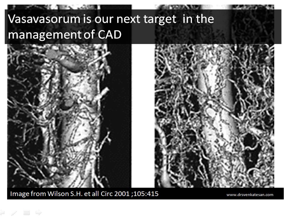
The easiest answer is, blood supply to coronary artery is taken care by a vast network of micro vessels called vasa vasorum(VV) . Of course, the inner layers of Intima and media do get some nourishment by the flowing blood as mentioned earlier.No one really knows the quantum of blood flow that perfuse within the planes of coronary artery.

*By the way , does the vasa -vasorum comes from extra-coronary source or from the same parent vessel ? (I think the answer is both ! will try to find out!)

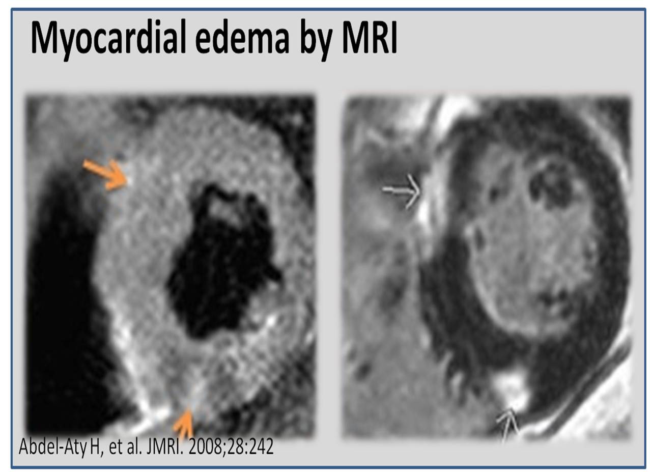
It should be noted Vasa vasorum is well developed only in large arteries. VV has one more important function ie to drain the metabolic excreta from the walls of blood vessels. This function could never be taken lightly as failure to do so will result in vascular wall edema in acute setting or thickening In chronic setting.

Does coronary arterial tree goes for necrosis in STEMI ?

There is some evidence , when acute total occlusion happens in an epicardial vessel , not only the myocardium is ischemic , the entire distal coronary vascular tree becomes vulnerable. The ischemic time and resistance of coronary macro vs micro vasculature is currently not known. It is expected to show significant variation . We know ,one of the important mechanism of no re-flow following PCI is due to microvascular damage(Non thrombotic)

Many times we fail to realise myocardial viablity and micro-vasculature integirty are two different things. ! This questions the concept of reperfusion based on the status of viable myocardium alone.This we have experienced in many patients as myocardial viability doesn’t guarantee you full recovery from LV dysfunction as microvasculature may recovery may lag behind or never restored (Permanent vasa-vasoral damage ?)

What is our knowledge base about exclusive pathology of coronary vasa vasorum ?

Do you know, ectasia, arteritis, aneurysms and external band like compression of coronary artery all are related to some sort of vasa vasoral dysfunction ? We are not yet clear whether atherosclerosis really involves the vasa vasorum.(Takayasu does it for sure ! )

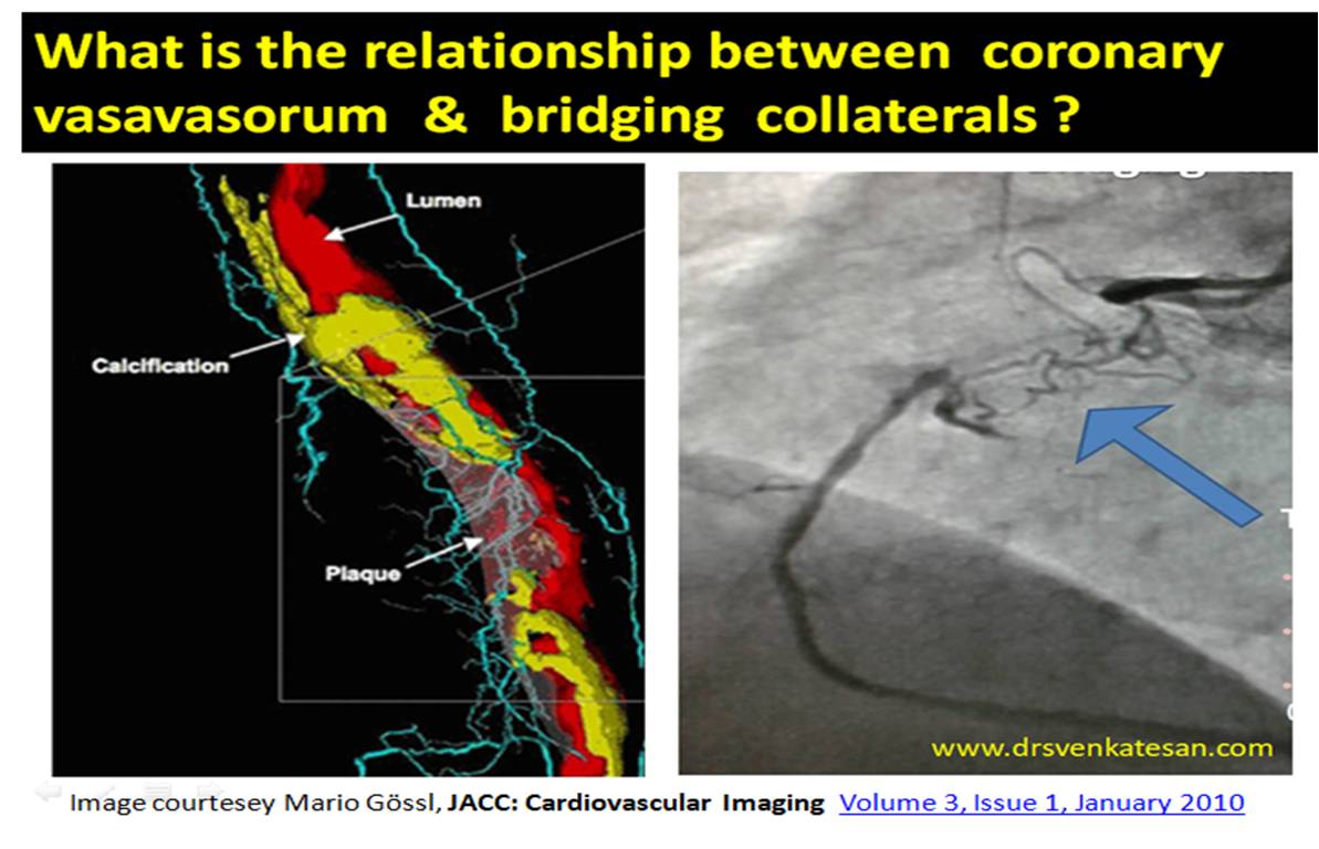
What is the relationship between vasa vasorum and coronary collaterals ?

It seems to me , many of bridging collaterals are nothing but extension of vasa vasorum and ultimately arise from epicardial coronary collaterals. (Some youngster’s take up this topics for research)

Why is high pressure post dilatation a double-edged sword ?

It’s often thought , larger the lumen its better. Need not be. These are all some questions which we don’t have an answer.

What is the radial pressure exerted by coronary stents on coronary trans -arterial perfusion ?

Does coronary artery go for Ischemic necrosis with high pressure Inflations ? As such there is no published evidence . By the time we wait for published evidence enough number of coronary arteries might get damaged. So try to use common sense .

Relationship between delayed Mal-apposition & vasa vasoral damage

It is very likely ,the so-called endo-leak which is quiet prevalent in aortic interventions is could be seen in coronary arteries. We are not recognising it. It could be same as Intramural hematoma in certain subsets.

Meanwhile, self expanding stents with good radial strength has made a come back .While it may prevent a mal-apposition ,has a potential to stress the vessel wall (Radially) and in the process interfering with perfusion.


Does Vasa vasorum promotes Atherosclerosis or negates it ?

hehttps://www.hindawi.com/journals/bmri/2014/701571


The irony is, while de-novo vasa vasorum is the life line for coronary arterial nutrition, neo-vascularisation is problematic .Then how to selectively promote good vasa vasoral growth and avoid the pathological network that promotes adventitial nodular degeneration ? This is were the curious basic scientists and casual cath lab guys need to interact.What is positive remodelling ? (Often referred to the famous concept of Glagov ) How can we promote it to maintain good luminal diameter inspite of large burden of atherosclerosis by manipulating the vasavasorum.


Final message

Cardiologists are ahead of others in many cutting edge technology. There is no two opinions about it. Who can repair a live beating heart without stoping it for a moment ? Still, there is a whole lot of coronary Ignorance waiting to be explored. Blood supply to coronary artery is one such area to be decoded.This will have larger implications as Vascular healing , plaque survival and growth depends upon vasa vasoral integrity as well as neo vascularisation.

While , metallic management of CAD seems to be the order of the day as it tends to give an instant fix .My guess would be medical sense would ultimately prevail one day with controlled vascular aging and natural ,pharmacological ,biological repair of cells will prevail over temporary patch work in cath labs.

Reference

What is the role of newer Imaging and OCT in visualising Vasa vasorum ?

It is going to open up new avenues in our coronary vision.

Vasavasorum review article

(Kensuke Nishimiya European Cardiology Review 2017;12(2):121–3)