Posted in clinical cardiology -Murmurs, opening snap | Tagged clinical cardiology, LV DP/DT and loud s1, what is the mechanism of loud S1 in mitral stenosis, what is the mechanism of opening snap in mitral stenosis |
Why VTs have wide QRS complex?
Brief answer: VT usually presents with wide QRS tachycardia because it originates in ventricular myocardium, travels muscle to muscle instead of the normal conduction system. However, VTs need not be wide always, if it captures the conduction system early and more proximally it can be as narrow as SVT.
Further reading: Only for cardiology fellows
Two empirical statements are made here. (The scientific chances of both being reasonably correct are fair)
It’s obvious, not all VTs are dramatically wide. When it is not wide, they test our knowledge and patience. Let us be clear about the factors that determine the QRS width during VT. Once we know this we can have our own inference.
What determines the width of QRS in VT?
1.Origin of VT
The focus of origin is extremely important. Pure myocardial focus distal to the conduction system is invariably very wide. We know VTs originating right over the fascicles are narrow.
2.His Purkinje breakthrough
The time taken to capture the normal septal conduction system is a critical determinant of QRS width during VT.This makes the VT from septal origin narrower.VT arising from the free walls obviously takes a longer time to engage the HIS Purkinje system. Imagine , If VT originates from the lateral mitral annulus, how much time it may take to reach RV free wall and lastly RVOT. Here the VT will become bizarrely wide.
3.The structural integrity of His Purkinje
It is important to emphasize a fact , even if the VT captures HIS Purkinje early, if they are diseased , still the VT will be wider.(Example bundle branch reentry in DCM in which VT keeps going around the conduction system still, it’s wider)
4.Course
Length of the re-entrant circuit. Macro reentry is expected to be wider. Focal or micro reentry will often be narrow, provided the distal circuit is not diseased.
5. Scars as barriers and boulders
If the VT circuit is interrupted by random scars en-route (from origin to exit) the VT width prolongs. (Evidence for scars is often visible in sinus rhythm ECG as notches /slurs or fragmentations in QRS )
6.Exit point of VT
This is a poorly understood term (at least for me) It is believed, VT can exit only epicardially. The line joining the focus of origin and the exit point is expected to decide the QRS axis. The problem comes when VT breaks out multiple paths and possibly sub-endocardial as well.
7.LV dysfunction
A severely dysfunctional ventricle can stretch the QRS irrespective of conduction system integrity.
8.The Ionic milieu of cells Interstitial resistance
We know, biological current is nothing but Ions in motion. So, no surprise it can alter the QRS morphology. The classical example is hyperkalemia , that can make ECG a wide and blunt sine wave. Local acidosis, hypoxia also influence the QRS duration.
9.Drugs
Any drug which has class 1C or 3 properties can slow the VT circuit velocity. Typically flecainide is well known to make QRS wider. Amiodarone may reduce the ventricular rate. in VT instead of reverting it. Apart from this these drugs depress the ventricular myocardium severely and prolong the QRS width independent to its action on the conduction system.
10.Mechanism of changing width
VTs can have varying QRS width as reentrant circuits change or experience slow conduction due to autonomic influences. VT with downstream aberrancy is also possible as the VT rate by itself influences the conduction property distally.(Just lie SVT with aberrancy)
A paradox about the width of QRS in VT
A curious phenomenon is often seen, when VT occurs in patients with baseline ECG which is already wide (As in an ischemic dilated cardiomyopathy with LBBB/RBBB). Here, the VT prematurely stimulates viable muscles distal to the diseased HIS Purkinje system (Which they are deprived of early activation of till then) .They seem to relish the early arrival of electrical impulse by brisk activation that converts wide QRS complex to narrow one. (This behavior is one of the principles of cardiac resynchronization therapy where we attempt to rewire the heart with multiple leads and shrink the QRS.)
*One more mechanism of wide QRS sinus rhythm becoming narrow during VT is due to a concept called source -sink relationship. The VT delivers enough energy overcoming His Purkinje resistance downstream. (This property is used in HIS bundle pacing )
Postamble
*Forget about wide vs narrow QRS debate. A significant chunk of VTs falls within intermediate width QRS(100-120ms) . Whether to label these as wide or narrow QRS squarely lies on whims of the reader. (Should we take the widest QRS in 12 lead ECG? Pre-cardial vs limb lead etc are not clear) Unfortunately, we don’t have a separate algorithm for this category. This issue demands a separate discussion.
Posted in Uncategorized | Tagged his bundle capture in vt, narrow qrs vt, SVT VT with abberrency, vt entry and exit point, what is narrow qrs vt, what is wide qrs vt, wide qrs vt |
Whenever a patient is getting discharged after a PCI, the treating cardiologist often faces this situation.
So, you fixed the block in my coronary artery doctor. Thank you so much. Now, I can have a peaceful life, free from future heart problems. “Am I right doctor”?
I wish I can answer “Yes” to your query but I can’t for the following reasons.
I have fixed only a lesion that caused maximum obstruction. Atherosclerosis is a diffuse disease and you have minor plaques scattered across your coronary artery. These can grow at its own will. So you carry a definite risk remote from the current problem. (Don’t get frightened, read further, you have definite solutions to reduce this risk.)
How common is the progression of native vessel disease?
It varies from 10 to 40%. Mind you, the exact incidence directly depends upon the compliance of medical management, risk factor reduction, and adaptation to a new life healthy lifestyle. In effect, you (the patients) decide the incidence.
One surprise phenomenon (though unproven) might happen. Since the tightest lesion is jailed with a scaffold the minor lesion is preselected to an accelerated process of atherosclerosis if medical treatment is not properly followed.
Dr.Zellweger from the university hospital, Basel, did an extraordinary study with 400 patients, meticulous 5 years follow up with SPECT and found remote lesions accounted for 40% of future events (Basel Stent Kosten-Effektivitäts Trial [BASKET]) The other study by Glazer and concurred with this. These studies reiterate the importance of taking care of the entire coronary artery instead of focused piecemeal care by scaffolds.
Does a proximal DES protect a distal lesion in the same artery by the drug effect?
It is a good thing to happen at least on paper. A proximal LAD with the latest generation Everolimus coated stent is expected to keep the distal LAD drugged for few months at leas.( with anti-mitotic activity) Thus preventing the progression of distal lesions.
No, I can’t believe this.In this era of momentary touch on sidewalls of artery by drug-eluting balloon (DEB) shown to do wonders, anything is feasible. Chacko’s (Ref 2 : JACC CV Interventions 2009)observation has a possible answer for this. It showed BMS vs DES didn’t make any difference in remote lesion progression.
Final message
These studies reaffirm one vital truth. Stents are temporary solutions to a permanent, systemic disease of the vascular system .Stents are indeed a major revolution in CAD, “if and only if” it’s used in a highly selected CAD population. Global attempts to project cath labs as a tool to control human atherosclerosis is a typical example of flawed science. The only effective way to tackle this menace is to faithfully follow overall healthy living, assisted by drugs.
This is the Editorial in response to Zellweger’s article
Reference
Postamble
One of my patients asked some time ago. If stents are the definite remedy for severe arterial narrowing, why not stent all my lesions (even the minor ones ) prophylactically doctor, so that it will not become tight at a later date?
That’s a good query. Your doubt is genuine , appear logical as well. But, unfortunately, it will be the most dangerous thing to do*. Metals are never friendly with the coronary arterial wall. We should use it extremely judiciously and only with tight flow-limiting lesions. These metals require annual (rather permanent) maintenance. Its taken care by multiple antiplatelet drugs. If for some reason your maintenance is erratic or the drugs fail to act you are at more risk of a future event.
(* This is what has happened (happening) in the past, that demanded urgent publication of appropriate usage criteria)
Now, the current belief among the “fair thinking cardiology community” is dramatically changing. It’s leaning towards non-stent management even with significant flow-limiting obstructions in otherwise stable patients(SIHD). This belief is accruing more and more evidence base (The COURAGE 15 year follow up / ORBITA/ISCHEMIA) All these studies confirm the emerging doctrine and bring back some semblance of sense into the cardiology community.
Posted in Uncategorized | Tagged COURAGE ORBITA ISCHEMIA, post ptca care, progression of native vessel disease after ptca |
Differential cyanosis classically occurs in PDA with reversal of shunt when raised PA pressures /PVR is able to supersede the systemic Aortic pressure and drive the blood from LPA to descending Aorta bringing down the lower limb saturation.
Of course, this can be undone by the presence of any other intra-cardiac shunts or aberrant left subclavian that arising from the desaturated descending aorta.
Other causes of reversed differential cyanosis
Where the upper body is cyanosed (desaturated) and the lower half is not. There is a conventional list of conditions.
(*This occurs due to streaming effect ) Highly saturated superior vena cava (SVC) blood into the right ventricle, reach MPA / through a PDA, and to the descending aorta, with streaming of more desaturated blood from the inferior vena cava (IVC) into the LA through PFO (Ref Yap S H Pediatr Cardiol. 2009 )
Now let us add one more cause for reversed differential cyanosis in the Modern Era
It is seen with ECMO in VA connection (Often reported in babies ) . The Aorta has high oxygen content entering from the femoral cannula going up into the Aortic arch., while deoxygenated blood from LV (because of failing lungs) reach antegradely to the Aorta. Ideally, the ECMO is expected to supply the entire aortic arch and hence oxygenation is uniform all over the body. It rarely happens as some amount of flow will come from LV unless its in asystole. However, If the severely dysfunctional heart tends to recover & lung oxygenation is very poor as well, the LV stroke volume competes with highly oxygenated blood coming from below ( femoral inflow ) into the Aorta , creating a watershed zone . This makes the deoxygenated blood perfusing upper half of the body and hyper oxygen saturation lower half. This is been referred to as North-south syndrome or (Harlequin syndrome the famous Italian comical character)
How to manage North-South syndrome?
Reference
Posted in Uncategorized | Tagged ecmo north south syndrome, harlequin syndrome, va ecmo side effects |
LV dysfunction is one of the most commonly used terminology by cardiac professionals.It can be systolic, diastolic or global, regional etc. But, before dysfunction sets in, the heart fights. The Left ventricle can behave in many different ways when confronted with stress. It increases the force of contraction, elevates it’s Intra cavitary filling pressure and still accomplishes its task of pumping adequately. Further, It can build fresh muscle (LVH). It can double up with more heartbeats. (All these factors are referred to as cardiac reserve mechanisms)
These reserve mechanisms can be activated in the short or long term. In the long term, autonomic activation with neuroendocrine factors joins the compensation process. These will work for some time till the circulatory system settles down to new homeostasis. However, they become counterproductive and becomes decompensated, ultimately heart failure sets in(Unless Intervened)

Mechanisms of cardiac reserve. Image source: https://courses.lumenlearning.com/suny-ap2/chapter/cardiac-physiology/
Is LV dilatation a mechanism of cardiac reserve ?
No one calls LV dilatation as a reserve or compensatory mechanism. (I wonder, why not ?) I think like RV , LV too has some potential to reversibly dilate . The quantum of which we are unable to estimate.This happens usually in response to chronic volume stress* like regurgitant valves or high output states. Though cardiomegaly and a huge heart convey a sinister outcome, many hearts shrink if the primary issue is corrected.(Typically in Anemia, Beri Berri. We also know LV may transiently dilate in response to some toxic /pregnancy-related cardiomyopathy.
* Mind you LV poorly tolerates acute volume stress as in Acute AR/MR
The critical gap in our understanding is about this question.
When does LV dilate physiologically and when pathological persistent LV dilation sets in (The absolute state of irreversibly lost cardiac elasticity.) We also know dilated LV will consume more oxygen due to enhanced wall stress (Laplace law) and hence its possible LV dilatation begets further dilatation. Optimal timing of mitral and aortic valve replacement in patients with AR and MR directly depend on this knowledge.
Final message
We need clarity in the following queries
Let us wait for the knowledge to evolve. Young cardiologists could take up this area for research.
Posted in cardaic physiology, cardiac physiology, LV dilatation is diastolic dysfunction | Tagged cardiac reserve, cardiology research papers, cardiology thesis topics, dilated left ventricle, lv dilatation is lv dysfunction |
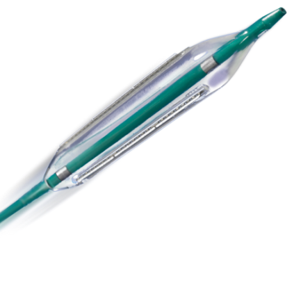
I asked some of my experienced colleagues, how much time they inflate the balloon to deliver a stent? Most answers were spontaneous and unanimous “It’s hardly 10 seconds, few said maybe up to 15s.
Can prolonged balloon inflation time reduce the need for post dilatation and prevent mal-apposition?
We know high-pressure Inflation( up to 20 atmospheres ) was a big revelation in the science of PTCA more than two decades ago. (Antonio Colombo JACC 1995 ) He proposed and proved high-pressure inflation eliminated the need for routine anticoagulation following stenting as approximation was better. He also pioneered the concept of dual antiplatelet therapy (DAPT) in the PCI arena.
Similarly, prolonged balloon Inflation (30 to 60sec) could be another trendsetting tip to prevent malposition. It delivers more sustained pressure. Its believed the imparted centrifugal force and the inbuilt radial forces add up to the stent vessel wall Interface and prevent mal-apposition.
Is there a downside to high-pressure Inflation?
There must be few. Potential new Ischemic events and arrhythmia. In calcium laden plaques( spur) risk of perforation may be enhanced.
Final message
I don’t know why this concept never took off. Many of us still fear to inflate the stent balloon no longer than 10 to 20 seconds? Adhoc post dilatation with short NC balloon appears mandatory in areas of mal-apposition. Meanwhile, we also understand sustained (30-60s) high-pressure initial inflation could deliver the stent in a more synchronized and smooth fashion with a perfect metal/vessel wall interface. Further , prolonged balloon inflation times could make a routine (By the way who does routine ?) IVUS/OCT redundant.
What do the experts say? What does science say? There is one meta-analysis that clearly says the advantage of long inflation time. This issue becomes much more relevant as it could avoid post dilatation which all of us know can be tricky. In fact, every balloon dilatation should be technically counted as another PTCA procedure and adds up to net total risk.
Reference
1.M. Saad, M. Bavineni, B. F. Uretsky, and S. Vallurupalli, “Improved stent expansion with prolonged compared with short balloon inflation: a meta-analysis,” Catheterization and Cardiovascular Interventions, vol. 92, pp. 873–880, 2018. View at Google Scholar
Posted in post dilatation, Tips and tricks in cath lab, Uncategorized | Tagged balloon inflation time in ptca, how to avoid post dilatation, malapposition and balloon inflation time, tips and tricks in ptca |
Assessment of LV diastolic function primarily depends on the Doppler flow profile across the mitral valve and also to be noted are the 2D features of LA and LV for associated abnormality like LVH, LAE etc.
Why diastolic dysfunction assessment difficult in AF ?
Since most diastolic doppler mitral inflow parameters involve analysis of atrial contraction A wave, atrial fibrillation makes it difficult to assess diastolic dysfunction. Since we have only early diastolic velocity to assess, the changes confined to this E velocity is of paramount importance. This E velocity again is subjected to cycle length dependent alteration in both its acceleration and deceleration time , making things still more complex.
However, the following features help diagnose diastolic dysfunction in AF
*LA dimension is a very good sign of chronic elevation of LAP and diastolic dysfunction in the absence of mitral valve disease. However, AF can dilate the LA making it a less useful parameter. But, it should be noted in AF both RA and LA dilate together.So, a disproportionate LA>RA (or if RA is normal size ) could still be a marker of baseline LV diastolic dysfunction.
Reference
Posted in Atrial fibrillation | Tagged atrial strain in atrial fibrillation, diastolic dysfunction, dual doppler probe in atrial fibrillation assessment, e velocity variation in atrial fibrillation, echocardiography, HFpEF, how to assess diastolic dysfunction in atrial fibrillation?, single beat doppler echo |
FFR is the ultimate hemodynamic test that measures the physiological Impact of lesions. Just pass a manometer tipped wire across the lesion and note the pressure drop (with or without Adenosine) All you have to remember is two cut off values .8 for FFR and .9 for IFR. Abracadabra . . . yes you got the answer , whether to proceed with PCI or not? It’s as simple as that. We are no longer blind to physiology to which many coronary purists often criticize us.

Coronary physiology simplified
Now , answer this question.
Is FFR heart rate dependent? If yes, how significant it is?
This simple question on coronary physiology caused the maximum distress to a large expert cardiologist group
Some of the answers
My Answer
It has to be yes, right, however minimal it may be. My interpretation of truth in FFR is, it can have a massive influence* . (*Unless you are sure (we can never be ) about achieving maximum hyperemia or this hyperemia is the same as physiological exercise.) In fact, the whole concept of FFR lies in the fact that it should induce enough HR raise that should be used as a surrogate marker for maximum hyperemia. Ideally, like stress testing, we need to test FFR at maximum heart rate and minimal heart rate. The difference could be documented as FFR max-min. This will throw new light into the physiology of microcirculation.
Should we need to create a heart rate corrected FFR?
Yes , I think we need to do it or else should report at what HR we are reporting the FFR. If FFR falls at a high heart rate and maintains at low it implies a significant lesion. So don’t get fooled with FFR of .9 measured at an inadequate heart rate.
IFR to replace FFR : On what basis?
Meanwhile, new generation coronary flow quantification tool IFR jettisoned Adenosine and simply measure diastolic instantaneous flow at resting state. This makes a mockery of coronary physiology, without a true debate about heart rate dependence of trans-lesional flow.
Impact on clinical practice
Even as we struggle to answer the fundamental question of the influence of Heart rate on FFR, many landmark studies had been done. They have ratified FFR as the most physiological modality to assess coronary lesion. Important guidelines have been written based on these studies. No one will ever know, the true impact on the current cardiology care, had we included heart rate adequacy /correction as an essential criteria in those FAMEd studies we hype about.
Counterpoint.
All is well with FFR.It has been tested with various heart rates.
FFR at peak hyperemia means there is no further HR rate induced potential microvascular reserve. So a properly administered optimal Adenosine augmented FFR should not bother the HR variability. (But its only theory)
If FFR is ok . . . IFR should not be ok is it not?, For the simple reason, there is no hyperemia in IFR , what is the use of knowing resting flow reserve (RFR)
Reference
Influence of heart rate on the FFR measurement – experimental and clinical study
Postamble with a slice of History
FFR is as old as the concept of PTCA. In fact, the original balloons used by the great Gruentzig’s * had a central port for pressure recording through which he measured both proximal and distal pressure curves to guess the significance of obstruction. After each inflation, he checked whether both curves are drawn together which he speculated to indicate a successful procedure physiologically.
Posted in fame study ffr, quantitative coronary angiogram | Tagged ffr, fractional flow reserve ffr, ifr, IFR QFR, instantaneous flow reserve, Limitations of FFR, pressure wire channel in gruentzig original ptca balloon zurich |