Feeds:
Posts
Comments

Posts Tagged ‘acute rheumatic fever’

Dr. Duckett Jones, the famed American physician, from Good Samaritan hospital, Boston would be a proud man in heaven, to find his criteria still being celebrated all over the globe. He will also be pleased to know his home country USA  is painted green on the world RHD map due to his untiring efforts that began in 1944. Of course, what the rest of the world has done in the last century has left us wanting (including the WHO).


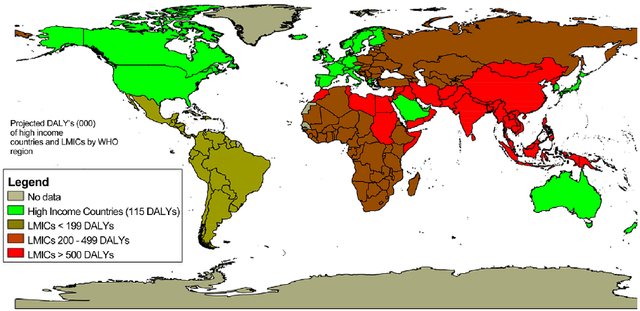
Global RHD map. Note the red and brown shading in south Asia and Africa. It is obvious, RHD is more about economics, equality, and poverty, rather than aggression from an otherwise innocuous microbe called streptococci which is omnipresent all over the world with equal concentration.

How to diagnose  Acute rheumatic fever (ARF)?

Simple. Apply jones’ criteria. Funnily, I found it can be a most difficult exercise to do, especially If we realize ARF can defy all the three components it carries. ARF  need not be acute, need not have rheumatic symptoms & curiously they need not have fever as well. Did you note this? The entire disease process can be subclinical in 50 % of children. Intelligent patients must realize, how scientifically quixotic conditions we, the doctors are expected to practice medicine.

There is one more ongoing confusion in many of us. Is Jone’s criteria meant for diagnosing the first episode of ARF, or second, or any subsequent episodes?  In the strict sense, it can be applied only for the first episode. But it may still help diagnose recurrent episodes. Dr. Jones was so precise in his observation when he suggested the in the later episodes .we may able to diagnose ARF only with minor criteria. But the lacuna here is,  recurrent episodes can be so atypical and carditis or chorea may be the only manifestation of that episode making the classical Jones triad redundant. 

Someone asked in my class Is there an entity chronic rheumatic fever? 

If you describe ARF  as a separate entity there must be Chronic RF? logical Isn’t it? . Do you think Jones wouldn’t have thought about this.  We don’t know,  echocardiography was not even thought of at that time. Better, we stop discussing Chronic RF. (Simply put,  all chronic indolent carditis with raised ESR  might fit into this imaginary entity)

How important is supportive evidence to Jones’s criteria? 

When we have trouble in fixing even the major criteria, where is the question for evidence for preceding streptococcal sore throat come in? By the time we see these children, a throat culture is no longer positive, though ASO titer/Anit DNAse might help. (It must be recalled that culture doesn’t differentiate carrier state from acute infection, a single value of ASO titer ahs little value) 

I asked a few of my senior pediatric professors how often they depend (or demand)  supportive criteria to diagnose ARF.  They agreed in unison, that they never felt the need for it except for academic or epidemiological reasons. When Jones wrote this criterion in 1944, he also never intended to include evidence for previous streptococcal sore throat. 

Final message 

Is the time nearing to revise Jones’s criteria again and restore with an original suggestion and get rid of supportive criteria?  Maybe Dr. Jones wouldn’t object to this as his aim was to tackle a global Pando-endemic rather than worry about few errors of overdiagnosis.

Counterpoint

* For the pure academics, there is exactly the opposite write-up demanding RTpcr to be included as evidence for streptococci sore throat in this site.  https://drsvenkatesan.com/2020/10/01/role-rt-pcr-in-the-diagnosis-of-rheumatic-fever-rhd/

Reference

1.https://www.acc.org/latest-in-cardiology/ten-points-to-remember/2015/05/08/15/22/revision-of-the-jones-criteria-for-the-diagnosis-of-acute-rheumatic-fever


2.

Rheumatic fever: Session 2 Preventive strategies 

Rheumatic fever and RHD can be prevented at multiple levels.

Primordial: Preventing all sore throats (that will include Streptococcal ) by promoting social and domestic distancing as we do now for the Novel C pandemic.

Primary prevention: It is about preventing the first episode of RF after getting a sore throat. A course of penicillin after sore throat and trying to interrupt the RF in its incubation period is the aim. I don’t understand why preventing sore throat after exposure to streptococcal droplets doesn’t come under primary prevention too.

Secondary: Preventing recurrent episodes of RF after an established diagnosis of the first episode. ( which of course can be subclinical) This is the classical prevention of monthly injection of benzathine penicillin.

* All levels of prevention activities at the level of the throat. None works in the heart directly.

*Tertiary prevention (Treatment ): It is treating the valve disease and trying to reset the rheumatic clock. Tackling the mitral valve disease with PTMC/MVR is the least economical and most expertise-consuming modality. (Of course more gratifying to both patients and cardiologist) It is all too common even in big tertiary centers do regularly PTMC but shrug off patients from monthly penicillin injections. There should be an in-house responsibility for the cardiologist, that  they should ensure at least 100 RHD patients get proper penicllin prophylaxis ( for every PTMC they do)

 Which is the best mode of prevention?

Primordial prevention is great. But the best yield will come from primary prevention.If you want to really avoid serious bites on the heart try to protect the heart from the first episode of ARF as the first bite is more intense. To make matters worse, the injury from the first bite is likely to continue irrespective of monthly penicillin.(Karthikeyan G, Mayosi BM. Is primary prevention of rheumatic fever the missing link in the control of rheumatic heart disease in Africa?Circulation2009120:709–

Can WHO enforce a world microbial order?

One real option that exists, which many feel is artificially stonewalled, is asking vaccine giants like Pfizer, Astra, or  BioNtech to fix a deadline and accelerate the process for a global Rhematic vaccine (Wating in the pipeline for 60 years you know)  with their newly accrued corona Intelligence. (We have few name suggestions Rhemavax or Rhemshield waiting  !)


Read Full Post »

RT-PCR: Real-time polymerase chain reaction, a sophisticated gene sequence-based biochemical test. Thanks to corona, this complex medical investigation has become a household name.

Jones proposed his criteria to diagnose acute rheumatic fever  in 1944, we still use it to diagnose with many modifications . Currently, AHA position statement – 2015 by Gewitz et all is  being followed. (Circulation 2015)

From Braunwald textbook of cardiology. Apart from this, there is one catch . Even if the child fulfills Jone criteria, there needs to be evidence for preceding streptococcal sore throat, either by culture or antibody. Now, can we include an RT-PCR as a new parameter to diagnose streptococcal infection is the question?

Why we Insist on evidence for preceding GAS?

It is for a the simple reason, many entities other than rheumatic fever may fulfill Jones’s criteria. (Still disease, HS purpura OR even simple viral arthritis etc) Some may even call it an essential criterion in the past. Practically it is not done is a  different story.

Tests for preceding streptococcal sore throat are ASO titer and anti-DNAase B. How about the now  glamorous RT-pCR for streptococcus ?  Though it was suggested as a useful test in the past ,the cost and logistics were  prohibitive so it was never considered to be included in the Jones scheme of things.

There could be three roles for RT-PCR testing in Rheumatic fever /RHD

1.RT-PCR as evidence for recent streptococcal sore throat.(GAS organism) Which still not practically used often but has big scope.(Ref 1)

2.To rule out co-viral (influenza-like) infections as a cause for fever and Joint pain (Used in population-based screening in a high endemic area (Ref 2)

3.There could be one more indication (experimental though) Micro-RNA detection by RT-PCR to identify children who are prone for progression to RHD. (Ref 3)

Final message

Now, we must introspect. While billions of dollars are going down the drain on  RT-PCR for diagnosing  a common cold pandemic which has no specific treatment. Will WHO and other cardiovascular preventive authorities  consider to include RT-PCR as screening  test for GAS in children. This will enable  early start of primary prophylaxis and prevent  RF/RHD  a century-long scourge of the third world.

Reference

1.

2 .Emmy Okello et all J Am Heart Assoc. 2020;9:e016053. DOI: 10.1161/JAHA.120.016053

3.Lu, Q., Sun, Y., Duan, Y. et al. Comprehensive microRNA profiling reveals potential augmentation of the IL1 pathway in rheumatic heart valve disease. BMC Cardiovasc Disord 18, 53 (2018).

Read Full Post »

The pericarditis  of acute rheumatic fever is not a true infective pericarditis.It is more of inflammation .It is primarily  T cell mediated  reaction . Neutrophils rarely take part  in this inflammation and hence  no significant  exudation . Hence , there is less  sticky and adhesive molecules inside the pericardial space .The most inflamed layer is epicardium which a nothing but visceral  pericardium .This layer lacks the tensile strength to constrict the underlying myocardium.

why rheumatic pericarditis does not go for constrictionFor constriction to occur the fibrinous  ( thick ) layer of pericardium need to be involved . In rheumatic fever  even though it is  pancarditis ,  fibrous layer is not  involved. Further the inflammatory gradient is thought to spread from within  (Unlike tuberculosis )

Note :  In chronic tuberculous pericarditis,  diffuse inflammatory process  invade from the exterior surface . Very often ,  one can not differentiate  layers. In extreme cases even myocardium and pericardium can not be separated .

Summary

The peri-cardial effusion of acute rheumatic fever

  1. Is transient ,non infective and resolving (Unlike valvular inflammation !)
  2. Less of neutrophil activation  (Less adhesion)
  3. It does not involve the thick , tensile  fibrous layer of pericardium hence lacks the contractile force .

Other lingering  questions

1.How common is tamponade  in acute rheumatic fever ?

2.How important is the mass of the effusion (Viz  a Viz  Intra pericardial pressure !) in causing tamponade ?

//

Read Full Post »

There is no primary rheumatic myocardial dysfunction .The LV dysfunction is related to the valvular lesion especially mitral regurgitation.While this is 100 % true in chronic RHD ,surprisingly it holds good even in acute rheumatic fever as well .(We have been thinking wrongly that acute myocarditis is responsible for most cases of cardiac failure in Acute rheumatic fever !)

( Incidence of LV dysfunction is still noted in 5% in RHD.This is may be due to chronic underfilling of LV in mitral stenosis, fibrosis secondory to hemodynmic stress as in Aortic stenosis or more commonly associated CAD.)

It is a paradox to note myocardits being a major component in acute rheumatic fever (ARF) ,still it does not persist long term .It invariably resolves and the injury to the valves goes on to result in progressive valve damage .
It is heartening to note this phenomenon in ARF as myocardial involvement behaves just like joint involvement.
Shall we modify the famous statement of the canadian Pathologist William Boyd Rheumatic fever licks the joint but bites the heart”
Though ARF bites the vlave , it simply licks the myocardium like the joints
Is there a chronic indolent myocarditis ?
It was Initially thought there could be process of chronic myocardial inflammation.But now it is almost proven there is no entity like that .
But , it is not uncommon some patients with RHD present with significant LV dysfunction which in all probability unrelated to rheumatic activity .
Assignment for cardiology fellows .
1.Where in the heart Ascoff bodies are densely found ?
  1. Mainly over the valve leaflets
  2. Atrial muscle
  3. Ventricular myocardium
  4. Pericardium
2.Does Ascoff bodies disappear in Chronic RHD ?
Reference

Read Full Post »

Rheumatic valvulits , Valvular inflammation and edema  is the traditional answer .A detailed Echocardiographic study from All India Institute of medical sciences New Delhi  ,India  which was published in circulation 1996 answers this question most authentically .

From a meticulous  Echocardiographic  study of about 70 patients  (with both first and recurrent episodes of carditis ) the following findings were observed.

After reading this article one should be able to answer variety of  questions in RHD  such as

  • How common is MVPS in RHD ?
  • How often MR dissapear with Aspirin etc ?
  • Echocardiographic correlates  for care -coombs murmur ?

Reference

http://circ.ahajournals.org/content/94/1/73/T5.expansion.html

Read Full Post »

Acute rheumatic fever classically involves large   joints of lower or upper limbs  referred to as  fleeting migratory polyarthritis .But this pattern is  not  exclusive.  In fact   acute rheumatic  fever commonly  present with atypical features .The incidence can be up to 25 % in various series .The most surprising thing is ,  it can involve spinal as well as hip joints . Mono arthritis is also possible.

The only contention is , atypical features are  frequently  labelled by  some  as post streptococal reactive arthritis instead of rheumatic fever .

It is  pure  semantics at play . Whether you agree with the terminology or not  ,  never hesitate to diagnose rheumatic fever when the  joint involvement is  atypical . If  you ignore this  you are bound to  be guilty  for damaging few hearts  later.

What are the unusual joint involvement in acute rheumatic fever  ?

  • Small joints of the feet
  • Small joints of the hands
  • Cervical spine
  • Wrist
  • Elbow
  • Shoulder
  • Hip
  • Thoracic  spine
  • Calcaneus
  • Lumbar spine

The involvement of above joint can be up  to 25%

Here is an excellent paper from Brazil about the huge variation in the pattern of joint involvement in acute rheumatic fever.

http://www.jped.com.br/conteudo/00-76-01-49/ing.pdf

Read Full Post »

Mitral regurgitation is  one of  the most common lesion of rheumatic heart disease .Mechanism of MR in acute rheumatic fever is different from chronic rheumatic heart disease.

Acute Rheumatic fever

The following mechanisms contribute to MR of acute rheumatic fever

  1. Edema of leaflets (Carey Coombs murmur )
  2. Valvulitis
  3. Small verrucous  vegetations (See Image )
  4. Acute LV dilatation in fulminant cardiac failure.

* Note  : Acute rheumatic fever in its first episode can never  cause stenosis  however fulminant the fever may be  .There is no acute mitral stenosis .But ,  during recurrence and reactivation some amount of stenotic process may occur.  Still ,  recurrence and reactivation are more often related to significant MR rather than MS. ( Isolated mitral stenotic lesions  rarely  give h/o recurrent rheumatic fever )

Chronic rheumatic  heart disease

As the mitral valve gets progressively damaged  any combination of MS or MR occur .The following mechanism are involved in  the genesis of MR. (Pathology of Mitral stenosis is not discussed here)

  1. Chordal shortening, tethering , pulling , prevent proper co-optation
  2. Chordal lengthening
  3. Chordal disruption (Minor > Major )
  4. Prolapse of either AML or PML (Not both ,unlike myxamatous MVPS)
  5. Infective endocardits  of  leaflet
  6. Perforations of  leaflet
  7. Annular  dilatation
  8. Fibrosis of posteromedial/Antero-lateral   pap muscle(Rare )
  9. Left atrial pathology

* The direction and the  width of MR jet is  related to the mechanism of MR.

If there is chordal shortening due to fibrosis  of mitral valve  co -optation plane is altered . The degree of chordal shortening , pap muscle fibrosis (rare)  symmetry of chordal involvement determine the MR.

Rheumatic mitral valve prolapse

  • This could be  more common than we realise.
  • It can be true or pseudo.
  • True prolapse occur due to chordal weakening or lengthening .
  • In chordal disruption the leaflet tips usually become flail

Since rheumatic process fixes the PML first , the AML   appear to overshoot the plane of PML and   appear as prolapse.(Pseudo )

The sail like AML commonly  directs the jet posteriorly and laterally .(Murmur conducted to axilla and back )

It is rare for PML to prolapse in RHD , if  it does occur ,  it directs the jet anteriorly (murmur conducted to aortic area mimic AS !)

It is rare to see a  perfect  central jet in RHD  . presence of  Central jet is a good sign to consider mitral valve repair.

Myocardial involvement in RHD.

Even though rheumatic fever is a classical  example for  pan-carditis , it is surprising   to note (Of course fortunately !)   how  myocardium escapes in the  chronic process of RHD.

Is it really true  ,  myocardium do not get involved in chronic RHD ?

Clinical cardiologists rarely discuss this issue. Pathogists indeed have documented significant lesions within myocardium  . Involvement of left atrial myocardium and  rarely  ventricular myocardium in the sub mitral  zone  can influence the  degree of  MR

* Even in acute rheumatic fever with fulminant carditis , myocardial involvement is  disputed by many  ! . My belief is ,  there will   definitely a subset  in   both acute and  chronic  forms of   RHD   , in which myocardium  gets  involved . In our institute LV dysfunction associated with RHD occur in  up to  5 % of  RHD population .

Importance of knowing the mechanism of MR

Two aspects  appear important

1. Is there a potentially  reversible component in pathology so that we can  wait  before intervention  ?

I have seen children referred for mitral valve replacement due to severe MR  . In due course   MR regress by the time they reach the tertiary center (waiting period included ) At least one child i remember,  the MV surgery was canceled  due to spontaneous regression MR.

It was later found the MR was  more of valve inflammation than degeneration .

* Always think about the possibility of reversible rheumatic MR  in every severe isolated  MR in children (Do not apply this rule in adults or in combined MS or MR  )  Do a ESR, ASO and start an  intensive anti inflammatory therapy  , aspirin with strict penicillin prophylaxis .With this  one can definitely postpone the surgery  in few cases  and  may avoid it altogether !

2. Surgical implication

If we could delineate  the  exact pathology of MR   it will facilitate  the   repair . Annular  reduction and  neo  chordae  etc . Of course ,the surgery could be  very  difficult in scarred mitral valves ,  Dr Sampath kumar *of AIIMS  New delhi , India  would  feel other wise !

*A pioneer in mitral valve repair in chronic  RHD (See reference 2 )

Questions  that need  answers

How is balloon/Surgery  related injury different from rheumatic process ?

Why is  rheumatic  mitral vale  prone for bacterial infection ?

What is the relationship  between  extent of  aortic valve involvement and  degree of mitral valve involvement in RHD ?

Reference

1.http://circ.ahajournals.org/content/94/1/73.full?sid=10599470-3563-4c38-b688-c5fc8c032f96

2. http://icvts.ctsnetjournals.org/cgi/reprint/5/4/356

Books

There two popular books exclusively  for cardiac pathology

1.Silver

2. Renu Virmani

Read Full Post »