Feeds:
Posts
Comments

Archive for November, 2026

Arterial grafting, i.e., LIMA to LAD, is one of the revolutionary concepts that redefined outcomes in CABG surgery. It is an artery-to-artery graft, and a perfect size and physiology match that made LIMA-LAD a success story. But the RCA graft choice is not that straightforward.
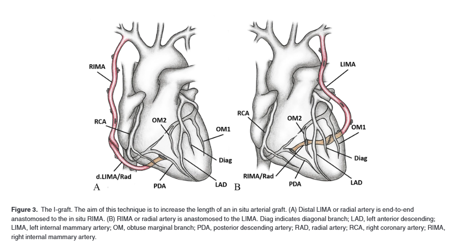
Image source : Prapas SN, Pangiotopoulos et al Open J Cardiovasc 2019

Why arterial graft for RCA is very often avoided ?

A. Anatomical, technical & logistics reasons

B.Coronary to the popular belief arterial grafts are not really beneficial over SVG in RCA.

C.Instituional and surgeon’s preference

D.No, it is never avoided Intentionally. (RIMA, Free radial or sequential LIMA is used whenever required )

Is Arterial grafts really avoided for RCA ?

Conduit selection in CABG need a fine balance between long-term biological durability and real world surgical logistics. Arterial conduits mainly LIMA or RIMA or radial artery possess an endothelium adapted to systemic pressures that resists atherosclerosis, yielding superior 10 year patency and lower rates of adverse cardiac events compared to saphenous vein grafts (SVGs), which undergo progressive intimal hyperplasia and vein graft disease. Consequently, major revascularization guidelines recommend arterial conduits over SVGs for significant lesions even for non LAD locations, especially a dominant RCA, in younger patients with low perioperative risk.

Despite a large cohort of patients meeting these favorable criteria, real-world practice indicate surgeons hesitate to use arterial grafts to RCA and SVGs remain the predominant conduit in over 80 % of procedures. This guideline – practice mismatch is more of technical and logistics .

Why ?

1.Surgeons routinely prioritize and reserve arterial conduits for big brothers LAD & LCX , leaving SVGs for the RCA rather than harvesting a difficult arterial graft with somewhat uncertain survival benefit.

2.Bringing RIMA near to RCA/PDA is not always easy , involves stretching .Free arterial graft is still possible, but need a fairly lengthty one.

3..Also, when RCA has only moderate stenosis (or diffuse distal disease) or where competitive flow from LAD can induce vasospasm in arterial grafts. In contrast, SVGs are larger caliber, lengthy, and most importantly user friendly without compromising flow.

4.With off pump , minimal access, Robotic CABG becoming more popular , an arterial graft to RCA is looks still distant wish.

5.Arterial grafts are still considered superior in specific subset of carefully selected lesions and patients.

Final message

Graft selection for the RCA is not a matter of strict science and superiority . It is more a matter of surgeon preference, convenience, institutional protocol. Most surgeons feel, it is not worth the time for a marginal benefit for arterial graft.(if at all. )

My understanding is,  SVG grafts have become the default option for the RCA. Note: This is in contrast to the LAD, where venous grafts are almost always avoided except in emergency situations , very poor LV function or when LIMA is injured.

Reference
1.Gaudino M, Benedetto U, Fremes S, et al. Association of Radial Artery Graft vs Saphenous Vein Graft With Long-Term Cardiovascular Outcomes Among Patients Undergoing Coronary Artery Bypass Grafting: A Systematic Review and Meta-analysis. JAMA. 2020;324(2):179–187. doi:10.1001/jama.2020.8228

2.Prapas SN, Pangiotopoulos IA, Leivaditis VN, Katsavrias KP, Prapa VS, Linardakis IN, Koletsis EN, Grapatsas K. The π-Circuit Technique in Coronary Surgery: Analysis of 1359 Consecutive Cases. Open J Cardiovasc Surg. 2019 Aug 27;11:1179065219871948. doi: 10.1177/1179065219871948. PMID: 31488952; PMCID: PMC6712752.

Read Full Post »

We know, atherosclerosis preferentially develops at arterial bifurcations because of shear stress, and other hemorheological forces.It is therefore no surprise that left main bifurcations and bifurcations at major branch points (diagonal, obtuse marginal, posterior descending artery) forms significant lesion subsets.(Estimated 20-30%)

Coronary lesions are treated according to their obstructive potential or, occasionally, their vulnerability to cause acute coronary syndrome (ACS). The former is the stronger indication, because the latter can often be addressed medically. PCI for plaque passivation of a non-flow-limiting thin-cap fibroatheroma (TCFA) remains a controversial indication, even after the PREVENT trial.

Returning to bifurcation lesions, interventional cardiologists have tied themselves into a debate over single vs two-stent strategies, as if no other means of treating bifurcations existed. Thanks to Drug-eluting balloons (DEB). They have added some fresh new thinking into the debate.

Scientifically and pragmatically, there are three non-stent alternatives for bifurcation lesions .

  1. Standalone Optimal medical therapy (OMT): Appropriate in a significant subset, often as part of the management of multivessel CAD.( Non flow limiting, Stress test/FFR negative)
  2. CABG: Is a default choice for many critical bifurcation or trifurcation disease.(Trifurcation lesions is seen in about 5 %). Evidende : Do not rely on RCTs, trust yourself. For complex BFLs , by passing the troubled terrain makes wisdom, rather than confronting them.
  3. Balloon-only strategy (POBA or DEB): simple kissing inflation with two plain balloons at adequate pressure (ATMs) can achieve an acceptable result. The fear of dissection at the carina and ostium is real but manageable, with bailout stenting if needed. Delayed dissection or delayed closure is uncommon when the acute result is satisfactory. Restenosis is the main limitation, so a final DEB touch-up over a POBA-kissed lesion is advisable.

*Note : Even Bifurcation lesions under medina1,1,1 need not frighten us as long as plaques a re stable and non flow limiting.

Evidence?

Surprisingly, major trials such as ISCHEMIA, COURAGE and ORBITA do not report the bifurcation subset of their populations. Nevertheless, it is reasonable to believe that a significant subset of stable, non-left-main bifurcation lesions can be managed with medical therapy alone. The true proportion such lesions is unknown and needs bifurcation-specific research , especially in methods to assess the true FFR in bifurcation lesions are required. (Note :Even a minimal ostial LCX or LAD can cross influence the FFR of either vessels diverting medical lesions to cath lab or vice versa)

Final message

If we are unable to think beyond , catheter based strategies in bifurcation lesion , there is something seriously wrong in they we are producing cardiologists.Bifurcation clubs across world to please take a note of this.

Read Full Post »

Dyspnea

Dyspnea is probably most common symptom encountered in the practice of clinical medicine. Although cardiac and pulmonary causes typically come to our mind first, the top two etiologies are actually neither of these. Skeletal muscle deconditioning is the leading cause, followed closely by anemia.The mechanism of dyspnea will remain complex , for the simple reason, it is perception of normal sensory stimulus.

Origin and mechanisim of dyspnea

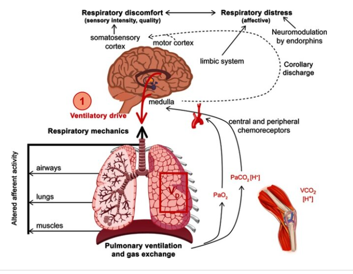
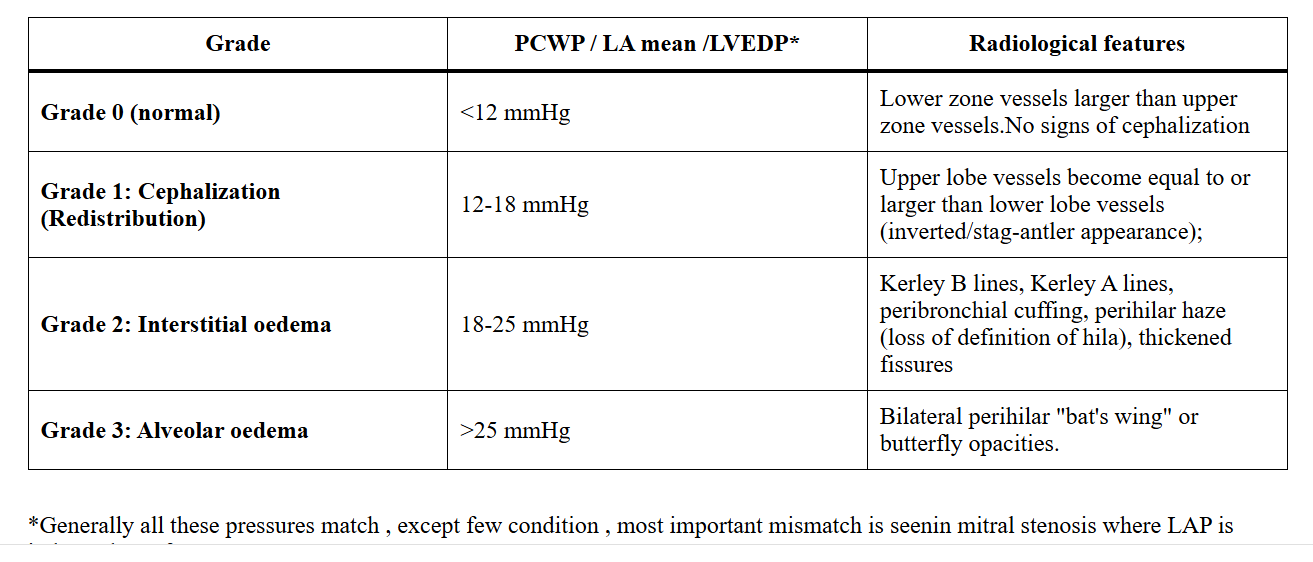
The Initial trigger can be either mechanical or bio- chemical(Chemo receptors) .Mechanical receptors are spread all over the body in muscle tendons and Golgi organs in every skeletal muscle. Contrary to the popular belief, signals from muscular triggers can exceed the J-receptors and cardiac stretch receptors even in cardiac failure. Whatever the initial trigger, dyspnea is a sensation ultimately perceived in the insular area of the limbic cortex. Note: Dyspnea can occur with a pure cortical trigger (Cortical emotional dyspnea).

Image source : Hanania NA, O’Donnell DE Int J Chron Obstruct Pulmon Dis 2019 24:14:1127-1138.

*The fact that a complete quadriplegic individual can still experience dyspnea demonstrates that the trigger can be entirely biochemical, mediated through chemoreceptors. Further, vagal afferents and efferents likely play a role, as this pathway completely bypasses the spinal cord.

Welcome a new class : Drug Induced dyspnea

Now we recognise some of the important drugs , we use can cause a sensation of dyspnea independent of cardiac ,or pulmonary pathology . Four commonly used drugs are discussed here. They can be called as dypneogenic drugs.

Adenosine: Two ways it cause dyspnea. Through A1 receptors (Gi-coupled) on pulmonary vagal afferents which lowers cAMP and through A-2 receptors (Gs-coupled) on arterial chemoreceptors, that raises cAMP . This dual activation stimulates localized nerve terminals result in dyspnea sensation without any truemetabolic alteration.(Note : Aminophylline can block this)

Ticagrelor: This again is related to Adenosine , but in an Indirect manner. It reversibly inhibits the ENT 1 (Equilibrative Nucleoside Transporter) By blocking cellular reuptake and clearance It also elevates endogenous adenosine in blood and lung tissues, activating vagal receptors to mimic a true suffocation state despite normal blood gas levels. Is Aminophylline useful here ?

Beta-Blockers ( Non selective like Propranolol etc ) : The adverse effect on airway is well known. It is due to Beta 2 -adrenergic receptor blockade on airway smooth muscle cells

NSAIDs (Aspirin) In susceptible individuals, NSAIDs inhibit the Cyclooxygenase-1 (COX-1) enzyme, that blocks prostaglandin (PGE-2) synthesis. This shunts arachidonic acid exclusively to lipoxygenase pathway, causing an imblance with Leukotrienes that result in both bronchoconstriction as well as inflamatory mucosal edema.

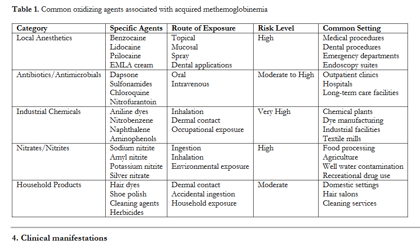
Note :Drugs that cause met-hemoglobinemia (A big list : Ref : Ramya Sri Bura et al Journal of Pharma Insights and Research, 2025, 03(01), 112-120) can be responsible for dyspnea,. It can be easily missed by routine pulse oximetry which shows normal saturation. Co-oximetry using multi wave length spectro photometry is required to confirm. Further , drugs like Amiodarone , Bleomycin Nitrofurantoin and Methotrexate can cause true pulmonary pathology like alveolar endothelial necrosis or pulmonary fibrosis. (They also qualify for dyspneogenic drugs technically)

Final message : Why learning about these drugs are important ?

It is important for two reasons.

1.When encountering patients with unexplained dyspnea please check the prescription drugs. There is a list of drugs ,  where the culprit could be hiding.

2.The fact that both pleasant and unpleasant emotions can evoke dyspnea , it is clear that some unknown neurotransmitters are clearly responsible for its modulation. This is important for further research , because the it gives us an pharmacological way to get it blocked.

Reference

Burki NK, Lee LY. Mechanisms of dyspnea. Chest. 2010 Nov;138(5):1196-201. doi: 10.1378/chest.10-0534. PMID: 21051395; PMCID: PMC2972628.

Further queries

Do we have drugs to block the sensatin of dyspnea ?

Opiods can do this .Curiously,it has been found Frusemide inhalation that sensitises pulmonary receptors

1.Grogono, J.C., Butler, C., Izadi, H. et al. Inhaled furosemide for relief of air hunger versus sense of breathing effort: a randomized controlled trial. Respir Res 19, 181 (2018). https://doi.org/10.1186/s12931-018-0886-9)

2.https://curiousclinicians.com/2021/05/12/episode-25-why-can-furosemide-improve-dyspnea-from-heart-failure-almost-immediately/

Read Full Post »

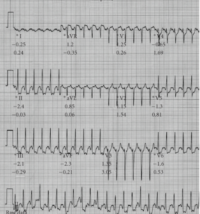
Peak exercise ECG in 48 year old women (Taken from from Ref 1)

What is the likely coronary lesion ?

Answer

Click here for diagnosis


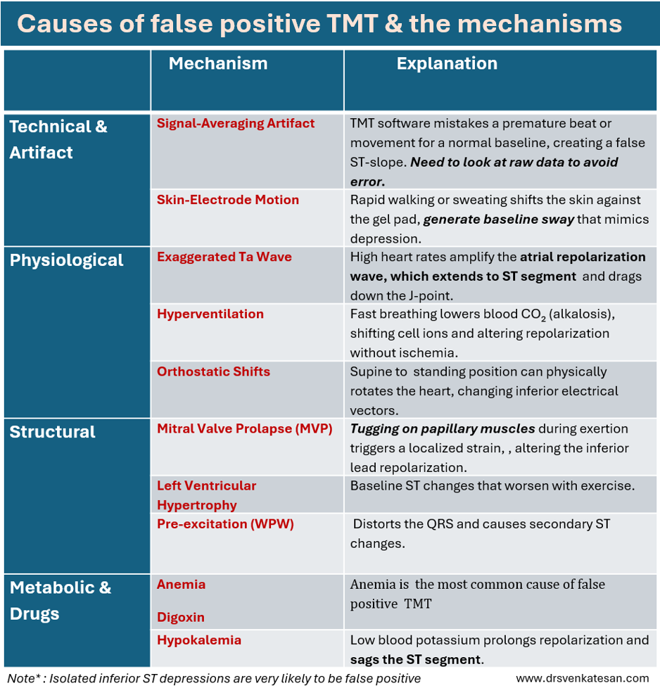
The mechanism of false positive TMT in MVPS is easy to understand.The pap muscle attachment zone of LV are under strain. In fact the commonest cause of ST depression in inferior leads are typical of MVPS. (Though it can occur in V5 and V 6 also as in this case) Both pap muscles are relatively posterior (Even the so called antero lateral) and the axis of strain is directed towards inferior leads.

The following table compiles few of causes of false positive stress test.

Final message

Never report TMT in Isolation. Not all the frightening ST depressions are left main. Unfortunately, no cardiologist can realsitically exist in this world , to report TMT as false positive, without doing at least a CT angiogram or a SPECT imaging

Reference

1.Michaelides AP, Liakos CI, Antoniades C, Tsiachris DL, Soulis D, Dilaveris PE, Tsioufis KP, Stefanadis CI. ST-Segment Depression in Hyperventilation Indicates a False Positive Exercise Test in Patients with Mitral Valve Prolapse. Cardiol Res Pract. 2010 Nov 14;2010:541781. doi: 10.4061/2010/541781. PMID: 21113438; PMCID: PMC2990857.

Read Full Post »