Feeds:
Posts
Comments

Archive for the ‘cardiology -ECG’ Category

In one of my classes , this ECG  was presented .  Controversy  erupted.It was about the  basics .

What is the QRS axis  of this ECG  ?

ecg north west qrs axis  indeterminate

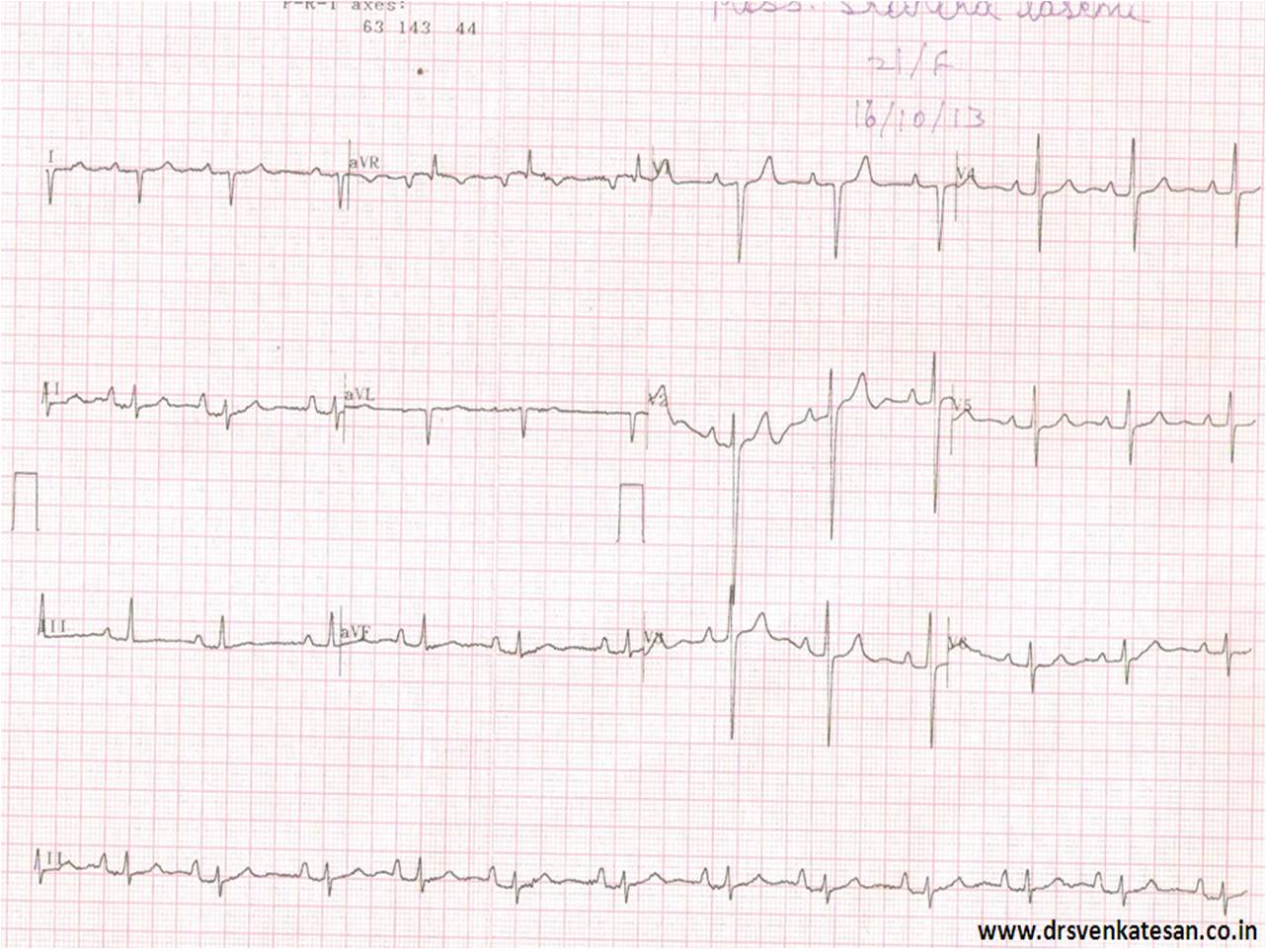
Not surprisingly there were  handful of answers .

  1. North west Axis
  2. Indeterminate QRS
  3. +150
  4. +180
  5. 0 degrees
  6. Extreme Right axis

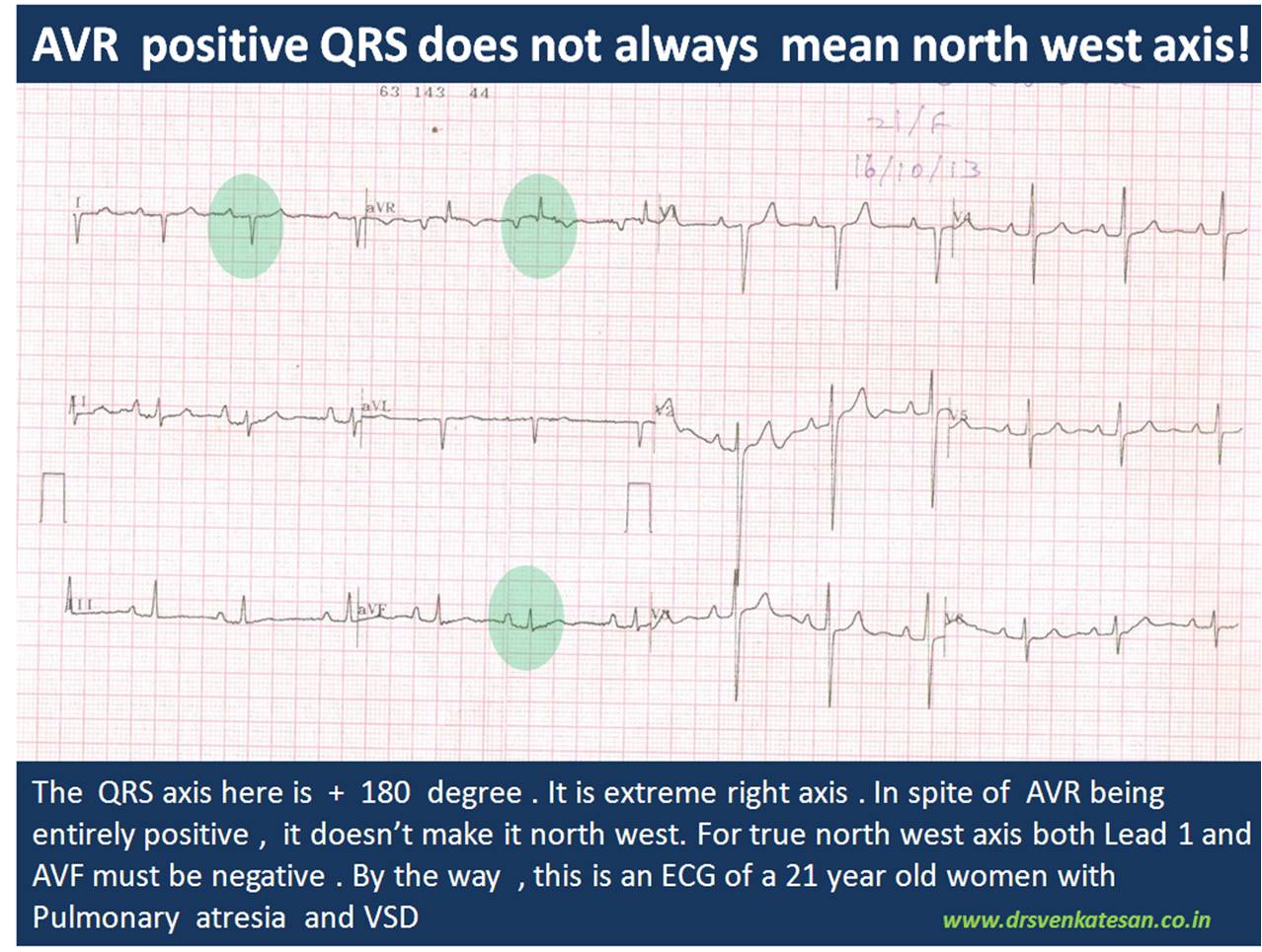
Which is correct ? My guess  is ,  it should be  closer  to + 180 .  Lead 2 is equiphasic and perpendicular lead is negative limb of AVL ie + 150 .If you  plot Lead 1 and  AVF in graph and calculate  we get + 135 . (In the strict sense , we are not  supposed to take one standard lead and an augmented lead for plotting ). Finally, the strongest argument was ,  since  AVR shows  only positive forces  it  would make  north west axis more likely .

ecg qrs axis north west indeterminate

Causes of North west  QRS axis

  1. Denova North west axis
  2. Extreme Left becoming NWA*
  3. Extreme Right  becoming NWA

*Left becoming NWA is much more common than other types.

Chamber enlargement alone is not sufficient to shift the axis to NW corridor. There must be anatomical distortion of  his bundle and it’s branches to shift the axis dramatically .This usually occur in complex congenital heart disease. In acquired heart disease the  an apical VT is probably  an important cause for NWA.

One word about indeterminate qrs axis .

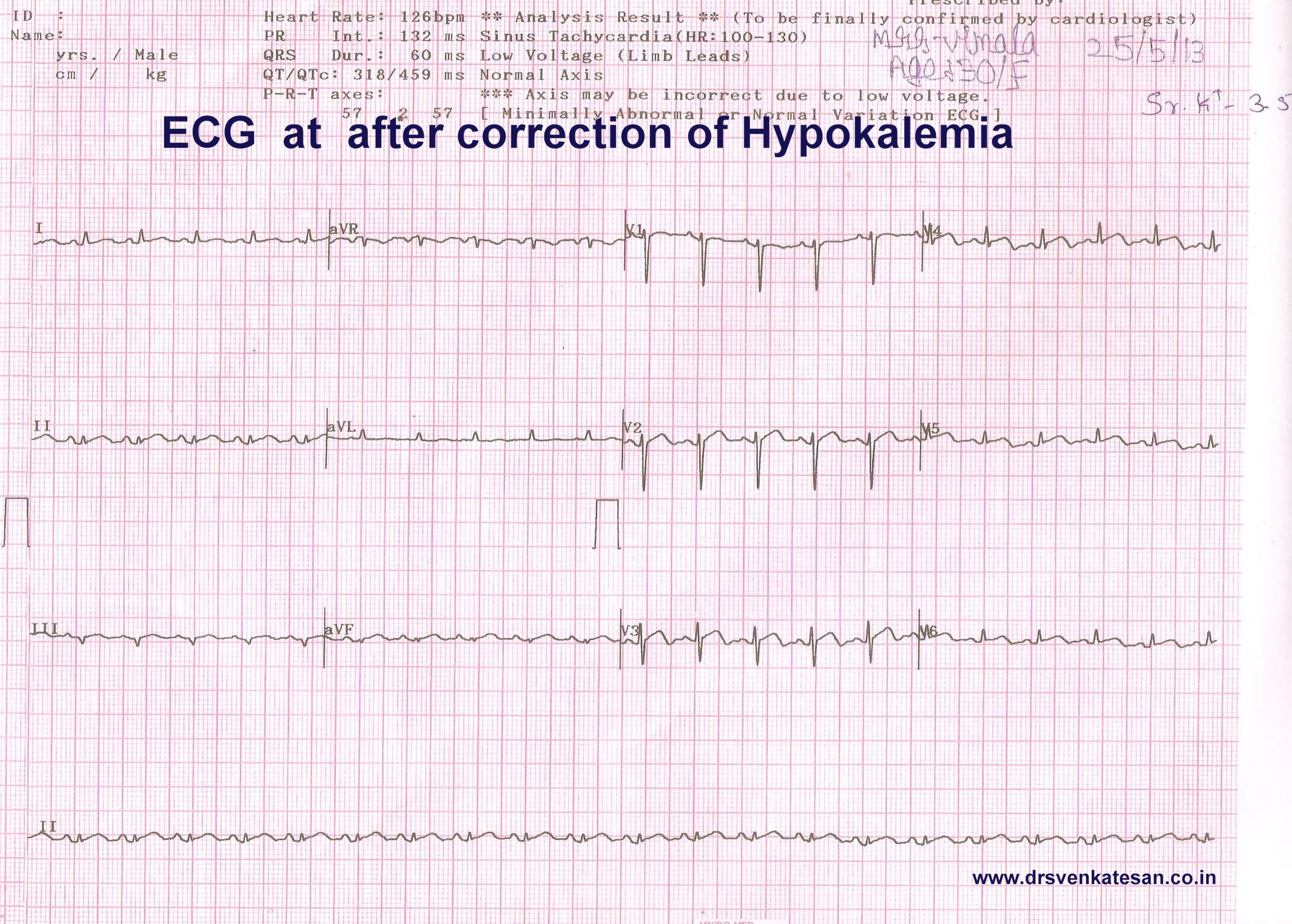
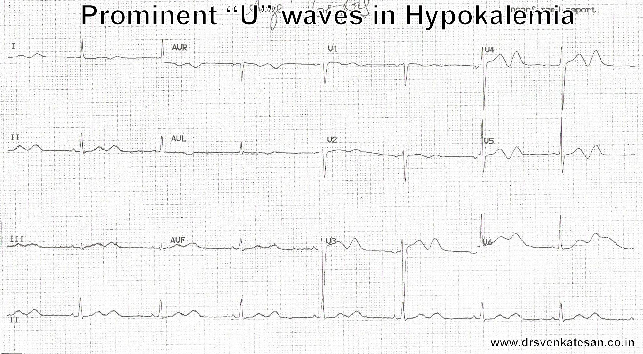
By the way , Indeterminate QRS axis  is not synonymous with north west axis. This term should ideally be used  if qrs complex is equiphasic  in all limb leads , when  qrs  axis  can not be truly determined .This situation commonly occurs when we encounter very very low voltage qrs  as in cardiac tamponade and severe hypothyroidism , constrictive pericardits, etc

If  the QRS is in north west  corrodor , How  to differentiate ,  whether it came from  extreme left axis  or  right axis ?

I am yet to find a correct answer for this.

  1. Pre-cardial pattern will help.
  2. A  q in V5/V6 would suggest  extreme left axis.
  3. May be we have to look the initial qrs vector in AVR lead for more clues

Traditionally , we talk about net qrs axis . We should realise net qrs axis is a combination of initial and late vectors .It can be dramatically different in different leads . QRS axis is  a two dimensional representation of three or more  (omni) dimensional electrical forces .That is the source for confusion. So,  do not unduly worry about the complexity .Blame it on the limitations of surface ECG !

Expecting some comments .

Read Full Post »

Human physiology can dramatically surprise us.Here is a situation regarding K+ ion and cardiac function.

Low potassium level is a well known cause for skeletal muscle  weakness and paralysis.While,in cardiac muscle usually the opposite happens.It is the high potassium levels that depresses and cause paralysis.(That’s why,it is used in cardioplegic solutions. )

But,the classical differences between skeletal and cardiac muscle need not apply in critically  low levels of K +

What happens when K + is critically low ?

We know, K + is the vital  ion that maintain not only the membrane potential ,(Recall Nernst potential ) but also keeps the action potential floating and dipping with every beat.

Imagine the intracellular chaos when these ion levels changes in dramatic fashion . (Of course,God has ensured very tight regulatory controls at various levels within each cell ! )

However , ECG changes are expected 100 % of time with falling K +  especially below 3meq.Surprisingly , low K + levels have little  mechanical impact.(Or is it our ignorance,considering the fact , cardiac electrical mechanical activities are tightly coupled?)We have to find answer from patients like this .

A  30 year old women came with breathlessness and fatigue and her ECG.

hypokalemia STEMI ECG changes

Can we afford to miss a diagnosis of STEMI ? With all our collective wisdom STEMI was diagnosed promptly . . . of course wrongly !

She was adviced  streptokinase.A shrewd fellow who reviewed  the old records spotted the past  history  hypokalemia , and Inj streptokinase was put on hold.(Lucky patient  . . . she was not shifted to cath lab )

Her  K + was 2.3 Meq. The LV function was significantly impaired with global hypokinesia, which  improved with correction of K+.

hypokalemia STEMI ECG changes 001

She was later referred for  nephrology work up , they had made a possible renal tubular disorder for the Hypokalemia.

Clinical Implication.

When potassium levels are critically low myocardial  function may deteriorate.Here is a patient with dramatic STEMI like ECG with extreme hypokalemia.

Our ignorance regarding electrolytes and myocardial function  remains unexposed .In critical care units  wide swinging metabolic and electrolytic  parameters are common.ECG is just a marker  for these .Similarly all LV dysfunctions are not primary myocardial disorders (Sepsis, Hypoxia, Extreme acidois , Uremia ,drugs,toxin  can lead to myocardial dysfunction.)

Experienced  physicians  do not form hurried opinion.Wait . . . allow things to settle down and assess again.After all ,there is long list of causes for ST elevation other than STEMI !

Reference

1.Chest. 1979 Feb;75(2):189-92.Cardiac dysfunction in a patient with familial hypokalemic periodic paralysis.Kramer LD, Cole JP, Messenger JC, Ellestad MH.
2.Acta Neurol Scand. 1978 Dec;58(6):374-8.Hypokalaemic periodic paralysis and cardiomyopathy.Schipperheyn JJ, Buruma OJ, Voogd PJ.

Read Full Post »

Scientific studies can be fun .In our spare time we often Indulge in rapid  fire sessions. We tested 30  wide qrs ECGs from our archives  (All proven VTs)  and  asked  our  cardiology fellows to apply Brugada criteria . They could   correctly  diagnose  VT  in 18* patients.The same ECGs were shown to the staff nurses of coronary care unit . 24  VTs were correctly identified  it.They did it by  their clinical sense and Instinct. (*12 vs 6  VTs missed)

wide qrs tachycardia svt with aberrancy and vt brugada verecki  griffith002

And now , four  clinical data was  provided. (Age , sex , Blood pressure , and  past H/o  MI were given )   The Nurses were able to predict it  28/30  VTs correctly.(97 % accuracy ) and the cardiologists  were able to equal the score now. So obviously clinical sense  was far superior .

Cardiology fellows were more likely to  mistake VT as SVT. This is far more common than SVT mistaken as VT. It is a strange academic  irony ,even the junior most nurses never missed a VT !

Summary

Simple sequence of history and clinical presentation is still far more powerful than ECG data in predicting wide qrs  arrhythmias . Nurses guess work is far superior than cardiologists  in predicting a wide QRS tachycardia as VT.

In fact , the  cardiology fellows are  preconditioned to   get confused   whenever they get a wide qrs tachycardia . Why  not aberrancy ?  In my  experience I have seen this question keeps  erupting inappropriately .Even  shrewd fellows suffer  from an  oscillatory  mind between VT and SVT .This is primarily because , every wide qrs ECG  is likely to  have at least two  criteria that fulfill both VT and SVT.

The implications are  genuine  and far reaching . While nurses  show a patient centric thinking  cardiology fellows  thought process revolves around ECG . Many modern-day cardiac physicians  are disconnected from clinical reality  and are obsessed with  complex EP concepts  and end up with a miserable face in the bed side !

This is not a new  revelation in 2013 . Masood Akthar told this  three decades ago.

Caution
Never try to glorify  guess-work . EP is a great science .The  pioneering concepts have made us understand how a VT emanates, travels , and exit from myocardium . We are able to localise it and ablate it .All credit goes to science . But , when it comes to bedside recognition of VT ,  clinical  sense  is a clear winner .With a  consistently > 90 % predictive value   it  can no longer be called as  a  guesswork   and becomes a hard scientific fact. Especially so , when the  intellectual  analysis of surface ECG   could predict  it  with paltry 70 %  accuracy (Read Reference 1)
Reference
This  analysis startlingly reveal  a fact .The over all accuracy  rate of predicting the wide qrs criteria  by  popular algorithms  is   between 66-77% ,  just 16 numerals   more than  gross   guess work  of 50 : 50 ( This  . . . or  . . . that )
Link to  Masood Akthar article

Read Full Post »

Cannon waves occur when Atria contracts against a closing tricuspid valve of  right ventricle .( There  would be a equivalent left atrial cannon which  goes into pulmonary vein as well  , it is discussed elsewhere !)

Cannon waves  happen only when P waves fall within QT interval in ECG as QT represents the electro-mechanical systole of  ventricles.  (Since P wave represents atrial systole , it is simple to understand when it falls within QT both atria and ventricular contractions collide to produce a cannon wave into the neck or pulmonary veins.)

The following two images of cannon waves  taken from the legend  Dr Paul woods own tracing  .

irregular cannon waves in jvp  complete heart block

regular cannon waves in jvp  svt avnrt  11 va conduction  002

Regular cannon waves

Occur during SVT  with 1:1 VA conduction.*

1 : 1  VA conduction  can be considered as  absence of  AV dissociation  (Rather  disciplined  VA association with every beat ) This is essential to create a hemodynamic  milieu for regular cannon waves.

* In AVNRT , VA conduction in strict  sense  is a misnomer  .It is simply a retrograde conduction thorough  the AV node .

Irregular cannon waves 

  1. Complete heart block .
  2. Multiple random VPDs
  3. Some patients with VT.*(Who are those patients ?  Those with AV dissociation when retrograde “P” wave falls  within QT interval cannon occurs. As expected this occurs in random fashion  which makes  the cannon fire irregular.

Can we get regular cannon in VT ?

Yes , but rare . As explained earlier this can happen only if AV  association occur on a retrograde fashion.

Further reading in this site

What-is-a-cannon-sound  , how is it related to cannon wave ?

Read Full Post »

hypokalemia U wave ECGhypokalemia U wave ECG 2

characters of normal U waves

It is usually recorded in the same direction as that of T waves

If it is opposite  it should be given significance .(Ischemic U waves ?)

More prominent in bradycardia

Mechanism of U waves

There are Three hypothesis

  1. Repolarization of papillary muscle
  2. Late repolarization of M cells in Mid myocardium
  3. After potential  to QRS  ie a  form of depolarization

Currently  the third option appears  closer to truth

How hypokalemia  result in tall U waves ?

If Hyperkalemia elevates T waves  it is natural  to expect  hypokalemia  to blunt , flatten or Invert the T waves .Of course , this happens in many . In others with severe hypokalemia an intact  T  is accompanied by a tall upright U  as well .This adds a mystery twist* to the exact genesis of this wave .

* During diastole cell membrane is closer to k +  if K + is deficient it tends to drift . (Some where i heard this   . . . but I do not understand it fully !)

.

ECG U waves 2

Reference

Read Full Post »

The other day my fellow got a call  from surgical ward for emergency ECG opinion for a  suspected Inferior MI .It later turned out to be an acute cholecystitis.

One of the important  anatomical mis-perception  among physicians ,  is to consider  inferior, posterior  and diaphragmatic surface  of heart  as separate entities .They are all  closely linked.In fact, they  more often  mean  the same  anatomical zones !

Heart is a dynamic suspended organ within the middle mediastinum .It  can assume a vertical or horizontal position due to number of surrounding anatomical  and physiological factors. (Diaphragm, Lung , being  important ).The ratio of intra thoracic vs Intra abdominal  volume  &  pressure determine whether the posterior surface of the heart is going to face the back of chest  or simply sit and  rest on the diaphragm .We know a horizontal heart is likely to inscribe q waves  in inferior leads .

acute abdomen diaphragm inferior wall mi cholecystitis pacreatitis

Courtesy : Basic image source from digitallab3d

The  diaphragm can be termed as an  anatomical causeway , that isolates   thorax  from the  abdominal  cavity .Close encounters between the organs separated by this delicate biological  membrane is  always possible .This is especially true for electrical signals  which show little  respect for anatomical barriers .

This is the reason there are too  many abdominal conditions that mimic  inferior MI during a painful  emergency (and vice versa  when inferior  MI mimics  acute abdomen .) In  our  department , we   have witnessed  the following conditions mimicking Infero-posterior ACS.

  1. Acute ascites with polyserositis
  2. Gross obesity with APD
  3. Posterior fat pad ( Necrosis ?)
  4. Thickened pericardium
  5. Minimal posterior pericardial effusion
  6. Diaphragmatic pleurits
  7. Esophageal spasm
  8. Fundal air  trapping and ballooning after a heavy meal !
  9. Acute duodenal ulcer perforation ( With gas under diapharam causing q waves)
  10. Acute cholecystits
  11. Diphragmatic hernia
  12. Achalasia cardia
  13. Pancreatitis

Final message

Do not rush to make a diagnosis of inferior wall MI when  you encounter inferior q waves  with  or without ST /T changes , especially  when the symptoms are atypical .

Read Full Post »

The humble looking strip of ECG  recorded  in millions ,  every day across the globe  has a complex definition.

And this definition  is  the most apt I have found.

An electrocardiogram (ECG) is a curve showing the potential variations against time in the whole body stemming from the heart, which is an electrochemical generator suspended in a conductive medium.

Einthoven(Dutch)  is the the man who invented the ECG  ,  got a Nobel price for not only inventing the string galvanmeter to record ECG,  but also  making us understand   the rules  of the  electrical wave front  that emanates from the heart.( Not to forget the  original concept  of who demonstrated electrical activity from heart by by capillary electrometer.  by Waller.(British). In my opinion waller should have shared the Nobel prize. I am  sure ,even Einthoven would agree to it.

Of course , do not ask  which  comes first  ” Ionic  flow” or  “the current”  that  comes with it !

Credit goes to  the creators of this  wonderful book  on medical physiology and put that free on the net . Three cheers to  them .

The book is from  University of Copenhagen  , Pannum Institute of Medical Physiology.

physiology text book paulev Zubieta sweden

Reference
British physiologist Augustus D. Waller of St Mary’s Medical School, London publishes the first human electrocardiogram. It is recorded with a capilliary electrometer from Thomas Goswell, a technician in the laboratory. Waller AD. A demonstration on man of  electromotive changes accompanying the heart’s beat. J Physiol (London) 1887;8:229-234
Willem Einthoven introduces the term ‘electrocardiogram’ at a meeting of the Dutch Medical Association. (Later he claims that Waller was first to use the term). Einthoven W: Nieuwe methoden voor clinisch onderzoek [New methods for clinical investigation]. Ned T Geneesk 29 II: 263-286, 1893

Read Full Post »

Pacemaker current is   strangely  referred  by physiologists  as  funny current (I f ) . I am yet to find the exact reason .  This is the current  that  sustain  our life right from the day 22  of  embryonic life when the  cardiac jelly beats for the first time.   SA node  solemnly  follow our  entire life  before  making  a  bid-adieu !

pacemaker  potential sa node 5


pacemaker current if funny current poential 002

pacemaker current if funny current poential 003

What is contribution of  If  current in the overall Pace-making  activity ?
This  has not been quantified . The fact that ,  Ivabradine induced  If  current  blockade does not result in serious bradycardia indicate  , SA node has alternate reserve currents as well . ( SA node  is a such a mystery  structure , it would never be a  surprise , if we  find many more  “not so funny”  currents !)

Read Full Post »

It is believed  T wave alternans  is a marker of impending ventricular fibrillation. Though it is not applicable in every clinical setting it is indeed true if we observe T wave alternans in an acute ischemic setting .Here is a patient with  ACS and inferior MI who developed T wave alternans after temporary pacing.

T waves alternans torades de pointes 3

t wave alternans

T waves alternans torades de pointes

went in  for a chaotic  T wave rhtythm  and ended up in VF that  required s shock.T wave  alternans is other wise known as repolarisation alternans .

Twist dance of Heart

Torsedes is twist around it’s axis.   Any   ECG wave  can twist in it’s axis .If T wave alternans  becomes gross it will twist 180 degrees   .Once this happens the heart can go for  fibrillation any moment !

Final message

Extreme form of T wave alternans would result in  complete twisting of repolarization vector which is a  harbinger of ventricular fibrillation

I wish  this can be referred to as Torsades  “T” pointes instead of  Torseades “de” pointes


Read Full Post »

ST segment elevation is the key parameter on which the fate of millions of infarct patients are recognised and managed. It is ironical we do not have standardised reference point for measuring the quantum of SR elevation .

This is especially difficult when ST segment blends with forward limb of T waves.

While we have reference point for measuring  ST depression  (Like during EST ). . . why we do not have one for ST elevation ?

Now we have adopted a rough criteria .Read below .

where will you measure st segment

How to measure ST elevation in ECG
How to measure ?
Measure The ST segment 40 ms from J point.
how to measure ST  elevation in STEMI 2
                        I lost track the source of this Image .(STEMI hand book 2012 ?)
Final message
ST segment  elevation  is  the key parameter in ACS.  Quantifying  it becomes  important in  assessing  the efficacy of reperfusion strategies and risk stratification. Fresh ST elevation can represent pericarditis, reinfarction or an early dyskinetic  segment . Unless we have  proper reference point there is  a room  for  error in this simple parameter.

Read Full Post »

« Newer Posts - Older Posts »