Feeds:
Posts
Comments

Archive for the ‘Cardiology -unresolved questions’ Category

Any new ST depression occurring during  EST is sine qua non for inducible ischemia.But,this rule does not uni-formally apply  in all 12 leads .ST depression occurring is certain leads is more important. While severe global ischemia can depress  ST segment  in most leads ,factually  only the leads V 5 and V6  predict true Ischemia.This because , bulk of LV muscle mass faces these two leads.

Isolated ST depression in inferior leads  during exercise

  • Is a frequent issue occurring at the peak exercise.
  • Is least predictive of significant CAD.
  • The exact mechanism is not clear.
  • Some continue to  believe it is indeed significant .
  • We have  observed  isolated  ST  depression > 2mm in inferior leads with significant CAD.
  • What really matters is the quantum of ST depression , symptoms, and exercise time and preexisting CAD .

Probable mechanism

  • Apart from true ischemia ,ST depression may indicate relative sub endocardial strain rather than ischemia.(By the way can simple stretch can cause ST depression ?)
  • The Infero posterior surface of heart represent  right ventricle .RV volume overlooked peaks exercise.Some think it represents acute raise in RV load during peak exercise.

How to report such EST ?

You can report it as such,  what you have observed.

  • ST depression noted in Inferior leads at peak exercise.
  • Mention whether it was angina free,
  • At what METS,
  • Total exercise time .

If you are statistically inclined  you can also mention the likely hood of CAD by positive predictive value (PPV) of the test (Low with isolated Inferior ST depression )

If you are really confused , and do not want to scratch your brain we have the most convenient terminology  invented by cardiac physicians ie Borderline EST, or Mildly positive EST “

Should we do Angiogram for such patients ?

In this era of catching normal people  who attend master health check ups  for a day care CAG  . . . it is not all  a crime to do angiogram in a  patient who shows suspicious  ST depression in three of his leads (2,3,AVF) especially if he also complains of vague chest pain.

Alternate investigation

Of course , we  always have the luxury of using  MDCT  that can stunningly  photograph the coronary arteries.

It is a mystery investigation, if it comes entirely normal every one is happy.Even slightest  defects in the photography  has a potential to confuse both physician and the patient .

What I do ?

I hesitate to  do routine CAG  if ST depression occur exclusively at  peak exercise beyond 10-12  METS , which disappear fast.(Many times we can apply this rule  to classical ischemic ST depression of lead V4 as well !)

ST  depression  in any leads (with any degree) following an episode of  ACS seems to be important.

Related topic

Read Full Post »

Following are revered  facts  . . .  among the  “Guardians of   Cardiology” !

myths-truths-300x300

When false truths are synthesized to conceal a true myth . . . where will the poor myth complain ?Following are revered  facts  . . .  among the  “Guardians of   Cardiology” !

  • Primary PCI  is a greatest innovation  in modern day cardiology .Without this modality  most  STEMI patients will buy Instant  tickets to grave yard !
  • A cardiologist who intends to  thrombolyse  a STEMI is considered as a low quality cardiologist .
  • Streptokinase should have  no place in the crash carts of modern coronary care units.
  • There is nothing called “Time window” for rescue angioplasty.
  • VVI pacemaker  will convert an electrical problem of heart block into a mechanical one by depressing LV function .
  • Digoxin is an obsolete  drug even in well established cardiac failure with dilated heart.
  • Beta blockers not only fail to control  blood pressure smoothly , it often converts  a hypertensive individual into a unhealthy one  by it’s prohibitive side effects !


Read Full Post »

Recently , I came across a   young women  who underwent the following three tests for one episode of syncope after witnessing her pet dog bleeding with  an Injury !

  1. Carotid doppler
  2. Holter monitoring and event monitors
  3. Brain MRI /MR angiogram

This was followed up  by Head up tilt(HUT)  in a premier hospital

After 1 week of investigation ,a diagnosis of  Neurocardiogenic syncope was made and she was reassured and no drugs were prescribed.

(The collective yield of the above three investigation in fixing  a specific diagnosis is  less than 10 % of all known causes of syncope )

Syncope  approach  evaluation

To diagnose  common syncope . . . we need common sense !

Syncope is a dramatic  symptom.It is one of the commonest symptom in ER as well . Life time incidence of syncope is at least one episode in 50% all human life ! The definition  of syncope until recently , was liberal .Any transient loss of consciousness with spontaneous recovery  was termed syncope.

This includes

  1. Hypoglycemia
  2. Anemia
  3. Siezure disorders
  4. Structural  neurogenic (Including ,  brain tumors , Dural hematomas etc )
  5. Panic attacks (psychogenic)

Cardiologists wanted to fix syncope as an exclusive disorder of  circulatory insufficiency.By bringing in a modification in the definition  , ie  syncope is  now defined as a transient loss of consciousness due to   reduction in cerebral perfusion  .

This definition helped cardiologists  to exclude the above entities . Still many would include all in single basket as patient should be seen as a whole and we can’t expect them to  land according to our convenience and classification.

Here is an incomplete* list about causes of  syncope (* 99% complete ?)

Vascular

  • Vaso- vagal syncope in young ( Neuro-cardiogenic , Common , Benign)
  • Autonomic dysfunction of elderly ( Including postural hypotension )

Cardiac

Arrhythmic ( Sinus node dysfunction /CHB/Idiopathic VT/Long QT syndromes)

Structural heart disease

  • Valvular  heart disease  (LVOT/RVOT obstructions)
  • Myocardial disease
  • Rarely ischemic heart disease

Miscellaneous

  • Severe pulmonary hypertension (Including PPH ,  pulmonary Embolism )
  • Paradoxical embolism.
  • Aortic arch disease -Takayasu related arteritis .

Investigation

We have a sophisticated array  of investigation for syncope .It can be a never ending exercise , ranging from  spinal cord evoked potentials to diagnose Shy-drager syndrome ,   . . .  to implanting long-term loop recorders to decode  heart beat behavior.

However , evaluation of syncope is the ultimate wake-up call  to all current generation cardiologists  . . . Why clinical cardiology  should  never  be allowed to die (and  it  will not ! )

Common sense begins with answering  few simple questions . Is it really syncope ?

If  you ask this question three times and with  specific leads to the patient  and the witness ,  truth will come out  . 90% of times it may not be syncope at all (Near syncope, accidental  fall, dizziness ,extreme blurred vision, drowsiness  etc)

If it is syncope , Is there a non cardiac cause ?

It may related to the Hypoglycemia / Anemia /Panic attacks.Get a neurologist opinion , it would be terrible mistake if you miss a space occupying lesion  within the brain. (Missing chronic silent sub dural hematomas is  frequent   in the evaluation of syncope of elderly !)

Ruling out  cardiac syncope is relatively easy

In the remaining  patients  basic investigation like routine blood tests,ECG, ECHO   will help us  rule out most serious cardiac disorders.Similarly  bulk of the electrical cardiac syncope can be diagnosed.(Holter , carotid study in selected few )

Need for neurologist -cardiologist interaction.

Syncope due to VBI,  transient Ischemia attack , Senile vascular dementia  is a grey zone . Many have complex neuronal -vascular mechanisms . What is Consciousness ?  and  What is LOC ?  :Is it the lack of blood or severely depressed nerve signal in the reticular activating system? Lots of interaction between cardiologist and neurologist is required to clear our ignorance.(I  have one such  elderly patient who is intermittently awake ! I call this chronic syncope !)  .

Undiagnosed syncope is not  a crime

Realise the most important lesson in Medicine . If you  have ruled out all serious  causes of syncope you should have the courage to be satisfied with that !

Scientific pursuits has a limit. Searching for the mechanism of a psychogenic  fainting attacks with intra cerebral electrodes is a clear case of  physician acquiring a psychotic  behavior !

Final message

Syncope is not only a dramatic symptom for the patient , it also unfolds a drama of costly  investigations  . .  . many  with  dubious value.

Talk to the patient personally for  10 minutes in a quiet room, try to apply that elusive  clinical sense  . . .   it would rarely let you down !

After thought

What is the true clinical value of * Head up tilt Test (HUT)?

Will be posted soon

Read Full Post »

Last week  there was a heated debate in our CCU regarding thrombolysis for  a patient with severe rest angina  and ST elevation in AVR  and ST depression in V2-V5  as it implies  Left main disease  Few argued left main disease is an exception where one can thrombolyse even with unstable angina !

One of my fellows argued ACC guidelines vouched for lysis in UA involving left main .( I do not agree )

A logical attempt to differentiate Left main NSTEMI//UA and STEMI

(In the strict sense Left main NSTEMI is misnomer as AVR shows ST elevation  isn’t ? )

left main disease

Final message

Such  patients with suspected LMD   are to be rushed to cath lab .  . . agreed . If it is not feasible , manage it as high risk unstable angina and do not thrombolyse .Let it be left main disease . Indications for lysis are clear. ST  elevation in AVR alone can not be taken as an Indication for lysis.For thromolysis to be effective there should be high thrombus burden with total occlusion . ST elevation in single lead (AVR ) is not a good  marker for left-main thrombus !

Read Full Post »

Oral anticoagulant usage has been steadily increasing for variety of  indications.Dengue fever is also  appearing in different avatars with  low platelet counts  and bleeding being a primary risk.

I was recently contacted by a physician , regarding a therapeutic dilemma .A young lady with mitral prosthetic valve and a febrile illness diagnosed as dengue . She has a platelet  count of 100,000 .She is on regular warfarin and aspirin .The physician  wanted to know , should he stop the OAC and aspirin ?

What are the options ?

  • Confirm if it is really dengue.
  • Look for clinical bleeding.INR, platelet function tests are not helpful.
  • Continue OAC.You can do that in most situations.
  • Stop OAC only if there is clinical bleeding  episode.
  • Anti-platelet drug usage  is more tricky .One may stop it if the trend of falling platelet is steep by at least two serial measurement.(or 50% fall from baseline)
  • Fresh blood and platelet infusions should be ready .
  • Finally and most importantly , Inform the patient and family about the difficult decision we are making.

*Is  OAC  safer than aspirin and clopidogrel in dengue ?

It is believed OAC has no major  Impact on platelet function .It may not  pose a threat of excess bleeding in the setting of  falling platelet levels .(*Evidence base -nil )

Another potential situation : DES and dengue

The number of DES in developing countries are increasing  where Dengue is endemic . It is not a surprising  to expect  both to  occur together.

Anti-platelet agents  can be problematic .It is better to withhold it during the active phase of dengue.(If the  stent has recently  been deployed you have no option !)

Final message

1.  Prosthetic valve , Warfarin Dengue .

2. DES, Dual antiplatelet agents ,Dengue.

They  extraordinary events  throw a complex therapeutic task .There are only two options .Continue or discontinue ! Whichever way you do , you explain to your clients (patients!)  the (un)reality games we play.

My personal option would be , with hold all hematological drugs during the active phase of dengue .

It is better to believe in the  natural thrombus fighting force . Leave the job of anti-platelet action  to the dengue virus for a week or two and give oral anticoagulants and dual anti-platelet agents a holiday

It may be foolish to rely on the dengue virus to guard against  prosthetic  valve  and DES thrombus , In reality we have to do that !

Reference

No reference exists.It is a statistical mind game.Individual assessment  should prevail. Either way, if something adverse happens court of law should protect us !

Read Full Post »

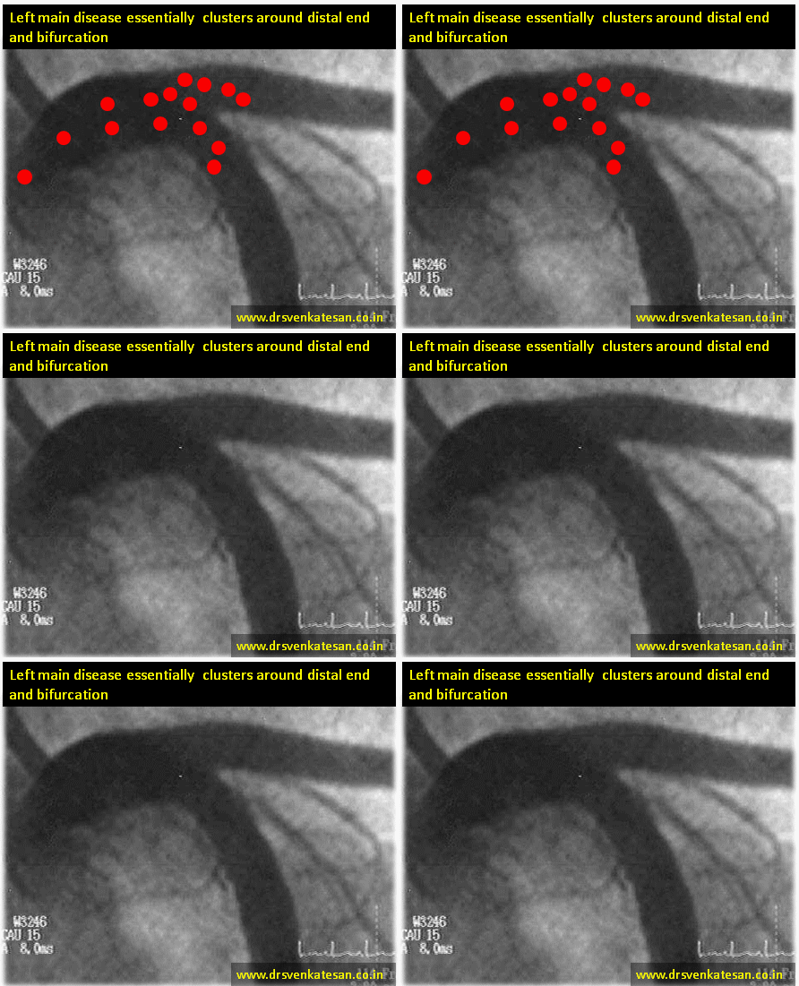
Distribution of Left main disease.

  1. Ostial
  2. Ostio-proximal (Within 1 cm of  origin )
  3. Shaft -Discrete  mid left main
  4. Shaft -Diffuse
  5. Isolated distal shaft( 1.0.0)
  6. Bifurcation ( Medina 1.1.0 -LAD)*
  7. Bifurcation (Median 1.1.0-LCX)
  8. Bifurcation ( Median 1.1.1)*
  9. Trifurcation ( With ramus )

* These three locations account for nearly 75% of all left main lesions.

left main disease coronary angiogram

We know atherosclerosis is  a branch point disease .Normal left main measures 1 mm to 20mm.The shorter the left main lesser is the the incidence of LMD. Short left main can not engage the atherosclerosis much (No left main = No left main disease ) However ,very short left mains  may increase ostial lesions .

  1. The commonest left main lesion is distal left main with one of the branch involvement (1.1.0.LAD is more common )
  2. Least common entity is discrete mid shaft lesion.

Simple strategy.

First dictum : All complex looking LMDs should be referred to a good  surgeon.

Final dictum : Remember medical management for left main disease is still an accepted strategy in stable , non flow limiting situations .

Interventional  Cardiologists  feel they have the exclusive rights   to indulge between these two  spectrum of LMD .May be true! But extreme caution is required as we are playing  our game in the most critical  coronary high way .

Some suggestions and thoughts.

  • 50 % diameter stenosis is significant. But significance does not mean we should tackle the lesion by aggression.
  • Symptomatic flow limiting lesion only to be intervened . (Flow limiting means both angiographic and a stress test .FFR <.8 is also an index for flow limiting .Symptom means Angina on exertion )
  • IVUS, OCT, FFR,NIR ,SYNTAX  are not path breaking tools .They essentially  add  more glamor  to left main disease than anything .
  • Most bifurcation LMDs are  managed by single stent with stent jailing the major side branch (Yes side branch can be LCX !)
  • However ,two stent strategies is not banished .It can be vastly  superior in some selected cases .(Especially with huge plaque load at carina )But needs expertise .
  • In very small vessels two stent strategies are risky .

Reference (2012 update)

left main disease  coroanry angiogram management  Fajadet

PRECOMBAT left main disease south korea everolimus
Link to related articles in this site

Read Full Post »

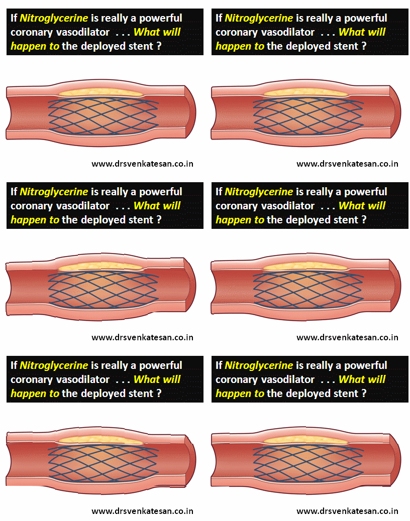
We know Nitroglycerine(NTG) as a most powerful epicardial coronary dilator  . We use it for instant relief during episodes of coronary arterial spasm in cath lab.

What will happen if we administer NTG over a stented segment ?

Does it dilate it with same vigor ? What will be the consequence  ?

A perfect setting for stent migration isn’t ?

Let us bust the myth around  NTG . NTG  rarely  show  visible coronary dilating effect except in the setting of coronary spasm .

NTG and coronary vasodilatation

Does a LAD with 3 mm diameter become 3.1 or 3.2  and so on with NTG ?

No .It won’t .It is my belief. It is well known , NTG’s action varies significantly in normal and diseased endothelium . Again , there is an irony .It seems , it can act only in normal endothelium , but  we need require it’s therapeutic action only in pathological segments.Further any stented segment would contain   clusters of  both normal and abnormal endothelium .

One more inference is that, stented segment exerts constant pressure on intima making any  pharmacological vasodilatation irrelevant .

Importance of  radial strength of a stent

This issue of vaso-dilator induced  stent migration may not arise in self expanding wall stent with high radial force.But we do not know how long these metals will carry this metallic property .Balloon delivered  stents ( currently used 99% of times ) do not have permanent radial strength .

Final message

I am yet to comprehend what nitrates are expected to do (and what it really does ?)  in a patient post PCI ? (By the way  . . . why we need to prescribe Nitrates it in the first place ? but  In real world most continue to take this for many reasons .)

We need to analyse the micro-vasomotion at the stent -coronary intimal interface.The dynamism in this  narrow space  can be critical  , and may make the difference between life and death !

After thought .

In the hind sight,  this post appears quixotic  for myself . But some one , some where , may generate a great idea  out of it , that will help our patients.

Read Full Post »

We expect LBBB in RV pacing . . .but if RBBB is recorded we are worried ! (Often times it may be neither  LBBB nor RBBB )

Is it really a panic situation ?

  • Not necessarily. The only issue is septal perforation .It is rare , can be recognised by echo or fluro .
  • In true RV apical pacing with tined leads , RBBB  is extremely uncommon .
  • If the lead  is fixed  in the septum and para hisian  area ,  there is  definite  possibility of  deviation from typical LBBB pattern . Screwing leads  that faces high septum  or outflow , RBBB  can be noted occasionally.
  • The commonest cause for RBBB pattern in RV pacing ,  is due to  screw tip going deeper into septal planes  and activating the fibers of left bundle early .
  • For LBBB pattern to occur right  bundle should be morphologically intact .In diffuse CHB  with bilateral bundle branch blocks  the relative contribution ( Impulse conduction ) will determine the QRS morphology . If right bundle is more damaged than left bundle ,RBBB pattern  may prevail  even in the midst of RV pacing !
  • In elderly men with sigmoid septum typical LBBBs are not observed.
  • Anther plausible mechanism would be , even though RV is paced , the pacemaker current’s  exit route may be from LV side .
  • Finally , always  think about coronary  sinus pacing .It is extremely common in blind temporary pacing.

What should we do if we encounter RBBB morphology after PPM ?

  • Analyse the ECG meticulosuly for capture or sensing failure .
  • Do an echocardiography in RV inflow view.
  • Screen the lead by fluroscopy
  • Check the pacing  parameters.
  • Do a holter if  you are really anxious .

If everything is fine , just forget the RBBB.Don’t split your hair for this apparent paradox. In medicine  impossibilities will always  galore !

This paper from Taiwan would vouch for this

RBBB during RV pacing safe ecg

Read Full Post »

We know LVH and SHT go together . Mind you , this is not an Intimate relationship.

Widespread utilisation  of echocardiography  has revealed  , definite  LVH occurs only in about 20% (A guess !) of  HT . (Do you know in the Famingham study the incidence of LVH  after 12 year follow up was a paltry 3 % .Will you agree with that ? Mind you , It was in 1969 when Echo was not there )

What determines LVH ?  The clear answer is elusive. It is easy to escape  from the issue by calling it  multi factorial !

Why don’t you try this question .

My guess would be ,  magnitude ( or  even duration of HT !)  is  less important than genetic predisposition  or  associated diabetes ,  renal involvement.Our analysis from  hypertension clinic reveals LVH is many fold common in secondary HT  when compared to primary HT !

I often used to provoke the students by saying if the LVH is gross in HT it can not be primary , 9/10 times  ! Invariably  we find some  other  association or reason for the HT !

Link to related topic in this site

Why-lvh-does-not-occur-in-all-patients-with-systemic-hypertension ?

How-diabetes-modifies-lvh-due-to-hypertension ?

incidence of lV left ventricular hypertrophy framingham study

Next  . . .

How does LVH regress with treatment ?

Read Full Post »

Regurgitant  lesions of cardiac valves  are often tricky for the heart . Myocardium shows “love- hate” relationship with these  leaky valves.  Some of them are  “sort of”  stress relievers for  LV . A mild MR will make the LV comfortable in terms of wall stress. When the wall stress is reduced the contractility increases and LV EF may raise a little.Hence EF is never going to help us to assess true LV function in MR .

LV end diastolic dimension(LVEDD) is  a preload dependent  parameter .A patient with 6.5cm LV EDD  may still  have good contractility  and he may reach even a  40mm LV ESD, implying an intact LV function.

LV function should be best  assessed  in systole .(After all ,  systole is the prime function of heart) .Further , it should be assessed when the LV is  free from  influence of the all  loading  conditions of heart .  (Note : The initial part of systole  depends on after load. As the systole progresses the influence  of after-load lessens .In the pressure volume loop* , the point at which loading conditions are least operative is end systole.)

* Please realise , heart is a dynamic organ there is no true load independent point in cardiac cycle  as pressure and volume are eternally coupled.

What happens in AR ?

The same rule applies for Aortic regurgitation, but the parameters worsen little later than that of MR. For same degree of regurgitant fraction MR will require early surgery than AR.The reason for better  tolerablity of  AR  is due to largely  intact LA function and compliance till very late stages of AR.(In AR- it’s single chamber volume overload , while in MR  it’s two chambers !)

Final message

LVEDD is not used in assessing MR  as it is a pre-load dependent parameter that will not reflect true myocardial  function /dysfunction. LV ESD is fairly accurate  measure of LV systolic function minus all loading factors .

Watch out  for next topic

Vasodilator therapy in MR and AR : How is it different ?

Read Full Post »

« Newer Posts - Older Posts »