Feeds:
Posts
Comments

Archive for the ‘cath lab tips and tricks’ Category

A patient with near 90% LAD disease who had a significant TMT/EST positivity with no clinical angina  was  subjected to FFR by a scientific  cardiac physician. Since FFR was recorded as  .9 , he was adviced against a stent and sent home with drugs.

Now , in the  physiological assessment of a coronary lesion ,  which one you are going to trust , TMT positivity or FFR ?

FFR  measures trans-lesional pressure drop  by creating a artificial exercise physiology  in a particular coronary bed by injecting just one of coronary vasodilators  namely Adenosine. FFR assessment can never be considered truely  physiological .There has been huge discrepancy in the amount , rate and route of administration and the hyperemic response to Adenosine.

Final message

In a single vessel disease population , if TMT is positive the lesion is to be taken as significant, irrespective of FFR.(Provided Anemia and other systemic factors are excluded )

*Read this and get ready to get  confused further , single vessel disease with TMT positivity  doesn’t mean medical management is never an option .OMT ,(optimal medical therapy ) even though a battered concept is not yet dead for SVD !


Read Full Post »

Every one talks about  coronary excesses ! It happens  both  in acute and chronic  fashion , not withstanding the inappropriately  understood  . . .   appropriately  released  guidelines  on inappropriateness ! The  burden  of coronary syndromes of the humanity, I am afraid would  include these man made excess as well !

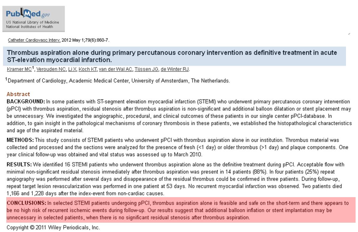
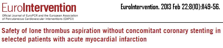
I stumbled upon two  small  “gems ” in this other wise wild dark  cardiology literature  .One from Kamaer , Netherlands and other from  Escaned from Spain.

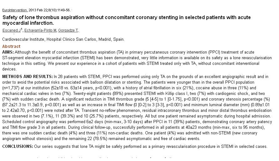
Both  talk about a  simple and logical modality in the management of STEMI . If bulk of the STEMI events are due to coronary thrombosis just tackle it  . No more  . . . no less” Stent only , if there is tight residual lesion.

1. From Amsterdam , Holland.

krammer thrombus aspiration alone priamry poba for stemi no stent

2.This one is from Spain.These studies I am sure , only a fraction of the interventional community would have read .Reason ? We are always hijacked by the moments of glamor ! I am just sharing them .hope few are benefited

primary POBA thrombus aspiration alone for stemi no stent stemithrombus aspiration alone for stemi no stent priamry pobaThese two studies with total number of 44 patients has a potential to redefine  the entire practice pattern of acute interventional coronary care.(Of course , if only , we are ready to make sense out of it !)

But , the concept will be heavily banished by strong visible and invisible forces   for the simple reason it suggests a true possibility  of knocking  out the role of  stent from acute STEMI arena.

When I discussed with my colleagues  for a large scale study  on isolated thrombus aspiration in STEMI , they told it  is not possible for ethical reasons !

I was amused , denying such a study is biggest ethical blow to the field interventional  cardiology !

Final message

Proof of concept does not require numbers .A study with less than 50 subjects  can be far superior than multi-centre ,multi-blinded , self steered ,peer reviewed largesse ! The truth of the study lies in the core consciousness  of people who do it , not in the numbers and exotic statistical methods !.

After all , one of the greatest medical study  was  done by James Lind  (Father of RCT) who discovered vitamin c as an antidote for scurvy,  with a hand full of sailors  while they crossed the Atlantic many centuries ago !

After thought

You say , thrombus aspiration is great , Why the hell , TAPAS , INFUSE AMI, and TASTE studies  confuse us regarding thrombus aspiration  ?

Don’t blame it on thrombus aspiration .We do it perfectly . It is because of what  we do after that ! We decorate the coronary lumen finally with a piece of metal cherry  undoing all the goodness of a great pudding !

Read Full Post »

The success of primary PCI  is defined on the basis of acute vessel opening and deployment of stent and wheeling out the patient out of the cath lab. Most PCI cath report promptly mentions TIMI 3 flow with Grade 3 myocardial blush.

I recently encountered 3 patients over a period of 2 months in my echo lab. All three had a recent primary PCI for STEMI found to have scarred IAS with moderate LV dysfunction.

These patients were medically savvy and asked a simple question after glancing at the report signed by me.

Doctor , If my myocardium has  been allowed to die a silent death  in a matter of 30 days and replaced by a  whitish scar , what is the point in calling the much hyped  primary PCI successful and charging me about 4 Lakh Rs . They wanted to know ?

I told them you have to direct this query o the supposed state of the cardiac Intervention team.I also told them it may not be proper to criticize anyone. The science of myocardial revascularisation is yet to be fully understood.

Why the myocardium goes for long-term scarring in spite of prompt early revascularization?

  1. Time window errors (What you think is early PCI may be in fact a late one!)
  2. Intermittent patency
  3. Re- occlusion
  4. Individual variation in hypoxia resistance
  5. Contra-lateral significant CAD.
  6. Recurrent  coronary events
  7. Poor compliance with medications.

Final message

Just because your patient has received a state-of-the-art primary PCI  in a high-end hospital does not negate the possibility of myocardium going in for scarring and resulting in significant LV dysfunction. There is something more hidden in coronary hemodynamics than it appears!

The so-called acutely successful PCI in your discharge summary actually may mean nothing. Unfortunately, we the pundits of cardiologists never bothered to include myocardial status as one of the criteria to define the success of the procedure.

Ideally, we may defer calling a primary PCI successful or not by at least a few months, when the true story unfolds ie how your myocardium has responded to the treatment and the after-effects of a stent.

Read Full Post »

Interventional cardiologist extraordinary  cath lab tips invasive great

Read Full Post »

Today , we  post cases for coronary angiogram , just like sending clients to breakfast table ! Close your eyes. Think for a moment. It is heartening to know how cardiac catheterization grew from a humble beginning . We know , Forssman , Cournand  and Richardson  who shared the Nobel price  for Inventing  cardiac catheterization in 1930s .

Soon after it’s  invention it was criticized by most, few ridiculed it outright , few others wondered about it . One man from the iconic  Grady memorial hospital  , attached to Emory silently  adopted this  procedure and almost single-handedly  did more than 1500 cardiac catheterization procedure. (Between 1940-50s)

How many of us know this man  from  Atlanta ,Georgia  ?

Some times history appears unkind. He is Dr Steads . . . to be precise Dr.Eugene Anson Stead Jr. ( 1908,  –  2005)

stead_eugene

Born in a humble background in the suburbs of Atlanta , became a great medical teacher , researcher and educator . He is one of the founding  fathers  of cardiac catheterization . Defined it’s usage in  clinical cardiology . The other major  achievement was his strong conviction that  medical science is indeed simple  but made complicated by complex concepts .This  thought transformed  in him ,  as he found the concept of physician assistant . He believed focused medical knowledge in young and enthusiastic  mind can make huge  difference in the way medical knowledge  is disseminated, applied and consumed .What a stunning truth even today !

grady_hosptial_pc

The legacy of Grady continues which is one of the largest public hospital in USA with special affinity to poor and low-income population.

The lab which Dr Stead worked was later taken over by Dr  Noble O Fowler* , another great cardiac physician continued the research and wrote the famous book on cardiac diagnosis and treatment.( * I think it should be in early 1950s when Dr Stead left for Dukes)

Final message

Invention of a concept is one thing . Accepting it , trying it ,  improving it ,  disseminating it , is an equally important  contribution to science. Dr Stead did exactly that .He remained  a positive force in  propagation of medical knowledge, made it  available for those  people who need it .

He passed away on June 12, 2005 at the age of 96 leaving behind a huge legacy .It will be  an  error if we don’t teach our  young students history of such great men , in medical  schools today  !

Read Full Post »

Cullotte is a trouser worn by 18th century French aristocrats.Now,in 21st century French interventional  cardiologists  found a striking similarity  of cullotte with  two stent strategy in bifurcation lesion .

Though this technique is in vogue for more than a decade , it has not been popular due to  perceived complexity .

It is making a come back in recent years , as we begin to negotiate bifurcation lesions with better expertise and hardware.

7  steps  in Culotte technique (From NORDIC study )

  1. Wiring  both main and side branch .

  2. Pre-dilatation of  main branch  and/or side branch,( Optional)

  3. Stenting of main branch

  4. Rewiring  side branch  through main branch stent strut,

  5. Stenting of side branch  through  main branch stent,(Now main branch is jailed by side branch stent

  6. Recross main branch  through struts of SB.

  7. Procedure completed with  final kissing balloon dilatation

culotte technique  stenting ptca pci 004

culotte technique  stenting ptca pci

culotte technique  stenting ptca pci 002

Reference

http://heart.bmj.com/content/90/6/713.full.pdf+html

NORDIC CULOTTE TECHNIQUE FOR BIFURCATION STENTING

Read Full Post »

Left main ostial lesion remains a  challenging task .A new stent design is  proposed here.

The lesion

Left main  ostial  stenting lesion003

The hardware 

left main ostial coronary stent drsvenkatesan

The technique

Left main  ostial  stenting lesion002

Final message

This thought came  when I  recently encountered a patient with a left main ostial  stent which was projecting well into aortic root .It is an open access patency ,whoever is capable of converting this idea  to a clinically applicable technique is welcome to proceed !

Read Full Post »

CAD is growing as an epidemic in most  parts of the globe. It  is  a major determinant of health status of any country .Great strides in diagnostic, treatment modalities of CAD  have been made in the last few decades. Still , the core principle of management of CAD resides in simple things like  risk factor reduction / optimization , life style changes and few essential cardio-protective medications  Aspirin, beta blockers and statins.

However , modern scientists have made a  firm statement that  knowing the coronary anatomy before starting the treatment is the only scientific approach . It is a huge assumption !

Is it practical ? or is it really required ?

CAD can be managed  by  means of medicines  ,  interventions or surgery. Revascularisation is required  only for  those , who have  critical , symptomatic lesions.

It is estimated , in only  a fraction of CAD patients ,  we would require to know the anatomy . We have set criteria to choose  patients  for CAG , who are  likely to have critical lesions.Physicians  are trained for that elusive wisdom to choose  such patients .Standard text books do mention clear-cut Indications for doing  CAGs. Unfortunately , it is  least respected and followed .

Cardiac physicians who  would boast  they  can’t treat a CAD without knowing  the coronary anatomy  are clinically handicapped  or poorly trained.

I am afraid such a class of  cardiologists are rapidly breeding in the country side. They are  encouraged to attend  CME on clinical  cardiology and basic principles of  clinical decision-making  .

We can’t  keep  on doing CAGs like ECG for every episode of  angina . In fact treating CAD without knowing  the anatomy remains (And it should  be ) the dominant theme contemporary  clinical practice . CAG is multi -edged sword

The most important side effect of routine  coronary angiogram  is , it  ends up in infinite number of inappropriate interventions ! 

I think , we should pray in Hippocratic  temples for sufficient wisdom  to choose our patients. We can also learn it from Neurologists , they  somehow  manage most  forms of cerebrovascular  diseases (scientifically too ! )  without asking  for angiogram of  circle of Willis !  Mind you. . . brain is equally a vital organ !

Final message

It needn’t be a crime to treat  CAD*  without knowing the coronary anatomy. Rather  . . . it would be so  , to ask for CAG indiscriminately  , in every episode of chest pain , without applying clinical sense !

* Emergencies included.

Read Full Post »

Click over the Image  for animation

ptca balloon for PTMC inoue 002

Read Full Post »

We do know about left main equivalent . Do we have a proximal LAD  equivalent ?

Proximal LAD lesions deserve a special attention and probably urgent intervention. Medical management is not an option in most  patients. (Proximal alone is not suffice , it should be critical as well )

But we have another issue on hand .What really is proximal LAD ? .

  1. First 5 Cm of LAD ? (LAD normally measure about  15cm so 1/3rd becomes proximal .How logical it looks  isn’t ?  )
  2. Before any  S1 or D1 ?
  3. Before any major S1 or D1 ?
  4. Septal branch is not considered at all  . A lesion is said to be proximal if it is before a major D1
  5. Some others may argue if there are  three major branch  distal to lesion it should be considered proximal.

Proximal LAD equivalents .

LAD first or second   bifurcation  lesions ( Medina1 1 1 , 1 1 0 , 1 0 1)

Mid LAD lesion with major D1 ostial  lesions

For  a super-dominant LAD  even  mid segment  lesion  can  be a   proximal equivalent (By area at jeopardy )

If LAD is giving collaterals  to LCX /OM  /  due to  associated  lesions ,   LAD lesion at any level  becomes  a left main equivalent .

Read the related article in this site

Read Full Post »

« Newer Posts - Older Posts »