Archive for the ‘Uncategorized’ Category
A hemodynamics quiz: What is the effect of AF on mean LA pressure?
Posted in Uncategorized on March 25, 2023|
ERS pattern in ECG : “Iatrogenic panic” is unwarranted !
Posted in Uncategorized, tagged ers pattern in ecg, ERS syndrome on March 23, 2023|
This is an ECG of a 25-year-old, recorded in master health check-up.
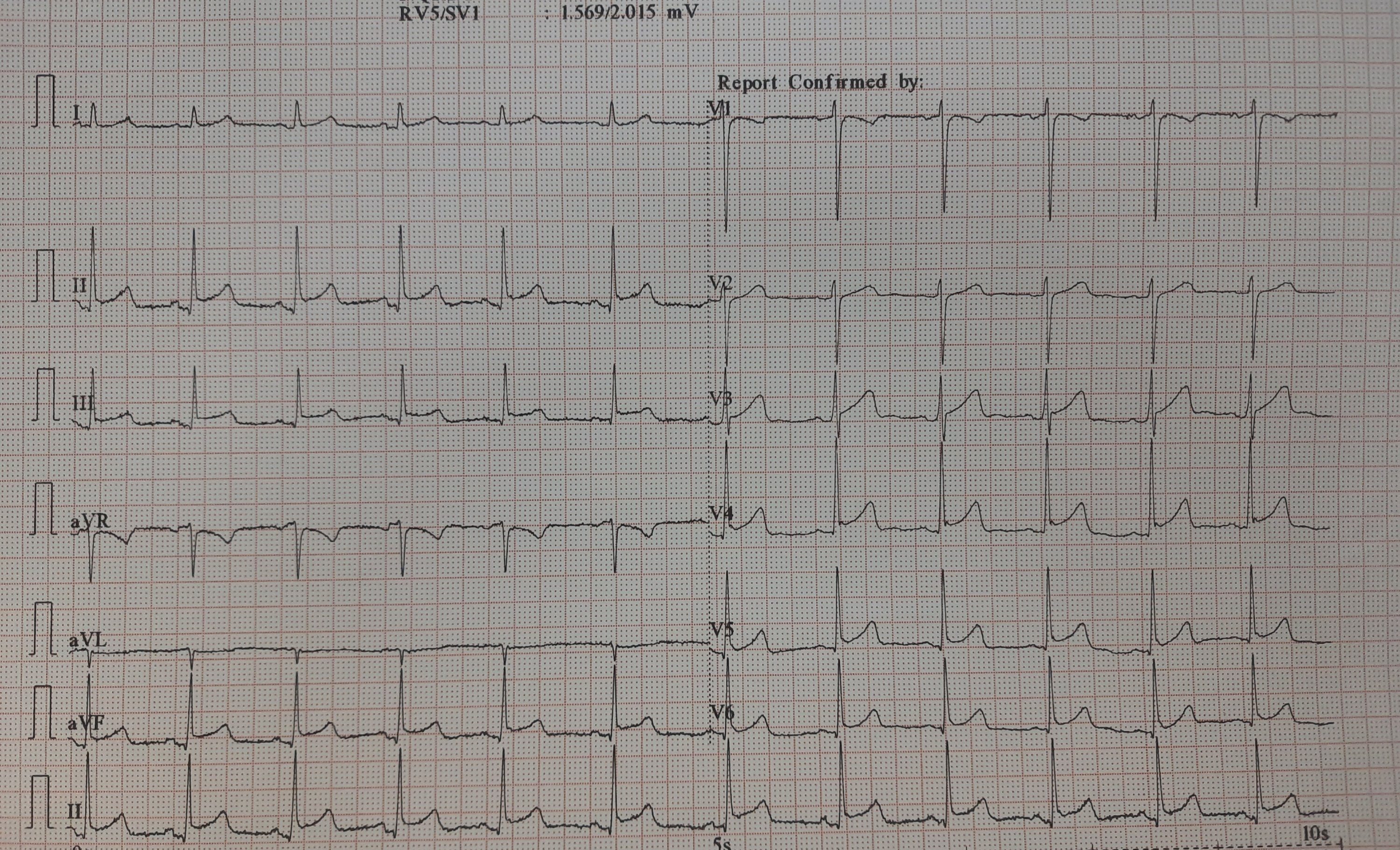
It would be mind-boggling to know the prevalence of such ERS patterns in the general population. One estimate suggests it could be anywhere between 3 to 13 % depending upon the criteria used. Let us assume the mean as 5 %. Then, it would be 30 crores of human beings in our habitat show this ECG pattern. If applied, in my city Chennai alone 5 lakh people could carry this tag.
While it is true, some forms of ERS and J wave syndrome can be markers of serious ventricular arrhythmias, either spontaneous or at times of Ischemia. Currently, It has become a fad, in cardiology academic circles*, to propagate the idea that ERS is no longer a benign condition. This is not acceptable at any degree of cognition. This happened mainly after few studies in powerful journals created some alarmist views. (*Maybe there is a bit of truth there. I still have doubts about whether we interpreted the Michel Haïssaguerre study properly)
Final message
ERS is a widely prevalent normal ECG variation with a minuscule risk. High-risk subsets need to be screened only if the J waves encroach and spill dangerously into the ST segment as well. Of course, this pattern is of serious concern if there is a family history of young SCDs has occurred.
Reference
Here is a good review of this topic by
Learning targeted IAS puncture in 20 minutes
Posted in Anatomy of heart, Uncategorized, tagged anatomy of heart, ias puncture, right vs left atrial anatomy on March 10, 2023|
The main reason for all those jitters, we cardiologists, get every time we puncture the IAS is not due to a lack of expertise and experience perse. There are two more reasons. First and foremost, it is still largely a blind* procedure. (Even in this era, where drones with HD vision shoot one-meter targets from a 1000 KM range ) *TEE and ICE are there, but they rarely give enough confidence.
The second reason is more important and is rectifiable. It is the perception error in our anatomical cognition, that is fed to us from first-year medical school. We are made to believe (at least to people like me ) The right atrium is aligned like a perfect box on the right side, sharing a wall called IAS, and the left atrium is obediently placed left of the right atrium. Please realize the heart is such a complex twisted single tubular organ, the venous end, in a stunning backward loop brings the LA most superior and posterior to the right atrium overriding the left-right relationship.)
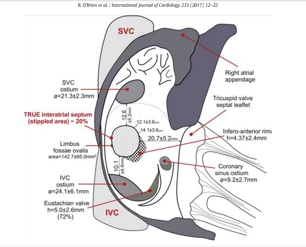
The right atrial terrain and IAS with multiple bumps and holes. Note the true IAS constitutes only 20% . This is where our punctures need to be.
Development of IAS
IAS development and the number of layers it sandwiches, the tortuous tracts of PFOs, the fossas, and its variable limbus is a big topic. Further, It is worth recalling, the true IAS hardly forms 20 % of the area of the interatrial contact surface.
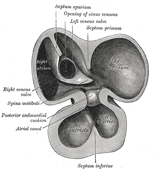
(the differential regression of sinus venous, along with infolding of the roof and along with curious septum spurium , the ubiquitous septum secundum make the texture, area & shape of IAS, a fascinating creation, though troublesome for the cardiologists ! ) Whoever named that part of vanishing IAS as spurious, (I think it is Henrry Grey ) has much fore-vision.
Forget about all this. Tell me how to cross this difficult terrain
Coming to the real world of interventions, we need to do targeted punctures in different spots of IAS in various interventions.(Mitra clips, LAA device, PTMC, PV abaltions, Mitral paravalvular leaks , TMVR etc) This has made this task even more tricky. Experts are always there to help us out. Like swimming, it can never be learned in books.
This 19-minute clip from. Seoul, South Korea is an excellent resource. Thanks to Dr. Sang Weon Park
Along with sound anatomical knowledge, improved hardware, and imaging like deflectable sheaths, TEEs, and ICE (intracardiac echo ), let us hope, it will soon become an easier task for everyone.
Final message
Understanding “attitudinal cardiac anatomy” with fluoroscopic overlay is the key. Again, it needs to be stressed, “Right is not right, and left is not left” when it comes to true atrial geo position. LA is equally posterior, superior, and of course to the left of RA. Some of my colleagues are blessed with a special 3-dimensional skillset (Inherited ?) I failed miserably to understand this, till very late. I am sure, Dr. Park’s video will help all our youngsters to cross the difficult gateway to the left side of the heart.
Reference
One more good read
B. O’Brien et al. / International Journal of Cardiology 233 (2017) 12–22
P wave spotting in AF is not forbidden
Posted in Uncategorized, tagged cardiology research topics for fellows, causes of absent p wabes, p vs f waves in af, p waves in atrial fibrillation, research topics in atrial fibrillation on March 8, 2023|
Fibrillation is a continuous, chaotic muscular activity. In AF, atrial muscle is expected to lose all coordinated contractions with fibrillatory waves replacing P waves. Have you ever spotted a suspicious P wave in a strip of otherwise explicit AF? If not, this write-up is not for you.
An evolving rare theme in Atrial fibrillation
Have a look at this ECG
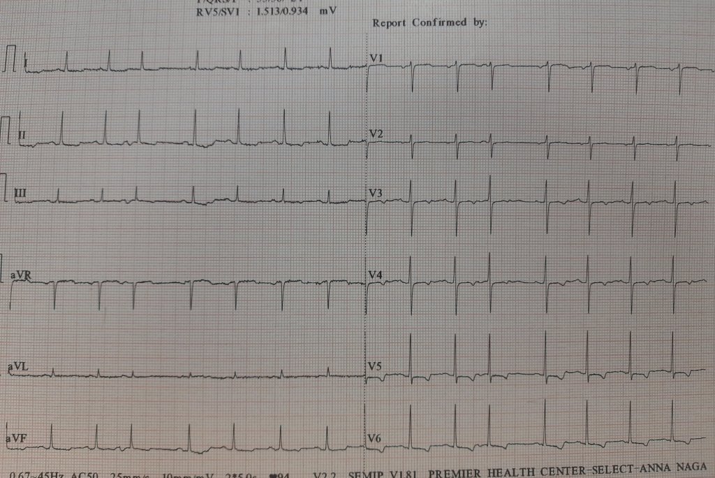
Here is an ECG, that was reported as AF, multiple APDs, or Possible AF, Pre AF. I suggested the term AF in transition. While few agreed, many said it is a straightforward SR with APDs, making it appear irregular RR.
But, the fact of the matter is, ECGs are insensitive to pick all fibrillatory wavelets. It can selectively pick a few coarse F waves and make them appear as P. I think, in this era, we should not diagnose AF by proxy, ie absent P waves. Rather, we need to look actively for fibrillatory wavelets. (Imagine all sinus arrests will qualify for f fine AF with a slow ventricular rate is it not ?)
The semantics of AF nomenclature is long. Intermittent AF, and paroxysmal AF, are well-known entities. It is now clear, AF can occur for a few seconds and vanish too. It seems we need to play some more linguistics with the most common cardiac arrhythmia. (Non-sustained AF, evanescent AF, etc )
Some thoughts on this hide & seek P waves
- Apart from the conventional list of absent P waves, one more example is repetitive APD can stun the atrial muscle for a few moments or minutes.
- Then, we always have the issue( eluded to earlier) of sinus node paralysis, with irregular junctional escape mimicking AF.
- Amiodarone can reduce fibrillatory rate, and (AF cycle length ?) Coarse F waves slow and stabilize it to mimic an organized P wave
- P on Ta waves (Like R on T ) can trigger a nonsustained AF for a few moments in a functional manner without real pathology in atria.
A funny memory brings back an EP truth
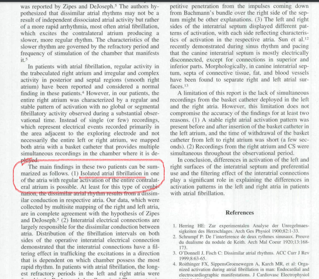
During our student days, my Professor used to trap us with this question, Which atria would fibrillate in mitral stenosis? Many of us blinked, and few had no hesitation to say, it is the LA that fibrillates. Now, after 50 years we realize, how fascinating the secrets AF has unfolded. Some organized activities are often in the right atrium, even as LA begins the process of AF. It is possible it may take variable time for the left atrial chaos to spill over to RA*. During these electrical uncertain times, some of the right atrial P wave activity refuses to die down. Even more dramatic one Atrium alone can permanently fibrillate and others completely insulated by blocking the signal in the Interatrial pathways. (Ref 1 ) Ndrepepa’s paper in the JCE 2000)
Final message.
True scientists rarely bother about questioning a dictum. The concept of non-uniform AF was first thought of by (Schrmp et al Ref 2) 100 years ago in 1920, and Zipes(Ref 3) hypothesized this in 1973. now, in the Year 2000, Ndrepepa confirmed it with EP studies. The spotting of occasional P waves is not forbidden in AF if the contralateral atria decide to block the incoming AF signals and keep generating their own P waves
Young EP guys, now that you are equipped with the sophisticated intracardiac GPS, please pursue this provocation in AF. One more piece of evidence we observed in the echo lab. Try to look at Tricuspid doppler A velocity waves in full-blown AF patients. You can see the surprise for yourself. This is very good research work to do. This is one of the ideas I gave to my fellows at MMC. Now, it is free for all to pursue whoever wants to do this. The clinical implication* will follow.
* A lingering query, how common is RAA clot in mitral stenosis with AF and the possible threat of pulmonary embolism?
Reference
2.Zipes DP. DeJoseph RL: Dissimilar atrial rhythms in man and dog. AmJCardioi I973;32:6l8-628.
3.Schrumpf P: De 1’interference de deux rythmes sinusaux. Preuve du dualisme du nodule de Keilh. Arch Mai Qwur 1920;l3:168-173
Postamble
The snapshot from Ref 1 . The term Isolated AF confined to one atrium could be a rare event, but, no one can deny we have plenty to learn from them
LA volume assessment : A critical parameter with a time trouble !
Posted in Cardiology Illustrations, cardiology physiology, Left atrial enlargement, Uncategorized, tagged LA end diastolic dimension, la end diastolic vs end systolic dimension, LA end systolic dimension, la pressure volume loop, LA volume by mri, normal LA dimension, normal la volume, When do you measure LA dimension volume on February 8, 2023|
LA dimension and volume have become vital parameters in recent times, especially, with the entity of HFpEF is becoming so common. LA not only acts as a live barometer, reflecting all that happens in LV, but it is also a chronic marker of LV diastolic function. (Funnily referred to as HBA1c of diastolic dysfunction)
What is normal LA dimension & volume ?
- Normal left atrial diameter < 4.1 cm in men or < 3.9 cm in women
- Normal left atrial volume indexed for body surface area (BSA) is 34 ml/m2 for both women and men
Which part of the cardiac cycle do we measure?
Ever since Wiggers introduced the overwhelming concept of LV systole and diastole, most of us ignored the fact that atria do have a separate contraction relaxation cycle, independent of what happens in the ventricle. Of course, atria and ventricles act as a single chamber in diastole. In reality, atria lack true boundaries when it acts as a conduit. The LA dimension varies considerably during the atrial cardiac cycle. Look at the LA pressure-volume loop, which can frighten anyone, with its horizontally lying figure of 8 pattern. During every cardiac cycle, the volume reaches atleast two troughs and one peak.
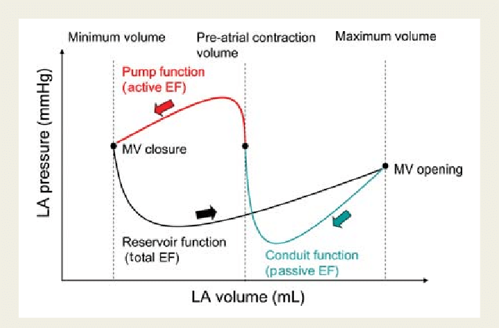
Don’t get frightened with this graph, spend some time, and you will get it right, Begin at “3” o clock position with the onset of diastole with a downsloping green loop, that continues as the red line of atrial contraction to end up in systole. The entire black loop, that happens during ventricular systole depicts the true reservoir function. with MV closed. ,
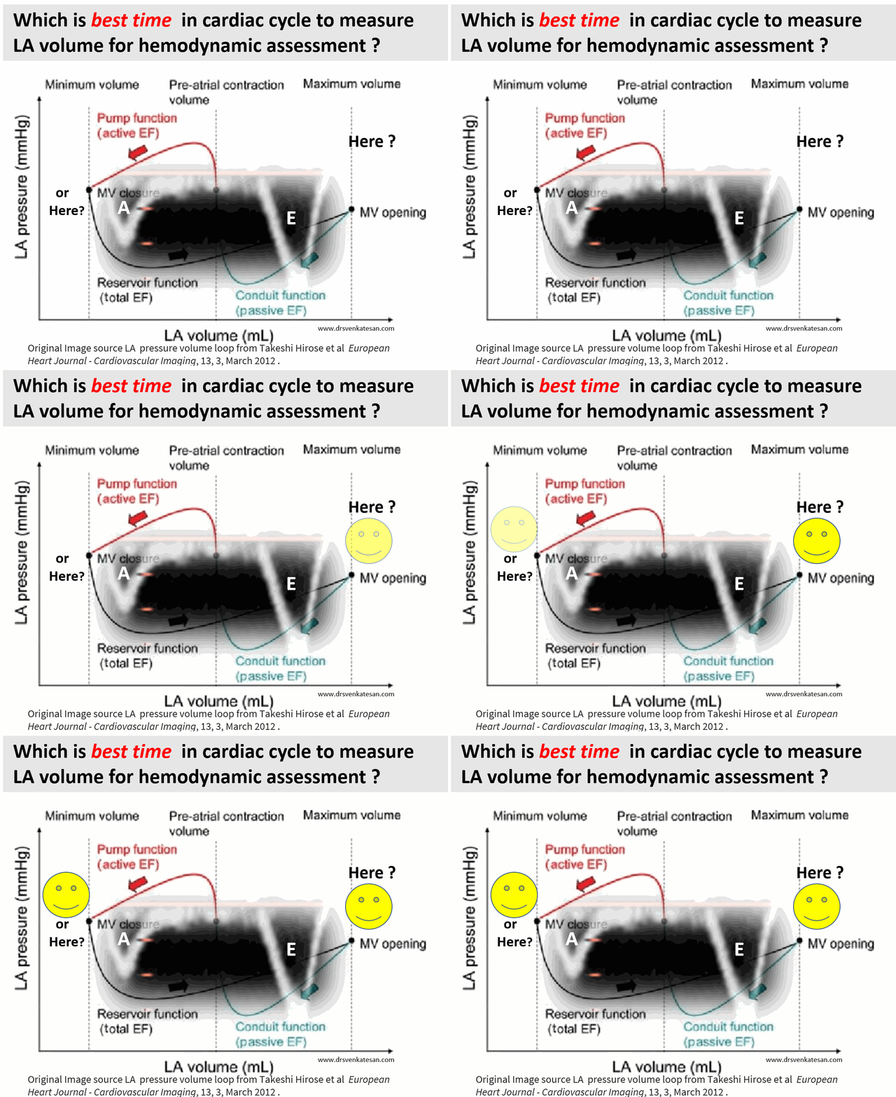
As of now, we have a consensus, LA volume is measured typically in LV end-systolic frame. ( Rather, we measure it at maximum LA volume ) However, we have 4 different LA volumetric components to assess, as this article excellently depicts. (Hoit BD. Left atrial size and function: role in prognosis. J Am Coll Cardiol. 2014 Feb 18;63(6):493-505.)

What could be the limitations of the traditional end-systolic measurement?
No single measurement will give an overall LA function assessment. But still, Somehow, we have measured the maximum LA volume as a reference of true diastolic function. This happens in LV end-systolic point where atria reach the maximum size. But, here is a catch, we assess the left atrial function before its main physiological function of emptying takes place.
How about assessing LA efficiency after it completes its job, ie end diastole?
In LV function end-systolic dimension has pride of place as it is devoid of influence from loading condition. If applying the same logic, the “end atrial” systolic dimension(Which is the same as LV end-diastolic point/or post A ) should be perfect. It can also help measure the residual LA volume after its systole.
A potential advantage of LV end-diastolic dimension (The Heart & Soul study )
Maybe, this is less affected in the presence of MR systolic jet will spuriously elevate LA volume. In AF also this parameter is less likely to be influenced by LA preload.
Final message
Suddenly, we are debating a fundamental Issue, ie timing of LA measurement. While the end-systolic size/volume is the current standard, the LA dimension in the end diastole also provides useful info. There are at least 4 different LA volumes, at different parts of the LA cycle that need to be studied for a proper understanding of diastology (Unlike LV which has only two).
Now, we may need to ponder, if there is a mean LA volume, measured with the 3D volumetric analysis or MRI, that could be representative of the global LA function.
Reference
Primary PCI and its silent encounter with “myocardial neutrophilia”
Posted in Uncategorized, tagged myocardial neutrophilia on February 1, 2023|
Myocardial infarction, a gross pathological entity renamed now as STEMI for clinical purposes, continues to be the most famous medical emergency that triggers a series of calls, right from 911 to the ER, that eventually ends up in CCU or a 24/7 cath lab. The heart, can’t wait for all these external responses when it is challenged with a vascular accident. The moment ATO occurs, two things happen. The endogenous fibrinolytic led by native tissue PA (Tpa) tries to get rid of the thrombotic plug by all its means. It succeeds in 15%. We call it spontaneous lysis or aborted MI. Many lives are lost in the remaining before they reach the hospital..
Meanwhile, the immune high-command deputes scores of neutrophils to the ground zero Fig 1) to supervise what is happening and try to heal the injury. It is worth, understanding, that activated WBCs, sort of convert myocardial infarction from a vascular event to inflammatory pathology, as the hour progress.
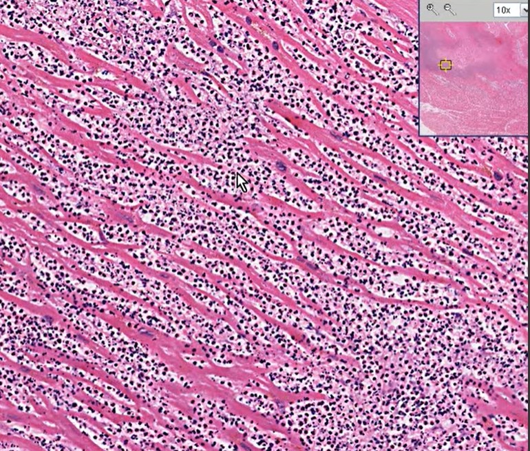
Fig 1 Infarcted myocardium Invaded by neutrophils
What do these neutrophils do?
It will be our ignorance, if we think, they simply crowd around the myocytes helplessly, that is hit by ischemia. They are sent with a specific purpose to protect and heal the myocardium. Very often it fails in its mission is a different story. These fighting neutrophils send a subtle signal of their presence with a mild increase in temperature during ACS ( Patel Prognostic usefulness of white blood cell count and temperature in acute myocardial infarction Am J Cardiol. 2005 Mar 1;95(5):614-8. )
Is neutrophil invasion good or bad for the myocardium?
These are pro-inflammatory cells. meant to promote healing and mitigate Injury. Interleukins and Leukotrienes do have a healing power as well. But, what happens, in reality, is, still a mystery. As of now, it is tempting to think, it does more damage than good. Its role changes over time.
Acute reperfusion Injury in Primary PCI
Neutrophils are quiet obedient cells generally, but once activated, their behavior can’t be predicted. It may start attacking the host cells, We know reperfusion injury is real and poorly understood. Delayed reperfusion, is well known to cause myocyte softening and lysis. What we know for sure is, primary PCI, induced accelerated flushing of these angry neutrophils is definitely related to no flow, microvascular plugging, and cardiogenic shock. (The fact that no-reflow and reperfusion injury is less of a problem in fibrinolysis demands introspection)
Diagnostic & prognostic value of neutrophilia
Neutrophilia is such a nonspecific response, faces ridicule for its clinical utility. But, In reality, it can be a worthy parameter, to time the age of the infarct (or even confirm* the ACS ) in otherwise equivocal clinical presentation.(Ref 2) More importantly, it provides prognostic information. One more potential use is Neutrophil count to guide the timing of surgery post-MI (as in VSR) A neutrophil count could help avoid the active phase of inflammation.
*I recall my surgical professor’s emphasis on leukocytosis to confirm acute appendicitis during the first clinical year.
Final message
There is no academic by-law, that forbids full-blown interventional cardiologists from having an affair with basic science. Unless the core science is irreversibly bonded to the bedside, we can never reap the true benefits of translational research. Hematological aspects in STEMI is one such underrated discipline. Also, we must encourage every postgraduate student in pathology/biochemistry/physiology to visit the CCUs or sit in the cath lab gallery more frequently. Watching the cardiology stalwarts plowing through the blocked coronary arteries, racing against time, is sure to kindle young minds, for a potential molecular breakthrough in cell survival and healing following myocardial hypoxia.
Reference
2. Thomson SP, Gibbons RJ, Smars PA, Suman VJ, Pierre RV, Santrach PJ, Jiang NS. Incremental value of the leukocyte differential and the rapid creatine kinase-MB isoenzyme for the early diagnosis of myocardial infarction. Annals of internal medicine. 1995;122:335–341. [PubMed] [Google Scholar]
3.. Cannon CP, McCabe CH, Wilcox RG, Bentley JH, Braunwald E. Association of white blood cell count with increased mortality in acute myocardial infarction and unstable angina pectoris. OPUS-TIMI 16 Investigators. Am J Cardiol. 2001;87:636–639. A610. [PubMed] [Google Scholar]
4. Bhatt DL, Chew DP, Lincoff AM, Simoons ML, Harrington RA, Ommen SR, Jia G, Topol EJ. Effect of revascularization on mortality associated with an elevated white blood cell count in acute coronary syndromes. Am J Cardiol. 2003;92:136–140. [PubMed] [Google Scholar]
Take a break from cardiology : How to debate a complex topic in a forum ?
Posted in Uncategorized, tagged evidence based cardiology, medical education, religion and god medical profession on January 16, 2023|
Pardon ,this video is nothing to do with cardiology. It is from the archives of the United nations library .This can teach some important lessons in art of communication , sharing to all folks, especially medical students. It was recorded in 1959 in Newyork, UN head quarters.Four 17 year old school girls & boys were invited for a debate on a complex topic. Does God exist ? How do you pray ? and what is the purpose of different religions ?
I keep wondering , how these youngsters accumulated so much wisdom and express it in such a polite manner too. Mind you, this was recorded , when learning happened with out any digital aids.The word Internet was unheard off. No ego, no bluntness, no diatirbes that has become a norm in many debates now. I got a punching lesson from this clip, be gentle when taking extreme views in any topic.
I wish, every medical debate in class rooms should happen this way.The key to succesful debate is, to accumulate knowedge, willingness to question the convention, and respecting the oppositie point of view.
The high point of talk show, was, when the Brazilian girl(due respects, she should be nearing 80 years now) tell us casually some things are not meant to be understood in life .I tell the same when some patients ask too many questions about their illness which may not have an answer.
Which is the Ideal site to ablate VT circuit ?
Posted in Uncategorized, tagged acc aha esc guidelines, cryoablation, exit site of vt, exit vs entry site, icd vs rf ablation, RF ablation, scar induced vt on January 12, 2023|
Management of recurrent ventricular tachycardia has developed a lot in recent times. Anti-arrhythmic drugs(AADs) were a cornerstone for most recurrent and refractory VTs till recently. Surgeries including CABG, repair of the aneurysm, and subendocardial resection has helped to control many post-MI ischemic VTs. Soon they became obsolete. Realistically, PCIs had little impact on post-MI VT for some unknown reasons. However, with the advent of ICDs and RF, ablation, a new dimension is added to this field.
ICDs, though an attractive device, don’t prevent a VT but vow to nullify the consequence of VT. This is problematic. ICDs in spite of their ability to prevent SCD effectively, it has little say in preventing non-sudden deaths of in any form of cardiomyopathy. (In fact, there is some evidence ICDs might increase myocardial damage with inappropriate shocks )
Hence, RF ablation can be called as true curative therapy by extinguishing hot spots of VT genesis. Still, we need to understand ablation is technically creating new dead myocardium (in alreadly damaged myocardium) and hence, can’t avoid a consequence. More importantly, ablation as a modality is technically more demanding. We have accumulated huge experience with the help of electro-anatomic imaging and cutting-edge hardware in the last few decades. Still, we find it difficult to zero in the target area of RF ablation, with all available techniques of mapping (Activation, substrate, entrainment, and pace mapping) The reason is, VT circuits can be really complex ones. Not only different loops but also it can travel deep into the myocardium (Intramural or epicardial) making a single approach endocardial often futile. Success rates vary between 60 -70% (Some may claim 90 +)
This post wants the young fellows to take on this fundamental issue and learn in-depth about the arrhythmia circuit.(Get Inspired by Dr. Samuvel Asirwatham of Mayo clinic work )
What is the best site to ablate ventricular tachycardia?
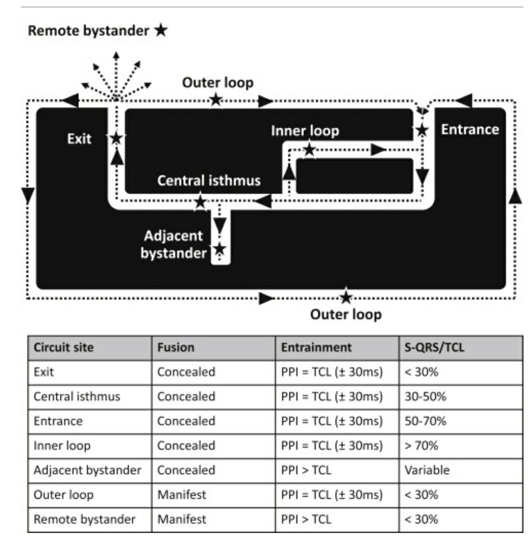
- Exit point
- Central isthmus
- Entrance
- Inner loop
- Outer loop
- Though logic would tell us we can intercept an arrhythmia by ablating anywhere in the circuit. But the issue here is we need to permanently ablate it. not just interrupting.
- The best site to ablate is the exit point or entry point. Maybe isthmus. I am not really sure. But, one thing is clear, the outer and inner loops are not important. Further, live tissues are the culprits and not the scars and infarcted zones.
- One more possible answer is to try to ablate all (or maximum) points in the circuits.
How to identify entry points and exit points?
Not a simple answer .EPs apply their life experience to do that, assisted by technology. Not everyone who has a Carto -GPS can do that.
One simple answer for the fellows is, in entrainment mapping, critical sites can be somewhat arbitrarily labeled as exit, central isthmus, or entrance on the basis of the Stimulus-QRS interval relationship to the TCL. Exit sites will show an S-QRS interval < 30% TCL (QRS onset shortly after pacing), central isthmuses will show an S-QRS interval of 30% to 50% TCL (some delay in QRS onset after pacing), and entrance sites will show an S-QRS interval of 50% to 70% TCL

Ruairidh Martin et all Circulation: Arrhythmia and Electrophysiology. 2018;11:e006569
Final message
Key to the successful ablation of VT is the accuracy in choosing the target site. Locating the target is meaningless if we can’t reach that site. Reaching the site is again futile unless we are able to deliver adequate burning or icing with sufficient contact.
Meanwhile, AADs can never be ignored in spite of the apparent side effects, for the simple reason they reach the VT circuits without any fuss. The Pharma industry has almost lost its interest in AAD research and the hyper-talented EP guys are squarely responsible for this unintended consequence.
Future directions
RF energy modification and newer catheter designs will help. While cryoablation shows a promising advantage over RF, it is still to prove its sustained efficiency. Meanwhile, other ablation modes are being tried. Transvascular ethanol ablation and stereotactic radio ablation have both shown promise for arrhythmias that fail other ablation strategies.
Some more questions
- What is the difference between arrhythmia focus of origin and entry point?
- Once the VT is set in, what is the relevance of its origin?
Reference
One of the very good reviews on the topic.
1.Gustavo S. Guandalini, Jackson J. Liang, Francis E. Marchlinski, Ventricular Tachycardia Ablation: Past, Present, and Future Perspectives,
JACC: Clinical Electrophysiology, Volume 5, Issue 12, 2019, 1363-1383,
2;,W.G. Stevenson, H. Khan, P. Sager, et al.  Identification of reentry circuit sites during catheter mapping and radiofrequency ablation of ventricular tachycardia late after myocardial infarction Circulation, 88 (1993), pp. 1647-1670
Reviewing top 10 Interventional papers of 2022 : A good lesson on NSTEMI management
Posted in Uncategorized, tagged acc aha nstemi nsteacs guidelines on January 3, 2023|
EHJ has listed the top 10 papers in cardiology in the year 2022 in its current issue.
Kindly go through this when free. While each of the 10 has its own importance, one meta-analysis, I thought was a real bright spot. Though the message it conveys is nothing new, it reaffirms an important management principle in ACS. Getting curious? Before going into the paper, a mini pretest
What is your take on these 4 statements on ACS? True or False
1. STEMI is an emergency, NSTE-ACS* is not an emergency
2. Both are true emergencies.
3. STEMI is definite, yes, but NSTE-ACS may or may not be (Mind the GRACE score dude !)
4. Even STEMI can be a non-emergency if the patient reports after 24 -48 hours.
(Remind you, NSTEACS = UA+NSTEMI , still often used interchangeably)
Hope we don’t have difficulty in identifying the wrong response. Whatever the answer to this somewhat insulting question to our intellect, forget it. Now, read this paper, which is listed as one of the most read last year. It is about the impact of early invasive strategy in NSTE-ACS.
Conclusion is pasted for the busy guys.
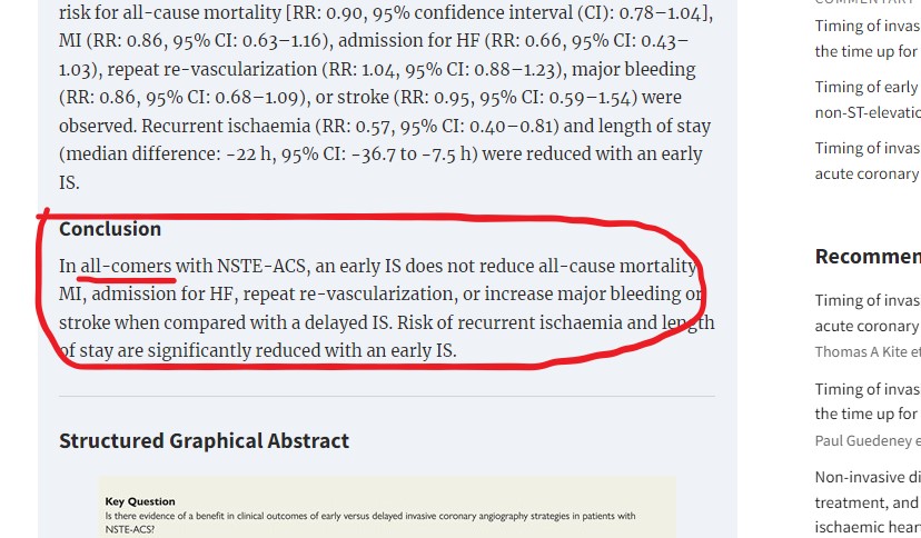
Post-test
- What is overall NSTEMI in-hospital mortality? (Everyone knows for STEMI it is around 4-8 %, no one seems to be sure about NSTEMI. I think it can’t be estimated accurately, guess it is 1 -2% )
- Is there a primary PCI equivalent situation in NSTEMI? What are they? (Left main UA with AVR ST elevation comes first on the list)
- Based on the urgency to treat how does the ultimate outcome change? (This is what this meta-analysis by Kite et all proved.It is not a new revelation though . Recall the landmark ICTUS study of 2010 which was largely kept in the dark )
Final message
One lay definition of STEMI is, It is a mass of myocardium under fire. True UA/NSTEMI is, a mass of myocardium, that is threatening to go on fire. When firefighters are able to reach a smoky building before the onset of a fire, no doubt, it looks like a great & awesome response, Isn’t it? Unfortunately, the myocardial landscape is different and NSTEACS is such a heterogenous entity that doesn’t welcome unsolicited aggressive, emergency rescue missions. That’s one of the messages we get (at least I got )from this top-read paper in 2022.
Reference
1.Emanuele Barbato, Margaret McEntegart, Tommaso Gori, The year in cardiovascular medicine 2022: the top 10 papers in interventional cardiology, European Heart Journal, 2023;, ehac778, https://doi.org/10.1093/eurheartj/ehac778
3.Damman P, Hirsch A, Windhausen F, et al. 5-Year Clinical Outcomes in the ICTUS (Invasive versus Conservative Treatment in Unstable coronary Syndromes) Trial. J Am Coll Cardiol. 2010 Mar, 55 (9) 858–864.https://doi.org/10.1016/j.jacc.2009.11.026
Happy new year & memories of 2022 !
Posted in Uncategorized on January 1, 2023|
Wishing every one of you an Enlightening New year. As we begin a new journey around the sun, yet another time, let us re-dedicate ourself, to use science, for the welfare of our planet & people.
Thank you , for visiting this site and make all its worth.
Categories
-

-
The contents of the this blog is being published as Kindle E book , as per the request of many of the readers. Every article will continue to be open source in this site. Again I shall reiterate the book format is not aimed at any commercial intent. It is only to facilitate learning in a single book format Here is the link to book
https://amzn.in/d/euhL5vu Archives
- October 2026 (2)
- September 2026 (9)
- August 2026 (8)
- July 2026 (6)
- June 2026 (9)
- May 2026 (6)
- April 2026 (11)
- March 2026 (10)
- February 2026 (8)
- January 2026 (8)
- December 2025 (11)
- November 2025 (7)
- October 2025 (8)
- September 2025 (7)
- August 2025 (9)
- July 2025 (10)
- June 2025 (8)
- May 2025 (9)
- April 2025 (7)
- March 2025 (10)
- February 2025 (4)
- January 2025 (9)
- December 2024 (11)
- November 2024 (8)
- October 2024 (10)
- September 2024 (5)
- August 2024 (5)
- July 2024 (6)
- June 2024 (5)
- May 2024 (4)
- April 2024 (7)
- March 2024 (4)
- February 2024 (8)
- January 2024 (6)
- December 2023 (8)
- November 2023 (13)
- October 2023 (14)
- September 2023 (5)
- August 2023 (6)
- July 2023 (10)
- June 2023 (5)
- May 2023 (5)
- April 2023 (4)
- March 2023 (5)
- February 2023 (2)
- January 2023 (7)
- December 2022 (3)
- November 2022 (5)
- October 2022 (5)
- September 2022 (4)
- August 2022 (3)
- July 2022 (9)
- June 2022 (2)
- May 2022 (1)
- April 2022 (2)
- March 2022 (1)
- February 2022 (3)
- January 2022 (7)
- December 2021 (3)
- November 2021 (5)
- October 2021 (8)
- September 2021 (4)
- August 2021 (6)
- July 2021 (6)
- June 2021 (7)
- May 2021 (5)
- April 2021 (4)
- March 2021 (3)
- February 2021 (6)
- January 2021 (8)
- December 2020 (4)
- November 2020 (5)
- October 2020 (7)
- September 2020 (7)
- August 2020 (10)
- July 2020 (6)
- June 2020 (9)
- May 2020 (9)
- April 2020 (5)
- March 2020 (7)
- February 2020 (3)
- January 2020 (4)
- December 2019 (4)
- November 2019 (6)
- October 2019 (3)
- September 2019 (6)
- August 2019 (3)
- July 2019 (1)
- June 2019 (3)
- May 2019 (2)
- April 2019 (2)
- March 2019 (2)
- February 2019 (4)
- January 2019 (2)
- December 2018 (2)
- November 2018 (2)
- October 2018 (2)
- September 2018 (1)
- August 2018 (2)
- July 2018 (3)
- June 2018 (1)
- May 2018 (3)
- April 2018 (1)
- March 2018 (3)
- February 2018 (3)
- January 2018 (1)
- December 2017 (3)
- November 2017 (3)
- October 2017 (3)
- September 2017 (2)
- August 2017 (2)
- July 2017 (2)
- June 2017 (2)
- May 2017 (4)
- April 2017 (3)
- March 2017 (3)
- February 2017 (5)
- January 2017 (3)
- December 2016 (2)
- November 2016 (5)
- October 2016 (4)
- September 2016 (3)
- August 2016 (5)
- July 2016 (3)
- June 2016 (4)
- May 2016 (3)
- April 2016 (6)
- March 2016 (4)
- February 2016 (3)
- January 2016 (5)
- December 2015 (6)
- November 2015 (5)
- October 2015 (8)
- September 2015 (2)
- August 2015 (5)
- July 2015 (7)
- June 2015 (4)
- May 2015 (6)
- April 2015 (5)
- March 2015 (7)
- February 2015 (15)
- January 2015 (8)
- December 2014 (5)
- November 2014 (9)
- October 2014 (7)
- September 2014 (9)
- August 2014 (5)
- July 2014 (11)
- June 2014 (5)
- May 2014 (4)
- April 2014 (5)
- March 2014 (8)
- February 2014 (8)
- January 2014 (5)
- December 2013 (7)
- November 2013 (7)
- October 2013 (14)
- September 2013 (12)
- August 2013 (15)
- July 2013 (15)
- June 2013 (15)
- May 2013 (15)
- April 2013 (15)
- March 2013 (15)
- February 2013 (15)
- January 2013 (15)
- December 2012 (15)
- November 2012 (15)
- October 2012 (15)
- September 2012 (15)
- August 2012 (15)
- July 2012 (15)
- June 2012 (15)
- May 2012 (15)
- April 2012 (15)
- March 2012 (15)
- February 2012 (15)
- January 2012 (15)
- December 2011 (15)
- November 2011 (17)
- October 2011 (17)
- September 2011 (17)
- August 2011 (21)
- July 2011 (20)
- June 2011 (17)
- May 2011 (15)
- April 2011 (17)
- March 2011 (25)
- February 2011 (20)
- January 2011 (20)
- December 2010 (18)
- November 2010 (21)
- October 2010 (21)
- September 2010 (25)
- August 2010 (20)
- July 2010 (10)
- June 2010 (11)
- May 2010 (19)
- April 2010 (16)
- March 2010 (14)
- February 2010 (22)
- January 2010 (18)
- December 2009 (20)
- November 2009 (20)
- October 2009 (3)
- September 2009 (21)
- August 2009 (19)
- July 2009 (12)
- June 2009 (12)
- May 2009 (11)
- April 2009 (15)
- March 2009 (21)
- February 2009 (4)
- January 2009 (12)
- December 2008 (13)
- November 2008 (9)
- October 2008 (22)
- September 2008 (20)
- August 2008 (16)
- July 2008 (14)
- June 2008 (7)
Blog Stats
- 6,752,248 hits
Please give your feed back .
Click below to see who is watching this website live !

- This site will never aim for profit. Still ,this donation link is added at the request of few visitors who wanted to contribute and of-course that will help make it sustainable .

Please Note


