Feeds:
Posts
Comments

Archive for the ‘Uncategorized’ Category

Identifying the culprit after a criminal event may be easy for the police.For cadiologists investigating the crime scene after a coronary event, it is a different story. (Of course, localization of IRA after a STEMI may not be really difficult.) But , when a patient is having UA  and coronary artery shows multiple lesions, we do have real diagnostic issue. The general dictum could be, tightest lesion or the complex eccentric ones with thrombus is likely to be the culprit. This has important therapeutic Implication,  as we are argued to address the active lesions first. The following study was done in 2009 trying to find the ARA solely by ECG features.

The conclusion was

The following ECG findings were helpful in localizing Angina related artery . ST depression in V3- V5 correlated  with  LAD  angina .Global ST depression was highly correlated with proximal LAD or Left main disease ( 6/6 patients). ST depression in V1 –V3 was associated more commonly with dominant LCX/OM disease. ST depression in 2 ,3 , AVF , or I, AVL  had  no significant correlation with either RCA or LAD  system.However multiple culprit lesions or diffuse inflammatory CAD should always be thought off. One more possibility is , its simply a demand ischemia or micro vascular angina were there is no true epicardial culprit lesion. 

A revisit to my 2009 IHJ article.

http://indianheartjournal.com/ihj09/nov_dec_09/509-523.html


IDENTIFYING ANGINA RELATED ARTERY (ARA) IN UNSTABLE 
ANGINA /NSTEMI BY ADMISSION ECG AND ECHOCARDIOGRAPHY
S.Venkatesan C.Krishnakumar .G.Gnanavelu .R.Subramanian.Geetha Subramanian B.Ramamurthy.P.Arunachalam.M.Somsundram.V.E.Thandapani.M.A.Rajasekaran.
S.Murugan , Madupraphu doss ,P.Pachiappan.
Madras Medical College. Chennai

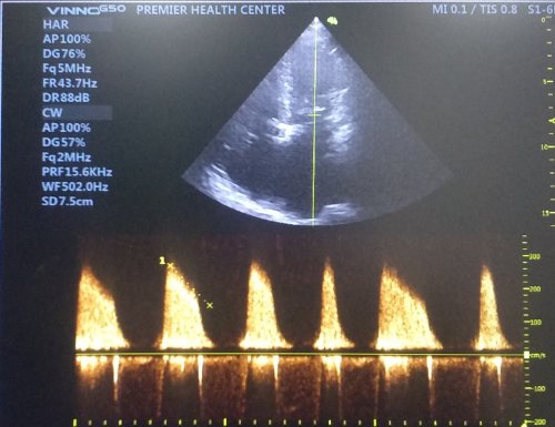
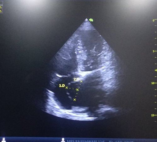
Unstable angina( UA /NSTEMI ) constitute a  heterogeneous  group of  patients with  lesions ranging from  normal coronary  artery  to severe multi vessel  disease. Even  though  multiple active plaques are documented ,  one  critical  lesion  would be   responsible  for  the  index  episode  of  angina..  Contrary to STEMI  there is no standard methodology   to identify  the  Angina  related artery.(ARA) in UA .We under took this  analysis  to find  whether  admission  ECG  with the help of echocardiography   could  predict  the ARA  in patients with UA

26  patients with  UA  admitted in  our  CCU  were  the  subjects of  study. Patients with   post  infarction angina,  CABG ,  PCI , old  MI , left ventricular  dysfunction  were  excluded. All patients  were treated  as per institutional protocol. Echocardiogrphic analysis   of  wall motion defects (WMD)  were  documented  between  2hrs  and  24hours of admission  .CAG  was  done  between  24 hrs and  7  days. The  coronary  lesion was considered angina related  if  the  WMD  detected   by  echocardiography matched with  the  myocardial  segments supplied by the  arterial territory  containing the lesion . After locating the ARA , the patient’s  admission ECG   was  compared  retrospectively   with  CAG  finding  to study  whether  it has  any  predictive  value  for identifying  ARA.  6 patients  who  had single vessel disease the ARA  localization  was straight forward. (LAD -4 , LCX -1 RCA-1 ). In 2  patients  there was  obvious  eccentric thrombus containing plaque indicating the culprit lesion . 18 had DVD or TVD with no clearcut culprit lesion.

The following ECG findings were helpful in localizing ARA.ST depression in V3- V5 correlated  with  LAD  angina .Global ST depression was highly correlated with proximal LAD or Left main disease ( 6/6 patients). ST depression in V1 –V3 was associated more commonly with dominant LCX/OM disease. ST depression in 2 ,3 , AVF , or I, AVL  had  no significant correlation with either RCA or LAD  system.

It  is  concluded  ARA  can be  identified  with  fair  degree  of accuracy   by admission  ST segment  profile. This  observation  differs with  the existing literature which  suggest little role for ECG to localize arterial lesion in UA. In patients with multivessel CAD  with  more than one  critical lesion  a  combination of ECG  and echo features  help  us to  fix the angina related artery and possibly the lesion. This has  important  therapeutic implication.

Keywords: Angina Related Artery, Unstable Angina/NSTEMI, ECG, Echocardiography.

Postample

I am reposting this abstract again because the same paper has been plagiarised in at least two occasions and got published in predatory journals. Now, we realise Journal article shopping and trading has become a scientific scam .

Reference

This paper  from Japan analysed this ARA concept in 1996 itself with SPECT Imaging


Read Full Post »

In the evaluation of syncope, history is most important to arrive at a diagnosis. Ofcourse, the first step is to confirm whether its truly a syncope or something else.(Metabolic/TIA or seizure.)We are easily carried away by the urge to order a Holter monitoring routinely. In reality, the yield is too low (<15%) .Even the utility value of Head up tilt (HUT) is being downgraded.

Paradoxically, resting ECG might give important clue in many. One need to specifically look into a set pattern of ECG. It generally falls in one the following in any patient with syncope.

This post specifically may not be exclusive but stresses the importance of resting ECG in the evaluation of syncope. by our urge (Stress testing is not included)

  1. Bradycardia( Sinus Node dysfunction/AV block) Note Brady cardias can per se cause syncope if pause >3-5sec Or it may lead to Brady (Pause) dependent escape VT.

    A pause can be a sinus arrest, Pause or SA block .If pause ends with a junctional escape it becomes a arrest.

  2. Look PR interval specifically(A bifasicular block shouldn’t be missed .It can be more dangerous than say a congenital CHB)PR interal represents condcution from SA node to Purkinje fibres in ventricel. The importance is directly linked to the location of the block than propably the degree of prolongation. Please note HV interval > 70 ms in any patient with prolonged PR is cause for concern,
  3. Preexcitation/Delta waves (Though Narrow QRS AVRT rarely causes syncope its very much possible during  Antidromic tachycardia. (AntiVRT), Antidromic AVRT or Accessory pathway with short RP <250ms need to be documented. Concealed paths are safe , but delta appearing during stress testing is extremely unsafe
  4. Post-excitation /Epsilon waves. (often noted in lead V 1, A marker of RVOT dysplasia as in ARVD. Also referred to as Fontaine wave who discovered it by bipolar cheat leads over V1 )

    Note the epsilon occurring after the qrs Indicating RVOT dysplasia

  5. Q waves (Markers of old MI -Scar Induced VT)
  6. High voltage QRS LVH /HCM /Aortic stenosis
  7. RV strain/S1Q3T3 pulmonary embolism.(Syncope is a common presentation with PE especially with minimal exertion or change in posture)
  8. Early coupled VPDS (R on T location a trigger for VT) Wedesky effect. The terminal portion of T which correspond to supernormal period.

    The significance of VPDs directly related to its prematurity than its focus of origin.The one that falls on the vulnerable period .Late phase 2 and phase 3 are more vulnerable as triggered activity

  9. Brugada (Type 1 with T inversion riskier, Camel hump less dangerous Joseph Brugada,

    Brugada syndrome -Note three types . Type 1 is typically risk prone. Please note it is the late ST declining component and the T iversion that confers the risk not ST elevation per se.The type 2 with a camel hump is confered with least risk

  10. Malignant ERS pattern (Most ERS or safe / Maligant forms infero lateral forms risky only at times of ACS not spontaneous risk
  11. J wave syndromes –Overlapping with Brugada /ERS Charles Antzelevitch,J Arrhythm. 2016 Oct; 32(5): 315–339

    ERS syndrome are so common. In the absence of sycnope, it should be ignored straight away. Recently it received too much hype among cardiologists increasing the anxietywhich is not warranted.

  12. Long QT Interval (Hypokalemia commonest, Congenital next Peter J. Schwartz,Long-QT Syndrome Circulation: Arrhythmia and Electrophysiology. 2012;5:868–877
  13. Fractioned QRS (Most often seen in DCM ischemic /non-ischemic confer VT risk usually with LV dysfunction, these are candidates for CRT-P/D as well)
  14. T wave alternans Fluctuating T waves indicate repolarisation alternans .It elevates risk of VT Narayan SM J Am Coll Cardiol. 2006 Jan 17;47(2):269-81
  15. Exercise Induced VT/ CPVT is to be considered seriously in all unexplained exertional syncope. Behere SP, Catecholaminergic polymorphic ventricular tachycardia: An exciting new era. Ann Pediatr Cardiol. 2016;9(2):137–146.

What next after ECG ?

After ruling out neuro cardiogenic syncope by history, one has to perform a good quality echocardiography that can clinch structural heart disease .In cardiomyopathies like ARVD or RCM MRI studies will be of immense value especially the LGE/DEMRI that picks up the scars and fibrosis as in sarcoid or tuberculomas etc. Event recorders are popular, may have a slightly better yield than Holter.EP studies are required in few as diagnostic or more commonly as a part of therapeutics.(Please note, EP lab Induced polymorphic VT has Zero diagnostic value as any normal human heart can be induced to VF by repetitive stimulation)

Management

The main purpose is to exclude serious primary electrical and or structural heart disease. However, fortunately, the most common cause of syncope is neurogenic or reflex mediated. It requires reassurance and fluid repletion Fludrocortisone,/Midoridine (Alpha receptor agonists are promising) Pacemaker/ICD is indicated in few with brady/Tachy -Brady .ICDs/RF ablation are Indicated in Ischemic VTs channelopathies with inherited VT/VF like Brugada. One important question still not clearly answered is when to refer a patient with syncope to Electrophysiologist. ? For me , it appears only a fraction may need it.

Further reading (2018 ESC guidelines)

Read Full Post »

A middle-aged man a Biotech engineer, who is just back from his annual health check, sitting in front of me with a deeply anguished face and said “Doctor my LDL is 130mg, and my diastolic BP is 90 mmHg and fasting sugar is 120 mg .I am very much worried about my future”

Wait , let me go through your file, I said ,

Isn’t a serious Issue doctor?

No, its not ,

But , doctor, I have read about ASCOT, SPRINT and HOPE-3 trials. I guess they tell us to keep the LDL, blood sugar and diastolic BP all these three parameters around 80. Isn’t doctor? He went on to add, that his old fashioned family physician has asked to continue the beta blocker. He said he is also aware of the fact, how JNC has ditched the beta blocker to the graveyard since they don’t do anything to central Aortic pressure. He continued, “Last year my routine coronary calcium score was beyond 300 . Shall I go for a regular coronary angiogram to ensure my SYNTAX score is around zero doctor” ?

I was quite shocked with his academic prelude, and asked him, by any way, he is a physician or a cardiologist?

No doctor, I am purely a non-medical man but follow all health related stuff from wall street medical bulletins. I am a busy man, still, work out regularly. I have important targets, ambitions to fulfill and lot to achieve in life. But, this LDL and BP is really bothering me doctor.

Yes, I got it . . . I understand your anxiety. Don’t get worried about all these biochemistry and hemodynamics. They are just numbers. Some of them will fluctuate to the tunes of your wall street as well.

Really Doctor?

Yes, we are all unlucky, in one sense you know. We are living in a man-made (scientifically) insecure environment. Great men in the past never had to bother with these silly numbers that currently define health. Alexander the great , neither had his Macedonian master health check nor he looked up for his lipid particles, (he was counting his horses Instead) Did Chengiskhan ever knew about his BP ?

Medical ethics master health check up holistic medicine life style nutrtion

If only these men were worried about these fancy number the world history would have been rewritten.

They didn’t even know an organ called the heart that is pumping 5 liters of blood every minute, until Harvey found the circulatory system 1000 years later. Still, they conquered the world. If we take world history millions of men and women have tasted the pinnacle success without really bothering to know their periodic Individual organ function status.

Here is one more story from my country, The Raja Raja Chola the great built this biggest Hindu monument called Brgadheeswara temple in Tanjavur ,Tamil Nadu , India in the year 1010 .

Raja raja cholan

A fictitious query – Who did FFR for Raja Raja Chola (947-1014AD) when he had vague chest pain from suspicious LAD lesion just after his war with Rashtrakuta empire .He went on to Live for 67 years conquering much of India without a single health check and ECG in his life time

It was an unparalleled kingdom of South India where millions of happy men and women who lived a healthy life with absolute faith and trust in their village healers who did the magic with Indigenous leaves, herbs, secret medical formulas based on ancient wisdom.

Longevity with a purpose

The anxiety to live long often keeps our lipids , sugar and blood pressure high . . . and sets a vicious cycle. Today ,this has become a perfect ground for the saviors of health care to trap us in a cartel who are conferred with an almost divine power of defining who is healthy and who is not.

Many times philosophers have felt longevity and the urge to live long, lacks a matching and meaningful purpose. Lack of purpose, as well as extreme obsession with a purpose, are equally dangerous. The purpose of life can never be equated solely on the longevity of our life. Life long fear and anxiety about possible illness and death is not welcome.

Human life span is mystical journey determined by genes as well the environment and its interaction with each other (Epigenetics) It’s destined to face challenges.Substantial of them can be managed without anyone’s help. I will be happy if you don’t ever need the help of cardiologist to get rid of fear and anxiety induced by general health awareness.

Isn’t prevention better than cure Doctor? I came for a possible coronary angiogram . . . but you have really confused me doctor!

No , I am not doing that Intentionally. From your angle its prevention of potential hidden disease. I am talking In a larger perspective, Master health checks many times end up as medical witch hunting. I am bothered about technological contamination that is all too pervasive among the health care system, especially manifesting as new non-existing diseases. (Skewed and tinkered normal curve )

We, the modern men . . . with all six senses intact, tend to make our life miserable with all these digitized biological data and deeply mined medical images from Innocuously good organs. Some times, we seem to more worried about artificial intelligence and least bothered to know the advantages of being naturally ignorant.

Life is not live data that is in motion. Have a good purpose in life, be physically active, think right, eat well, life shall be lived with peace. Please realize many pockets of the world had been more peaceful, healthy, and cultured in the past, than the current glorious and glamorous times. Of course, life expectancy has definitely prolonged with breakthroughs medicine but It’s not clear it has any positive impact in terms of overall global well being.

Please wake up , you are in the middle of patient consult story … Doctor!

Oh yes , thanks. As a parting advice, I sermonized, homo-sapiens are generally programmed to live for about 100 years except in a fraction who have either true incurable disease or those who succumb to a bad fate.

I realized , what should have been a simple prescription for an ARB +Thiazide + Statin and a stress testing , turned out to be an unsolicited compulsive lecture on life’s purpose, and philosophy etc I said sorry to my patient.

He silently got up. His body language was clearly not convincing enough to suggest, he has accepted my confabulations. He left the clinic with a humble thanks probably looking for a more saner physician!

.

Read Full Post »

Somehow the concept of  Evidence based medicine (EBM )never excited me in spite of great strides it has made. Probably the main reason for this is, EBMs origin, quality, and credibility is currently severely compromised. (Though It appears to ooze science 24/7 and make us believe in it too !) Herewith, sharing some of the forbidden thoughts(with lots of pun)  for a (un)successful practice of EBM. This is definitely not meant for young and novice medical professionals. Strictly for the ones who can segregate sense from non (S)

Evidence-based Doubting 


Reference

Read Full Post »


Yes, Its “evidence-based fun”. Forget all those anti-platelet trial dramas … showing in the cardiovascular theatres near you . There is only one genuine drug , that’s the good old humble Aspirin . Mind you ,none of other  actors can ever be imagined for primary prevention.

By the way , there is absolutely no controversy for the role of Aspirin in secondary prevention after established CAD.(We know , how Aspirin has taken up a critically  Integral role in saving the life of the stents  as well as  patients,  post PCI)

Oh , what a disgrace for this drug when it comes out of the glamorous cath lab zones. Its use is often frowned upon for preventing simple CAD. (All due to a single factor, fear of bleeding ? No , its exaggerated in most studies)

Overlap between Primary and Secondary prevention 

In primary prevention of CAD , what do we attempt to prevent? How do you differentiate established CAD from  “Established coronary atherosclerosis  but Non-established CAD ?”

The fundamental flaw in this perceived controversy is in our inability to define what is significant CAD in the asymptomatic population.Do we need a clinical event to say, established CAD?

For the attention of  evidence-based script writers , a long query  . . .

“How much evidence we have to conclude , that a  patient with manifest clinical CAD carry more risk for a recurrence than an asymptomatic  high-risk patient who is likely to develop the first clinical event (which happens with a  bang that could be a major ACS ) due to underlying silent Atherosclerosis.?  

Reference 

Click  here to for more  unscientific review on primary prevention of CVD.


Read Full Post »


*When I tried to condense three decades of my learning into the medical profession in three lines, I scribbled this. Sorry folks, if It doesn’t sound scientific for some of you! 

By the way, What is successful medical practice? Success for the Doctor, patient or both?  The answer to this question is never simple. 


Read Full Post »

Pericardial effusion can be detected in many normal pregnancies. The Incidence is up to 40%.The normal fluid within the pericardial sac is around 25ml. A thin sheet of echocardiographic fluid collection in diastole up to 5mm is considered mild.

A trace  or minimal effusion may be a better terminology that describes most physiological pericardial fluid compartment. They have no physiological significance.Mechanism is due to overall increase in size of vascular compartment and especially the right heart volume overload.

Pericardial fluid drains through systemic pericardial veins and lymphatic channels also drain into venous system through thoracic duct . In pregancy these drainage pumps work overtime at its peak capacity. It’s natural it might get fatigued and show some residual fluid collection which should never exceed mild.

We also know thyroid hormones is one of the housekeeping hormones within the pericardial space.Physiological hypothyroid state is possible.Effusion in true pathological Hypothyroidism causes secondary dyslipidemia. Here, some unknown lipid sub-particles clog the lymphatic and cause pericardial effusion which is actually a part of widespread systemic edema. (Myxedema)

https://www.ncbi.nlm.nih.gov/m/pubmed/12756478/

Reference Hurst’s The Heart Valentine Fuster  Mcgraw Hill, 14th edition Page 2347

Read Full Post »

Significant MR is a contraindication for PTMC. However,  If MR jet is central , and mild (some times little more than mild as well ) PTMC can be safely done. The MR may not worsen .It may even disappear.

Note: Eccentric MR jets are indirect evidence for sub valvular disease. Its very likely to get worsened and may require a mitral valve replacement .

Here is patient with severe mitral stenosis, the MR is in all probability safe.

Angle of eccentricity 

One must realise , the eccentricity of a jet is not very objective .What may appear as central jet in long axis may be wall hugging in 4 chamber view. This is very important to recognise. Further , even central jets can reveal a invisible eccentricity detected only on 3D MR jet reconstruction.

One simple way to ascertain central jet is to  check whether the MR jet align in the same angle as diastolic color jets of MS into LV inflow . (Looking carefully ,the diastolic color jets also provide us info about sub-valvular disease )

More anatomical distortion in this patient 

Incidentally , this patient also had another anatomical adversary  ie ,the bulge of IAS into right atrium. This can happen two ways .Septal aneurysm or a normal septum bulging to RA due to raised mean LA pressure.


img_20190214_184238254084131.jpg

Miral stenosis with Atrial fibrillation showing the changing mitral inflow jet .


img_20190205_163719776750314.jpg

The radius of curvature of IAS bulge Indicates its more of septal weakness that contributes than the raised pressure. There is a small risk in these patients the IAS flap may give way during the procedure and a small ASD may be created .(Hemodynamically may not be significant though)


How does the MR disappear after balloon dilatation ?

If you ask this question , it means your are a thinking cardiology fellow , good.

Guess your answer. Its all about physics of  MVOs behavior  in systole and diastole . The mitral valve tissue attachment and adhesion causes not only a  diastolic narrowing but also a fixed systolic regurgitant orifice.Once you relive it the leaflets begins to co-opt normally without a systolic leaky orifice.

Final message

Though there are clear contraindications ,suitability of mitral valve for PTMC is more of a personal experience and confidence. A MR jet of grade 1  may be acceptable. A huge LA, Distorted IAS anatomy, a clot confined to LA appendage are relative contraindication only. The puncture site on IAS , minimal manipulation guide wire within LA, a gentle over the wire technique to cross mitral valve or some of the tips for success.

Never hesitate however to refer complex cases of mitral stenosis to the surgeons. Of course , you can’t insist them to do a valve preserving OMV .It is unfortunate(They have good point of argument as well)  most of the surgeons have have made Mitral valve replacement as a default modality

Post ample :

When we were cardiology fellows , we used to have a mitral valve scoring system for suitability for PTMC. Its called Wilkin’s score. Its a purely an anatomical score. (I guess still its expected in Board exams) What we need is comprehensive anatomical and physiological assessment of mitral valve. With due respects to  published literature this scoring system lacks  two  vital parameters we look before PTMC , namely the extent of commissural calcium  and degree of MR.


Read Full Post »

Let me see how many find sense in this Nonsense !


Read Full Post »

Cath labs are probably the best place to practice preventive cardiology . . .

Practice of medicine is primarily guided by Infinite Information , plenty of Intuition, little bit of Intelligence and unquantifiable amount of Ignorance.The science of coronary reperfusion is standing example for variable mix of the above.The term no reflow is a jargan used liberally in cath labs right from first year fellow to super consultant without knowing what exactly they mean by it.

What really is No-reflow then ?

The academic definition :According to Kloner no-reflow is defined as suboptimal myocardial reperfusion through a part of coronary circulation without angiographic evidence of mechanical vessel obstruction.( Kloner RA, Ganote CE, Jennings RB. “The “no-reflow” phenomenon after temporary coronary occlusion in the dog.” Journal of Clinical Investigation, 1974; 54: 1496–1508.)

With due credits to his seminal paper, I always wonder what exactly Dr Kloner meant when he labelled it as No-reflow!

No is ok , Flow is ok , What does the adjective “re” mean to you ?

Re stands for repeat ? recurrent ? Yes , it may mean any one of it. It may mean nothing in many patients as their post PCI flow is same or found to be no better than pre PCI flow with no significant forward flow at all ! In this situation No-reflow is same as No-flow (Never flown ) in physiological terms irrespective of epicardial patency.

Literally, the term no re-flow tell us, there has been a dramatic procedure related*( or preexisting ) destabilization in distal blood flow. It can be any downgrading of basal TIMI flow. ( Example : TIMI 3 becoming Zero , or TIMI 1 becoming 3 then back to 1 or Zero.) The terms slow flow, low flow, stuttering or trickle of flow all fall under the common  category No reflow. In a crude manner one may conclude no reflow to be  masquereding term  for failed PCI.

*Mind you , No reflow can also occur with pharmacological lysis as well.(Less common)But personal experincee suggest it has a less benign course.

Mechanism of No- reflow

Many mechanisms has been proposed and disposed by experts. However , all suggested mechanisms end up in the final common theme ie micro vascular obstruction.

Can no-reflow precipitate a fresh ACS ?

Could be yes . How ? The distal thrombus migration clogs the active collateral channels at its entry point.This is probably the most unrecognised concept which is difficult to prove though.The problem is , we may not realise this as it could be silent or may just present as LV dysfunction , Infarct extension, or Ischemic cardiac failure.

Why is treatment of no reflow is so dismal ?

I think, by now you can guess the answer and get it right too !

Is no flow better than No-reflow?

It may seem a foolish question one could come across in coronary hemodynamics. The prevailing coronary doctrine, as we understand is , all ATOs need to be opened in STEMI in an emergent fashion. (Other wise patients or their myocardium can’t be salvaged )But, we also realise ATO do get converted to CTOs in a safe manner following a STEMI in a significant number. It’s the ultimate myocardial mystery when we realise even the ATO fails to damage the myocardium significantly in some patients.

Presence of acute collateralisation to IRA from non IRA is observed instantaneously and spreads rapidly towards myocardium in distress.It is observed in atleast 40% of all patients.(Ref 2,3).

The anatomical and physiological codecs of acute collaterals in ACS is secretly located close in the God’s domain.But, Interfering with it, is definitely in human domain.

Should we need to worry about the impact of PCI on these acute collaterals ?

It is estimated up to 5 grams of thrombus could spilled over to the distal coronary bed. Mind you thrombus formation is not a one time process.Can you guess where these thrombus lodge in ? We don’t know what is thrombus clearing capacity of LAD/LCX/RCA vascular bed. But. we have observed naturally formed thrombus is less likely to disseminate and migrate than the catheter and wire induced.My presumption is , coronaries show their dissent and disapproval in the only way they know, ie non stop generation of thrombus (Not withstanding our DAPT/2b 3a /Bivaluridins etc)

Acute collateral shutdown : A new concept

We are not sure if there is a collision between two streams of reperfusion that happens after a STEMI. One spontaneous from the collateral and other from antegrade(Either spontaneous or man made)

Is it benefitial, detrimental or neutral ? We don’t know the answer for sure. My understanding is some of subset of critical STEMI are heavily dependent on this life line however miniscule it may be.

It doesn’t require a double blinded study to prove what would happen when a hurried cardiologist attempt hurried PCI who often has to a change his target to thrombus instead of myocardium .

When aggression is shown on the thrombus, it’s more likely you end up in no reflow . One possible new mechanism(Proposed by the author) of No reflow is distal dissemination of thrombus debri that plugs the lateral entry points of collaterals.

This is the time , no reflow shows its violent face. Invariably hemodynamic deteriation occurs and the entire reperfusion team would seem to count their luck than expertise.

Can we perceive, predict and prevent this ?

We should , we need to , but how ? Since we know the true success rate of no reflow is miniscule, serious introspection to be done. Funnily (but realistically) one can take a famous cue from the most underrated medical specialty Social and preventive medicine .

Yes it’s “Prevention is better than cure” and mind you, if there is no cure, how important prevention becomes.Strangley , preventive cardiology is meant for lesser professionals , who and talk about diet, excercise and lifestyle.

No , it’s not . Preventive medicine needs a new defintion , rather new understanding .Its’ all about preventing an expected or unexpected adverse event anywhere.

How many of us really believe there is no effective cure for No-reflow and it is directly related to aggressive thrombus clearance strategies .

Should we argue new age Interventionists to practice preventive cardiology right inside the cath lab and do away with non-academic temptations.(Surprise , this is exactly mega trials on thrombus aspiration told us (Class 3 Indication for routine thrombus meddling)

Final message

Stable and comfortable, late ATOs need not be opened like defusing a time bomb. We will never know which side of the bomb the cardiologist is sitting.

The incidence of new onset No-reflow can be higher than what we presume.Further it can trigger a fresh ACS by whipping up the injured and resting myocardium.(Mostly attributed to late reperfusion Injury and the acute collateral shutdown.)

Postample and Counterpoint

As an interventional cardiologist, No-reflow is one among the expected complications , which are part of the profession.Never bother about these unscientific utterrings . That’s the job of critics. You go ahead and fight with the coronary artety in every case of ACS. Only weak minded unprofessionals would love to sit on a case of ACS and play a waiting game in CCU. True professionals shall look for multiple criminal targets beyond thrombus, myocardium, IRA , non IRA, doesn’t matter . Do it with confidence.Hope for the best, don’t bother too much about the endpoint.

Mind you, that’s what , we are trained and paid for and possibly respected too in this most glamorous subspecialty of Medicine.

Coming next

*Is Catheter, Guidewire Induced thrombus radically different from natural denovo thrombus ?

*How common is angiographically blind No reflow.(TIMI 3 with good and bad blush included)

*What are the residual defects and long term myocardial sequale ? (Inspite of successful tackling of No reflow )

Reference

1.Claire Bouleti et all The no-reflow phenomenon: State of the art Le no-reflow : état de l’artArchives of Cardiovascular Disease (2015) 108, 661—674
2.L YJ,Masuyama T,Mishima M, et al Effect of pre-reperfusion residual flow on recovery from myocardial stunning: a myocardial contrast echocardiography study. J Am Soc Echocardiogr 2000;13:1825. doi:10.1016/S0894-7317(00)90038-5

3.Ha M, Sakata Y, Nakatani D on behalf of the OACIS Investigators, Sakata Y, Nakatani D on behalf of the OACIS Investigators, et al

Impact of coronary collaterals on in-hospital and 5-year mortality after ST-elevation myocardial infarction in the contemporary percutaneous coronary intervention era: a prospective observational study

Read Full Post »

« Newer Posts - Older Posts »