Feeds:
Posts
Comments

Archive for the ‘Uncategorized’ Category

Professional medical practice demands to put always the patient’s interest first. Unfortunately, current practicing methods threaten doctors to yield to patient’s whims & wishes which are influenced by significant non-academic forces. The principle of Informed consent is gradually losing its true meaning. Who is informing what ? and to whom?  is becoming a hazy conundrum in complex two-way confabulation based on severely battered evidence-based medicine.

pateint empowerment

Some of the conversations not heard in silent corridors of big hospitals

Why did you stent his LAD ? , He had triple vessel disease Is’int CABG Ideal?

“Yes , I agree. What  can I do , the patient  chose to get stented”

Why did you replace his keen joint, it was not that bad isn’t ?

“Yes, I agree but the patient chose it.”

Why did you do the cesarian madam? the pelvis was fine, she was contracting well  Isn’t?

“Yes, I agree, what to do. The patient decided it”

Why did you do the endoscopy, you are sure it was simple dyspepsia right? 

“I agree. What to do? The patient wanted it”

Final message 

We all agree patient empowerment is a critical component of health care delivery and management. I am afraid it can very easily go wrong and take a bizarre direction. Many times I felt it has seriously Interfered with professional decision making.

Still, I am not able to come to terms with this awkward situation. “How can  patients  (or their health care provider) enforce me to do a procedure on them , which I feel is Inappropriate or Injurious to them !

Missing you Dr.Hippocrates

Read Full Post »

Bernoulli equation is the most critical equation on which the foundation of clinical Doppler echocardiography is built. Bernoulli equation tells about fluid mechanics. Bernoulli’s principle states that the sum of potential and kinetic energy of fluid per unit volume flowing through a tube is constant.

A more detailed explanation regarding Bernoulli equation is linked in this video

Applying Bernoulli in Echocardiography

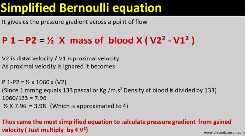
So, if we can somehow measure the velocity gained across a point inside the heart we can deduce the pressure gradient. Here comes the Doppler principle that helps us calculate the velocity. Doppler is based on the reflection of sound and the Doppler shift. With the Doppler shift, we can arrive difference in velocity across a valve, or conduit. When fluid flows across a narrowed orifice (Valve /Outflow) it accelerates and builds up velocity. This gain in velocity is equal to the pressure lost ie as given by the Bernoulli equation. Since potential energy is related to height and gravity same intracardiac zones it cancels out on either side. Hence, essentially the Bernoulli pressure gradient is equal to the difference between the kinetic energy on either side.

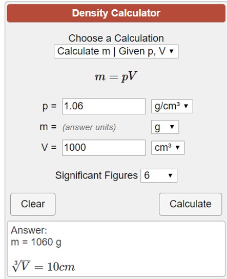
Let us see how this 1/2 of mass becomes 4. We have to convert density of blood which is 1.060 to mass.

mass to density

Note : Mass = ρV . Density is mass per unit volume. So the “m” in the equation is some times referred to synonymously with the density of blood.

modified simplified bernouli equation doppler pressure gradient mass density velocity drsvenkatesan madras medical college echocardiography 2 tr jet lvot gradient

Application in clinical echocardiography

There has been pioneering work from Holen, Hatle and Angleson who proved the value of this equation in the clinical situation in the late 1970s. Of course, Gorlin and Gorlin worked on this similar concept in the cath lab derived pressure gradients

Reference

1.Gorlin R, Gorlin SJ. The hydraulic formula for calculation of the area of the stenotic mitral valve, other cardiac valves, and circulatory shunts. I. Am Heart J 1951:41:1-29.

2.Holen J, Aaslfd R, Landmark K, Sknonsen S, Ostrem 1. Determination of effective orifice area in mitral stenosis from noninvasive ultrasound Doppler data and mitral flow rate. Acta Med Stand 1977;201:83-88.

3.Hatle L. Noninvasive assessment and differentiation of left ventricular outflow obstruction with Doppler ultrasound. Circulation 1981;84:381-
4.Hatle L. Brubakk A. Tromsdal A. Angelsen B. Noninvasive assessment of pressure drop p in. mitral stenosis by Doppler ultrasound. Br Heart J 1978:40:131-140.

Read Full Post »

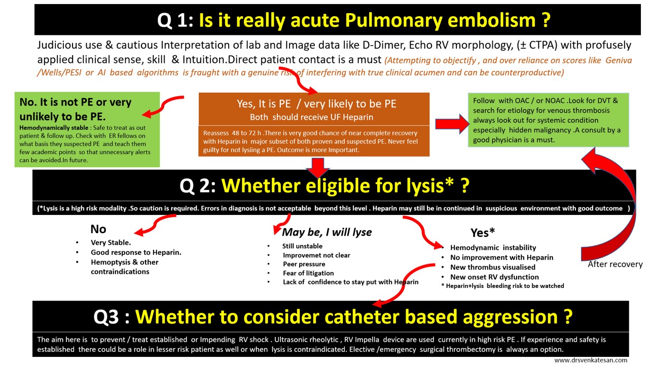
Here is a 3-minute algorithm for the management of acute pulmonary embolism. Just need to ask 3 questions.

Caution: User discretion is advised. Tainted with reasonably acceptable levels of non-scientific content.

Click over the image for a high-resolution slide

Some more critical  questions need to be answered.

What is hemodynamic stability?

It is purely based on clinical signs and judgment.(One need to be doubly sure to rule out hypovolemia and sepsis-related hypotension)

Is RV dysfunction equivalent to hemodynamic stability?

No, it is not. Clinical instability must be associated.( The dogma is,  if the patient is stable even if there is significant RV dysfunction by echo , that RV dysfunction is not attributable to the current episode of PE)

Can we diagnose and proceed with lysis without CT pulmonary angiogram confirmation?

Yes, you can, provided your suspicion is too strong or you have the extraordinary talent to argue/defend even a fatal bleed ( with your boss or in medical audit ) in a patient who was subsequently proven not to suffer from PE .

How to switch over to Lysis from Heparin alone protocol?

Occasionally one may require to do it. There is an added risk of bleeding here. It can’t be avoided in some situations as Initially, it appear as low-risk PE later on becoming more Intense. Generally, high-risk unstable patients should receive lysis straightaway.

Is 60/60 sign is really useful in deciding lysis?

60 /60 sign tell us if Pulmonary artery acceleration time (PAT) and the TR jet both are less than 60 the likely hood of PE is high in a patient with suspected PE.

  • This sign recently got popular not because of its utility, rather because of its simplicity and attractive caption.
  • It may be very specific but least sensitive (<20%) So it can never be used as a screening test.
  • It also fails to differentiate chronic RV dysfunction from acute RV dysfunction.
  • The PAT is strongly influenced by RV dysfunction (It pulls it down below 60 as PAT is dependent on RV Dp/Dt and falsely diagnosing PE
  • 60/60 sign adds up to the value of  Mconllels sign and can confirm PE with almost 100% specificity.

Read Full Post »

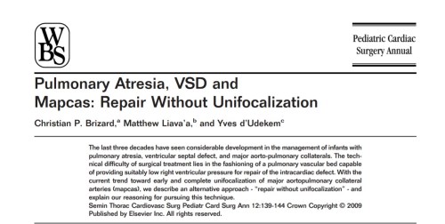
Pulmonary atresia with VSD is one of the complex CHD subsets that requires a meticulous understanding of anatomy, physiology of pulmonary circulation. It can be termed as TAPAC -Total anomalous pulmonary arterial connection in extreme cases. Should we attempt to reverse this total chaotic pulmonary blood supply is the question?

It demands a highly focused cath study(hands & brain) and CT Imaging which might actually throw more light. Post-study Interaction with surgeon and team of cardiologist are vital. The decision to take up the challenge of surgery or abandoning poses equal intellectual stress. Continuous and critical decisions need to be taken. Repeat surgeries and cath based Interventions are often a rule.  Very few centers have mastered this surgery.

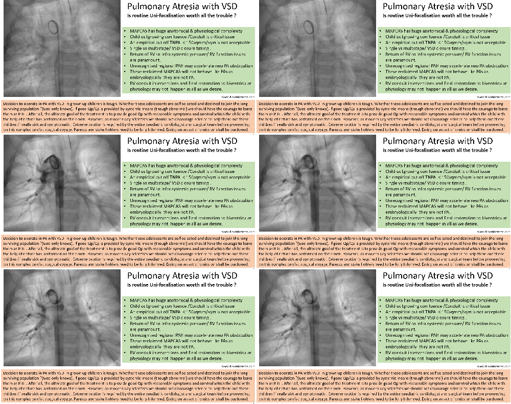
A single slide presentation


pulmonary atresia 4

In spite of all technological developments in pediatric cardiac surgery, there is considerable variations and expectation of the surgical outcome. The major surprise is the original Melbourne group(Ref 1 )  that advocated the uni-focalization as a  concept has almost abandoned this. Stanford and other groups still continue to use this technique more often as a single-stage procedure to improve the outcome.

Let us hope these children get the best of the right mix of technology and natural survival power and more importantly we must ensure the former do not interfere with the later

Reference 

1

Post ample

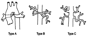
Surgery has definitely  revolutionized the outcome in neonates and children in less severe forms of PA with good central pulmonary arteries ( Most of the Barbero Marcial Type A and many type B) The perceived negativity in this post regarding the outcome of surgery is primarily belong to some of the  Barbero Type B and many of  C.

gr1

Barbero-Marcial M , Jatene A Semin Thorac Cardiovasc Surg. Surgical management of the anomalies of the pulmonary arteries in the tetralogy of Fallot with pulmonary atresia.1990 Jan;2(1):93-107. 


Read Full Post »


Read Full Post »

Science is a journey in pursuit of truth. Hence, we search for it again and again.  (Thus, recurrent search becomes Re-Search)

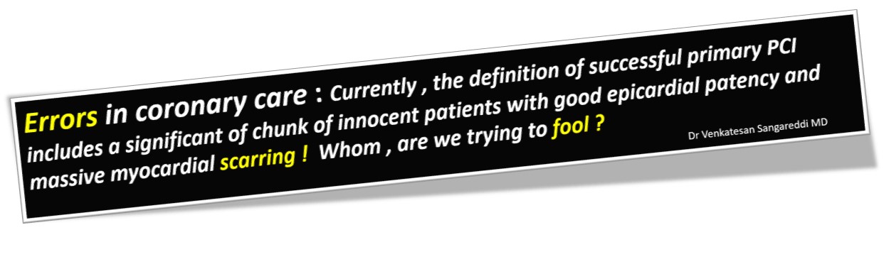
As we try to progress in our knowledge towards absolute truth,  we need to admit our errors first. I think, one such error is blinking right in front of us in the vibrant corridors of coronary care and cath labs every day!  It is about the definition with which we deal the success of primary PCI. (A supposedly revolutionary acute coronary therapeutics  this century)

Waiting for the day . . . when all those fancy primary PCIs that leave the myocardium hurt (& retire ) with significant LV dysfunction to be reclassified as clear cases of primary PCI failures.

Read Full Post »

Exertional dyspnea disproportional to the effort is the most common (cardinal)symptom of heart disease. Whenever we discuss the mechanism of cardiac dyspnea , we primarily attribute it to left heart disease, elevated LVEDP and the resultant pulmonary congestion.Conventional teaching in the past (may be in the present too !) doesn’t implicate raised RVEDP in the genesis of dyspnea.

It’s good to recall , the sensation of dyspnea is felt at the peri -Amygdala nuclear zone after complex processing with various cortical and sub-cortical level .It is subjected to as many afferent triggers other than J receptors in pulmonary micro circulation. (Eg Exercising skeletal muscle). It is believed, mechanical stretch receptors exist within the walls of heart along  the sub-endocardial aspects of chamber.

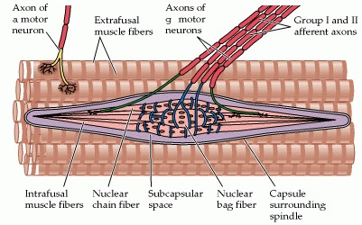
(Muscle spindles which are the sensors of muscle tension are extensively noted in skeletal muscle that contribute to the origin of dyspnea .We are not yet accruing enough evidence  whether cardiac muscle do have the same muscle spindle or it’s equivalents to cause dyspnea when stretched. However, we clearly witness in the practice of clinical cardiology , isolated elevation of RVEDP ( also RVSP ) to cause significant dyspnea in specific clinical situations.

Potential causes for Isolated Right ventricular dyspnea

  • Pulmonary hypertension  (COPD included* where in it could be a combination of lung and cardiac dyspnea)
  • Acute pulmonary embolism
  • RV Infarction
  • Acute rupture of sinus of Valsalva aneurysm (RSOV) Here RVEDP is often > LVEDP and dyspnea is due to the acute stretch of RV
  • Isolated normal pressure TR(RVEDP is low still cause dyspnea  due to volume related RV triggers)
  • Any RVOT obstruction (Classically valvular pulmonary stenosis)
  • Does RV dilatation without elevated RVEDP cause dyspnea ?  Though right ventricle is developmentally and hemo-dynamically better suited to handle volume , still, it  struggles to manage sudden increase in volume .(Another clinical example is seen in patients who are on dialysis)

*RV diastolic dysfunction is still a Infantile hemo-dynamic concept .Whether it can raise RVEDP significantly during exercise and Independently contribute to dyspnea is at best a hypo-science.

Role of muscle spindle and mechno-receptors


Muscle spindle

structure of skeletal muscle spindle. Though we don’t have a highly developed spindles in smooth muscle and cardiac muscle we have evidence to suggest cardiac neural ending do have mechano-receptors with afferent connection through visceral neural plexus that can trigger both heart rate and respiratory centers Further reading : Neuroscience. 2nd edition. Show details Purves D, Augustine GJ, Fitzpatrick D, et al., editors. Sunderland (MA): Sinauer Associates; 2001.

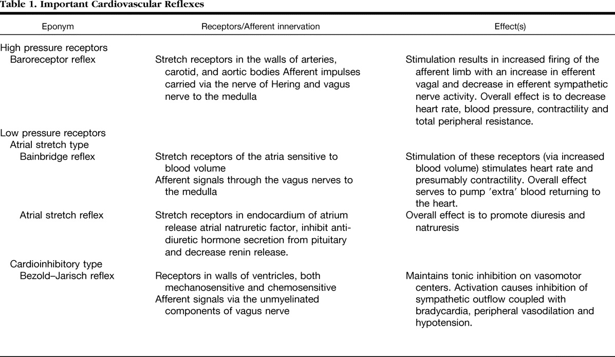
Bain-Bridge reflex: The hidden link in right heart dyspnea

Bain-Bridge reflex is a 100 year old concept. still helping us to understand the basics of right heart hemodynamics and how adjustments with acute volume loading take place.He proposed that  veno-atrial stretch receptors are located  primarily in great veins as it enter ,right atrium (RV as well).

This gets activated through vagus and stimulates  in brain-stem sympathetic system and increase the heart rate to handle the excess blood reaching the heart. How often we feel the symptom of palpitation  whether due to this reflex ( when it is operating) is not really tested. But, what we can infer is , the surge in sympathetic tone perceived can be perceived as  dyspnea.

*Clinical Relevance of the Bezold–Jarisch Reflex and its possible interactions with Bain Bridge reflex is a different topic.

It is interesting to note many of these reflexes cause hypo-tension, bradycardia and hypopnea (Even near Apnea.) The word dyspnea is surprisingly not used .It is highly plausible many of the unexplained dyspnea we see in otherwise healthy population is attributed to acute or chronic volume overloading or under-loading of right heart.

Role of PFO in right heart dyspnea

PFO is a natural decompressing orifice in the IAS guarded by a flip-flap safety valve which is a remnant of septum primum .Though it can flow either way , since the flap of the valve is larger in LA side,  it gets closed when  LA pressure raises but opens up , if RA pressure raises making it more often a right to left shunt at times of elevated RA mean pressure. In isolated right heat pathology , this communication shunts  right to left and  adds a new dimension to cardiac dyspnea (Now, It becomes a hypoxic /biochemical dyspnea over and above the right heart stretch related dyspnea )

Other mechanisms in right heart dyspnea

Pulmonary arterial stretch and altered QP : Role of ventilation perfusion mismatch should also be considered as a cause for dyspnea in isolated RV pathology. The term V/Q mismatch is a poorly understood term fro me. My Inference is, since RV contraction  provides the Q in the equation V/Q .Whenever Q falls V has to fall to maintain neutrality causing net hypoxia and dyspnea.

Final message

Dear fellows, never hesitate to attribute the origin of dyspnea,  to elevated RA mean pressure /RVEDP. It is due to RA/RV stretch secondary to volume and pressure overloading with a perfectly normal pulmonary capillary wedge pressure or LVEDP. As in the left heart ,this occurs both in pathological as well as perfectly exaggerated physiological times.

Reference 

1.Bainbridge FA. The influence of venous filling upon the rate of the heart. J Physiol. 1915 Dec 24;50(2):65–84. [PMC free article] [PubMed] []

2..A J Crisp, R Hainsworth, and S M Tutt  The absence of cardiovascular and respiratory responses to changes in right ventricular pressure in anaesthetized dogs. J Physiol. 1988 Dec; 407: 1–13(This paper actually undermines the importance of RV receptors. It is still perplexing to note both the inflow into RV (ie RA  and the out flow  pulmonary artery circuit has richly innervated by receptors , its difficult to accept why we  have failed to get much evidence for RV stretch receptors) Its potentially great area of research for cardiac physiologists. That will be a tribute to the greats like  Bain Bridge and Bazolds Jarich.)

Read Full Post »

Why VTs have wide QRS complex?

Brief answer: VT  usually presents with wide QRS tachycardia because it originates in ventricular myocardium, travels muscle to muscle instead of the normal conduction system. However, VTs need not be wide always, if it captures the conduction system early and more proximally it can be as narrow as SVT.

Further reading: Only for cardiology fellows 

Two empirical statements are made here. (The scientific chances of both being reasonably correct are fair)

  1. 80 % of wide QRS tachycardia by default is VT. That means 20 % of wide QRS is not VT. We all accept that.
  2. 80 % of narrow QRS tachycardia is SVT. It may also mean, up to 20 % of VT can be narrow QRS.

It’s obvious, not all VTs are dramatically wide. When it is not wide, they test our knowledge and patience. Let us be clear about the factors that determine the QRS width during VT. Once we know this we can have our own inference.

What determines the width of QRS  in VT?
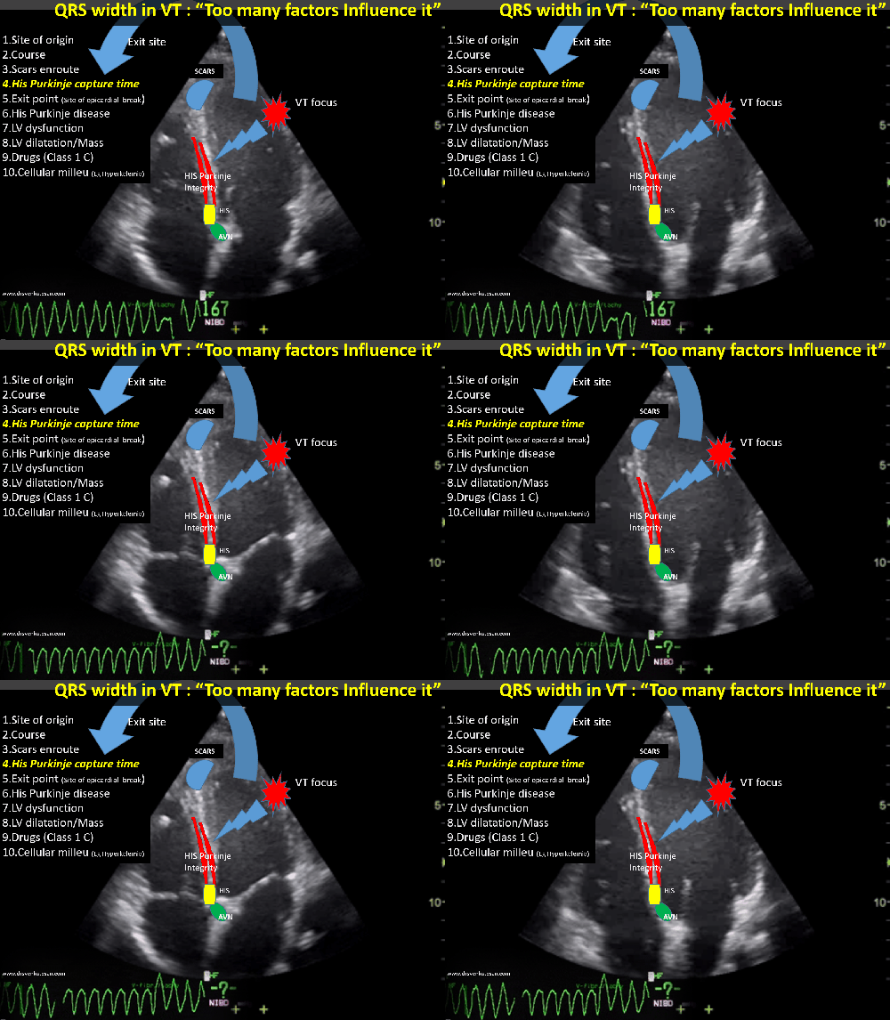
1.Origin of VT 

The focus of origin is extremely important. Pure myocardial focus distal to the conduction system is invariably very wide. We know VTs originating right over the fascicles are narrow.

2.His Purkinje breakthrough

The time taken to capture the normal septal conduction system is a critical determinant of QRS width during VT.This makes the VT from septal origin narrower.VT arising from the free walls obviously takes a longer time to engage the HIS Purkinje system. Imagine , If VT originates from the lateral mitral annulus,  how much time it may take to reach RV free wall and lastly RVOT. Here the VT will become bizarrely wide.

3.The structural integrity of His Purkinje

It is important to emphasize a fact , even if the VT captures HIS Purkinje early, if they are diseased , still the VT will be wider.(Example bundle branch reentry in DCM in which VT keeps going around the conduction system still, it’s wider)

4.Course 

Length of the re-entrant circuit. Macro reentry is expected to be wider. Focal or micro reentry will often be narrow, provided the distal circuit is not diseased.

5. Scars as barriers and boulders 

If the VT circuit is interrupted by random scars en-route (from origin to exit) the  VT width prolongs. (Evidence for scars is often visible in sinus rhythm ECG as notches /slurs or fragmentations in QRS )

6.Exit point of VT

This is a poorly understood term (at least for me) It is believed,  VT can exit only epicardially. The line joining the focus of origin and the exit point is expected to decide the QRS axis. The problem comes when VT breaks out multiple paths and possibly sub-endocardial as well.

7.LV dysfunction 

A severely dysfunctional ventricle can stretch the QRS irrespective of conduction system integrity.

8.The Ionic milieu of cells Interstitial resistance

We know,  biological current is nothing but Ions in motion. So, no surprise it can alter the QRS morphology. The classical example is hyperkalemia , that can make ECG a wide and blunt sine wave. Local acidosis, hypoxia also influence the QRS duration.

9.Drugs 

Any drug which has class 1C or 3 properties can slow the VT circuit velocity. Typically flecainide is well known to make QRS wider. Amiodarone may  reduce the ventricular rate. in VT instead of reverting it. Apart from this these drugs depress the ventricular myocardium severely and prolong the QRS width independent to its action on the conduction system.

10.Mechanism  of changing width 

VTs can have varying QRS width as reentrant circuits change or experience slow conduction due to autonomic influences. VT with downstream aberrancy is also possible as the VT rate by itself influences the conduction property distally.(Just lie SVT with aberrancy)

A paradox about the width of QRS in VT

A curious phenomenon is often seen, when VT occurs in patients with baseline ECG which is already wide (As in an ischemic dilated cardiomyopathy with LBBB/RBBB). Here, the VT  prematurely stimulates viable muscles distal to the diseased HIS  Purkinje system (Which they are deprived of early activation of till then) .They seem to relish the early arrival of electrical impulse by brisk activation that converts wide QRS complex to narrow one. (This  behavior is one of the principles of cardiac resynchronization therapy where we attempt to rewire the heart with multiple leads and shrink the QRS.)

*One more mechanism of wide QRS sinus rhythm becoming narrow during VT is due to a concept called source -sink relationship. The VT delivers enough energy overcoming His Purkinje resistance downstream. (This property is used in HIS bundle pacing )

Postamble

*Forget about wide vs narrow QRS debate. A significant chunk of VTs falls within intermediate width QRS(100-120ms) . Whether to label these as wide or narrow QRS  squarely lies on whims of the reader. (Should we take the widest QRS in 12 lead ECG?  Pre-cardial  vs limb lead  etc are not clear) Unfortunately, we don’t have a separate algorithm for this category. This issue demands a separate discussion.


Read Full Post »

Whenever a patient is getting discharged after a PCI, the treating cardiologist often faces this situation.

So, you fixed the block in my coronary artery doctor. Thank you so much. Now, I can have a peaceful life, free from  future heart problems. “Am I right doctor”?

I wish I can answer “Yes”  to your query but I can’t for the following reasons.

I have fixed only a lesion that caused maximum obstruction. Atherosclerosis is a diffuse disease and you have minor plaques scattered across your coronary artery. These can grow at its own will. So you carry a definite risk remote from the current problem. (Don’t get frightened, read further, you have definite solutions to reduce this risk.)

How common is the progression of native vessel disease?

It varies from 10 to 40%. Mind you, the exact incidence directly depends upon the compliance of medical management, risk factor reduction, and adaptation to a new life healthy lifestyle. In effect, you (the patients) decide the incidence.

One surprise phenomenon (though unproven) might happen. Since the tightest lesion is jailed with a scaffold the minor lesion is preselected to an accelerated process of atherosclerosis if medical treatment is not properly followed.

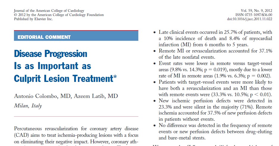
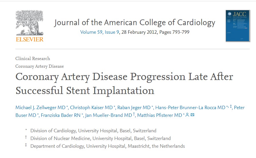
Dr.Zellweger from the university hospital, Basel, did an extraordinary study with 400 patients, meticulous 5 years follow up with SPECT and found remote lesions accounted for 40% of future events (Basel Stent Kosten-Effektivitäts Trial [BASKET]) The other study by Glazer and concurred with this. These studies reiterate the importance of taking care of the entire coronary artery instead of focused piecemeal care by scaffolds.

Does a proximal DES protect a  distal lesion in the same artery by the drug effect?

It is a good thing to happen at least on paper. A proximal LAD with the latest generation Everolimus coated stent is expected to keep the distal LAD drugged for few months at leas.( with anti-mitotic activity) Thus preventing the progression of distal lesions.

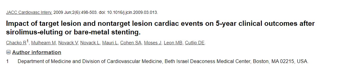
No, I can’t believe this.In this era of momentary touch on sidewalls of artery by drug-eluting balloon (DEB) shown to do wonders, anything is feasible. Chacko’s (Ref 2 : JACC CV Interventions 2009)observation has a possible answer for this. It showed BMS vs DES didn’t make any difference in remote lesion progression.

Final message 

These studies reaffirm one vital truth. Stents are temporary solutions to a permanent, systemic disease of the vascular system .Stents are indeed a major revolution in CAD, “if and only if” it’s used in a highly selected CAD population. Global attempts to project cath labs as a tool to control human atherosclerosis is a typical example of flawed science. The only effective way to tackle this menace is to faithfully follow overall healthy living,  assisted by drugs.

This is the Editorial in response to Zellweger’s article


Reference

1.Glaser R, Selzer F, Faxon DP,Clinical progression of incidental, asymptomatic lesions discovered during culprit vessel coronary intervention. 2005 Jan 18;111(2):143-9 2004 Dec 27
3.

Postamble

One of my patients asked some time ago. If stents are the definite remedy for severe arterial narrowing, why not stent all my lesions (even the minor ones ) prophylactically doctor, so that it will not become tight at a later date?

That’s a good query. Your doubt is genuine , appear logical as well. But, unfortunately, it will be the most dangerous thing to do*. Metals are never friendly with the coronary arterial wall. We should use it extremely judiciously and only with tight flow-limiting lesions. These metals require annual (rather permanent) maintenance. Its taken care by multiple antiplatelet drugs. If for some reason your maintenance is erratic or the drugs fail to act you are at more risk of a future event.

(* This is what has  happened (happening) in the past, that demanded urgent publication of appropriate usage criteria)

Now, the current belief among the “fair thinking cardiology community” is dramatically changing. It’s leaning towards non-stent management even with significant flow-limiting obstructions in otherwise stable patients(SIHD). This belief is accruing more and more evidence base (The COURAGE 15 year follow up / ORBITA/ISCHEMIA)   All these studies confirm the emerging doctrine and bring back some semblance of sense into the cardiology community.

Read Full Post »

Differential cyanosis classically occurs in PDA with reversal of shunt when raised PA pressures /PVR is able to supersede the systemic Aortic pressure and drive the blood from LPA to descending Aorta bringing down the lower limb saturation.

Of course,  this can be undone by the presence of any other intra-cardiac shunts or aberrant left subclavian that arising from the desaturated descending aorta.

Other causes of reversed differential cyanosis 

Where the upper body is cyanosed (desaturated) and the lower half is not. There is a conventional list of conditions.

  1. Transposition of the great arteries (TGA) with patent ductus arteriosis (PDA) and elevated pulmonary vascular resistance
  2. TGA with PDA and pre-ductal aortic interruption or coarctation
  3. Supracardiac TAPVC* + PDA
  4. Anomalous right subclavian artery connected to hypertensive ductus through RPA

(*This occurs due to streaming effect ) Highly saturated superior vena cava (SVC) blood into the right ventricle, reach MPA / through a PDA, and to the descending aorta, with streaming of more desaturated blood from the inferior vena cava (IVC) into the LA through PFO (Ref Yap S H Pediatr Cardiol. 2009 )

Now let us add one more cause for  reversed differential cyanosis in the Modern Era

It is seen with ECMO in VA connection (Often reported in babies ) . The Aorta has high oxygen content entering from the femoral cannula going up into the Aortic arch., while deoxygenated blood from LV (because of failing lungs) reach antegradely to the Aorta. Ideally, the ECMO is expected to supply the entire aortic arch and hence oxygenation is uniform all over the body. It rarely happens as some amount of flow will come from LV unless its in asystole. However, If the severely dysfunctional heart tends to recover & lung oxygenation is very poor as well, the LV stroke volume competes with highly oxygenated blood coming from below ( femoral inflow ) into the Aorta , creating a watershed zone . This makes the deoxygenated blood perfusing upper half of the body and hyper oxygen saturation lower half. This is been referred to as North-south syndrome or (Harlequin syndrome the famous Italian comical character)

How to manage North-South syndrome?

  • Try to Improve the oxygen perfusion with high-frequency ventilation(This is logical first step , to improve the native lung function)
  • ECMO flow rate may be increased and overdrive the LV ejection .(This can be counter-productive as we are hitting a recovering ventricle)
  • Converting to VV ECMO if the hemodynamics allows. This is possible as North-south syndrome is a sign of recovering cardia function VV ECMO will convert it into a primary lung support

Reference

ECMO review article

Read Full Post »

« Newer Posts - Older Posts »