Internet has revolutionised in sharing our knowledge . But , the freedom it gives has a trade off .Identifying genuine knowledge in the vast cyberspace is like searching for lost treasure in the ocean bed.
Archive for the ‘Uncategorized’ Category
Great medical web sites : How to innovate medical education ?
Posted in Uncategorized, tagged great cardiology web site, great medcial videos, great medical web site, innovations in medical education, top medical sites, university of wisconsin on September 23, 2009| Leave a Comment »
Medical pharmacology is not simply learning how a drug acts , it is also about “How a drug company acts*” !
Posted in Uncategorized, tagged bio ethics, mechanism of drug action, pharma industry, pharmacology on September 19, 2009| Leave a Comment »
Pharmacology is a major discipline in medicine where we learn how a drug acts in our body for various ailments . Now in this era , doctors need not only how a drug acts but also how a drug company acts ! This has become vitally important for the welfare of the mankind .
In this context one of the best books on medical pharmacology is from the
Famous Editor of New England journal of medicine
A must read for all genuine medical professionals
Watch Marcia Angell talk http://www.youtube.com/watch?v=ouF3ISihHLM
Full lecture of Mercia Angell http://videos.med.wisc.edu/videoInfo.php?videoid=940
Click to buy/read the book http://www.amazon.com/Truth-About-Drug-Companies-Deceive/dp/0375508465#reader
A Review about the book http://calitreview.com/176
Should we not clarify what exactly we mean by wide qrs tachycardia ?
Posted in Uncategorized, tagged avnrt, brugada criteria, ECG, ep study, infectious disease, pace journal, podrid, supraventricualr tachycardia with aberrancy, svt, svt vs vt, ventricular tachycardia, vt vs avnrt, vt vs svt, what is wide qrs tachycardia, wide qrs tachycardia, width of qrs, zipes on September 18, 2009| 1 Comment »
Wide qrs tachycardia has a unique place in clinical electrocardiography .It is a much fancied and glamorous entity for the simple reason , it continues to be the cardiologist ever solved puzzle .For over three decades of research, clinical debates , symposiums , seminars have effectively failed to take away the uncertainties in decoding the wide QRS tachycardia . (Specifically , VT vs SVT with aberrancy)
Some wondered , should we really waste our efforts in differentiating the two . In emergencies it never matters , in fact one need not attempt to do this often futile exercise !
Few dedicated criterias like Brugada etc have helped us .
While the difficulties in differentiating between VT and SVT with aberrancy remain over the decades .A less reported , but more common issue is confronting us .
It is the big question of differentiating a wide QRS tachycardia from a narrow QRS tachycardia
This occurs more often than we realise ,because we define wide QRS tachycardia in a vague manner
- Normal qrs width between Up to 80 / up to 100 ms acceptable ?*
- Narrow qrs tachycardia 80 ms?
- Wide qrs tachycardia i> 120ms ?
- Definitely wide qrs >140msec
* The confusion is mainly because 20ms difference between limb leads and chest leads .
In reality one may not be able to all tachycardia into narrow or wide .
There is big overlap zone that need to be labeled a intermediate qrs tachycardia
If we can triage the tachycardias into three instead of two it may help us arrive fast , to the correct diagnosis
Narrow QRS tachycardia ( qrs 80ms)
- Sinus
- All svtS (avnrt etc)
Intermediate QRS tachycardia 90-120
- Most of the SVT with aberrancy ( Except antidromic SVTs which are really to wide !)
- Septal VTs*
- Fascicular VTs*
- VT in PPM and ICD /CRT patients **
* Any VT that arise near the major conducting system of ventricle conduct fast and hence qrs are relatively narrow.
**These are rare entities where base line wide QRS getting narrower with the onset of VT . (Ref : http://europace.oxfordjournals.org/cgi/content/full/eun254v1)
Wide qrs tachycardia >120ms
- Most of the genuine VT (Ischemic , myocardial origin)
- Post MI VTs
- SVT aberrancy especially AVRT
- Any SVT with preexisting BBB
- Marked electrolytic disorders
Unresolved questions
- Which lead we should look for measuring the width of qrs ?
- Should we take the narrowest qrs or widest qrs or should we take the average ?
- Should we calculate how much the tachycardia has widened the qrs from the baseline width of a given patient ? Is it not possible , what is wide for some may be normal for another !
- If there is no isoelectric line and ST segment blends with qrs complex how to mark end of qrs ?
- If limb leads show a narrow qrs and chest leads shows wide qrs what is the significance ?
- In precardial leads if one lead alone shows a narrow qrs , what is the significance ?
- Can a narrow qrs VT conduct with aberrancy and making it really wide ?
Final message
When we are able to solve complex electrophysiological problems , we must also realise even simple tasks can be demanding in medicne ! It is proposed to create a new group “Intermediate QRS tachycardia “that can help solve the issue where we have difficulty in labeling these tachycardias which fall in the greyzone .We can try & apply the modern EP based VT criterias to this group and find out the hidden truths !
What is protected left main disease ? How good is the protection ? If good , why should it need another protection ?
Posted in Uncategorized on September 15, 2009| 7 Comments »
Left main disease is an important subset of CAD , and it has special interest for the interventionist. Traditionally cardiologist have a fear to touch this lesion , as they thought a sudden occlusion within this vessel is life threatening . Later on as they gained experience it was thought we could intervene safely at least in protected left main . Subsequently it was realised this fear was largely unfounded , after all the proximal LAD is equally dangerous and we spend hours together inside an LAD ! .Now we have technology and expertise to do successful PCI any where in LM. And unfortunately , the same expertise is not applied in selecting the ideal patients who will benefit the most . LMD has become a glorified indication for PCI.
The terminology of protected and unprotected LMD is in vogue for many years . Unfortunately it do not convey a uniform meaning . In next few minutes , I shall share my views on the nuances of protected and unprotected LMD .
The term protected was not coined by cardiovascular physiologists but by interventional cardiologists . Hence it connotes a anatomical meaning rather than physiological. Protected LMD meant there must be a at least one graft to either LAD or circumflex . And this graft should be functional . The presence of this graft is supposed to increase the comfort levels of the interventionist as well as the patient.
A left main coronary artery disease angiographically can be classified as
Common types of Left main lesion
- Asymptomatic , non flow limiting , angiographically insignificant disease(< than50%)
- Ostial
- Ostio proximal
- Shaft : Mid, distal or diffuse Left main
- Bifurcation
Unprotected left main
- All the above lesions
- Non functional GABG grafts ( eg: LIMA occlusion makes LAD unprotected)
Protected left main
- Post CABG with atleast one functional graft to LAD /LCX
- ? Left main with total LAD and very good LAD collaterals from RCA /LCX
Partially protected Leftmain
It could mean any of the following, Left main Plus . . .
- Incomplete occlusion of single LIMA graft
- Occlusion of SVG-LCX and patent LAD-LIMA
- Occlusion of LIMA- LAD graft but patent SVG-LCX graft
- Patent LIMA-LAD but a critical LM / LCX bifurcation lesion with no grafts for LCX*
The above 3 situations may demand a PCI .But logic would suggest one would try to open up the partially occluded graft rather than open the left main . Of course the decision involves status of RCA .
*The only indication for a PCI in protected LMD could be 4
Unusual ( Crazy !) questions about left main disease
Can left main be protected by collateral circulation ?
It is very common to find Left main bifurcation lesion with LAD having very good collaterals from RCA sometimes filling up to proximal LAD .This can be considered “protected left main equivalent”
As on today , cardiologists would rather believe a surgeon’s graft rather than a naturally grown collateral from RCA however extensive it may be !
But logics and real case experience would indicate in a patient with LMD and an extensively collateralised LAD can in fact be considered a protected left main.
If a left main is well protected by a functional LAD graft , why should we do a PCI for left main at all ?
This question was risen in one of our cath conferences , a patient who had functioning LIMA to LAD graft.His RCA had a functioning venous graft and his circumflex had a partially functioning graft.The left main had a near total obstruction and the proximal LAD was faintly visible .
Since the patient had class 2 angina Options were discussed .He satisfied the current criteria of protected LMD .Just because he fulfils the criteria of protected left main , he does not become eligible for left main PCI . After all he is having this LMD for many years. Protecting again the left main which is already protected is not a big deal in terms of outcome . Double protection is waste of resource at additional risk. It was decided to attempt a PCI to SVG graft to LCX. If it does n’t work leave him with medical management.
Does every patient after a CABG has a high chances of developing LMD ?
What is accelerated atherosclerosis of Left main following LAD /LCX grafts ? It is true left main has high risk of accelerated atherosclerosis and it undergoes gradual obstruction once the LAD and LCX is grafted.This is due to low flow across the native left main as distal grafts maintain the flow . This is all the more likely in good bulk of patients who had undergone CABG where LMD was the indication .
A typical scenario
A left main patient who undergoes a CABG a follow up for a suspected angina angio after 5 years show the totally or near totally occluded native left main . Sudden Visualisation of worsened leftmain disease makes this patient eligible for a PCI as he fulfills the criteria for protected leftmain .
Final message
A well protected left main with a good functioning graft especially to LAD most often do not require a fresh revascularisation procedure irrespective of the tightness of left main disease . Most of such patients will be candidates for medical therapy .Contrary to the popular belief , left main intervention could be confined to ” unprotected LMD rather than well protected LMD” as the potential benefits are more .Further interventional resources need not be wasted in giving second alternate protective channel for an already protected vessel !
Of course it should be remembered in any given patient with protected or unprotected LMD the indication for revascualrisation is based on the severity of lesion , symptoms, LV function , residual ischemia, viability etc .
Suggestions , comments and corrections welcome
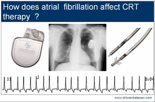
How the presence of atrial fibrillation influence the effectiveness of CRT therapy ?
Posted in Cardiology - Electrophysiology -Pacemaker, Uncategorized, tagged cardiac resynchronisation therapy, care chf, companion, CRT, madit chf on September 12, 2009| Leave a Comment »
Atrial fibrillation and CHF are close companions. Either it precipitates CHF or follows it.In advanced heart failure of any etiology the incidence of AF can be up to 40% .Medical therapy of AF is fairly effective in patients with normal LV function .But when associated with refractory cardiac failure it becomes too complex to control .
Currently CRT with ICD is becoming the standard OF care for advanced CHF. The efficacy of CRT is being rigorously being assessed . Even as the controversy about the wideness of QRS is being settled , the issue of optimal timing of CRT has risen . Now , the MADIT-CRT has answered this issue “Earlier it is better , it can be indicated even for class 1 patients”
While MADIT -CRT will increase the number of CRT implants , we have no clear cut answer for the efficacy of CRT in patients with AF .( Of course , the MUSTIC and CARE HF sub group analysis suggested AF has no significant impact on CRT efficacy )
Why is AF important in CRT ?
There are two issues that need analysis
- A patient who has chronic AF at the time of CRT
- Development of new onset AF after CRT implantation.
Impact of AF during CRT
- Inter atrial synchrony is lost. ( Significance not clear . . . makes AF permanent)
- AV synchrony is lost
- Rapid AV conduction : May trigger too much of Bivi pacing if sensed by LV lead
Presence of AF at the time of CRT gives us an opportunity to tackle this issue.
How to tackle sudden AF induced CRT response ?
There are variety of algorithms available to
- Ventricular sense response
- Conducted AF response
- Atrial tracking recovery
In dual chamber pacing mode switching converts DDD into VVI .This happens at the cost of loss of AV synchrony .This may have profound implication in CRT .
Then the big question comes . What is the use of having Intraventricular and interventricular synchrony without AV synchrony ?
When nothing works .The best strategy is ( Rather deemed to be best ! )
- To ablate the AV node pace the atrium and ventricle (RV & LV) .
Note : Ablation of AV node and putting a dual chamber pacing can never guarantee a physiological pacing as the atrium continues to fibrillate and AV synchrony is rarely there .
Final message
For CRT is to be successful , there should be maximal Bi-Vi capturing , of course this capture has to optimally timed , and must reverse the three pathological asynchronies , namely intraventricular , Interventricular and atrio ventricular asynchronies.
It is obvious , presence of AF complicates the issue as it demands constant monitoring and programming of the device (Of course now most of them are automated) . It may require knocking down of AV node , which not only carries a risk of SCD * , it also make these patients permanently dependent on the RV pacing . This adds on , another risk , for an acute complication if the RV lead fails for some reason.
Reference :
EP experts generally take too much liberty in adopting this strategy for the simple reason it solves the nuisance of atrial impulses interfering with ventricular leads function that result in inappropriate ventricular capture fusion or ultimately poor BiVi pacing . But it is not an easy decision atleast for the patient ! This article , emphasises the dangers involved in ablate and pace strategy for uncontrolled AF.
Further reading
- Fung, J. W H, Yip, G. W K, Yu, C.-M. (2008). Does atrial fibrillation preclude biventricular pacing?. Heart 94: 826-827 [Full Text]
- Khadjooi, K, Foley, P W, Chalil, S, Anthony, J, Smith, R E A, Frenneaux, M P, Leyva, F (2008). Long-term effects of cardiac resynchronisation therapy in patients with atrial fibrillation. Heart 94: 879-883 [Abstract]
- Buck, S., Rienstra, M., Maass, A. H., Nieuwland, W., Van Veldhuisen, D. J., Van Gelder, I. C. (2008). Cardiac resynchronization therapy in patients with heart failure and atrial fibrillation: importance of new-onset atrial fibrillation and total atrial conduction time. Europace 10: 558-565 [Abstract] [Full Text]
Why ECG evidence for AV dissociation does not occur in majority of patients with ventricular tachycardia ?
Posted in Uncategorized, tagged av dissociation on September 10, 2009| 2 Comments »
AV dissociation is the specific marker for diagnosing VT. Evidence for AV dissociation manifest in many ways in ECG. *Random p waves unrelated to qrs complexes , fusion beats , capture beats are the common features that help us diagnose AV dissociation. Unfortunately these occur only in about 40 % of patients with VT.(Fusion beats in VT are also called as Dressler’s beat)
For clinical features of AV dissociation follow this link
What is the normal AV association ?
In normal physiology , even though atria are passive , powerless chambers in terms of mechanical activity , it reigns supreme control over ventricle and dominates electrically . In fact , the atrium and the AV node together , dictates when the ventricle has to contract and at what rate . So, in normal human beings in sinus rhythm , there is a complete AV association where both chambers live in a perfect harmony.
What is VA association ?
The atrium and ventricles are not only related antegradelyy it also has a concealed retrograde relationship , (which is often pure electrical ! ) called VA conduction .The conduction velocity and the refractory period of VA junction is variable .The AV junctional refractory period is determined by the penetrating power of both atrial and ventricular impulses .
What is complete AV dissociation ?
For complete AV dissociation to occur there should be no physiologically or electrically linked relationship between the atria and ventricle.For it to occur the atrial impulse has to get blocked in AV junction .
This block can either be functional or organic, partial or total , persistent or intermittent
This occurs in primarily in AV junctional pathology like CHB etc, that result in complete AV dissociation . The next major cause for AV dissociation , is by an interference from an accelerated lower pacemaker as in ventricular tachycardia or accelerated idioventricular rhythm .
What does the atria do when the ventricle starts contracting rapidly and independently as in ventricular tachycardia ?
When the ventricle , starts firing independently at a rate of > 200 each of the impulse and tries to traverse the AV junction retrograde . At the same time , the sinus impulse which does it’s normal routine job by beating around 70/beat , faces an unusual interference on its normal downward journey by the pathological bombardment from the upcoming ventricular impulse .What happens when both these wave fronts meet head on . (The hither to perfect harmonical relationship becomes a rivalry for the electrical control of heart.)
Sadly , the ventricle mostly succeeds in the race and most of the ventricular impulses retrogradely enter the AV junction and colludes with the incoming atrial impulses. When this happens , the AV dissociation is said to occur. The important point here is, many times if the retrograde VA conduction is fast and optimally timed , it can cross the AV node without difficulty and reach the atria and subsequently even depolarise the SA node and reset it . If the VT is persistently conducting retrograde it can suppress the SA node as long as the VT is there. This makes a P wave becoming totally absent. (Note of caution : If you say VT as one of the causes of absent P wave you may be failed in your cardiology board , but this remains a fact !)
So the atrial depolarisation and contraction During VT is a complex one. It depends mainly on the intensity of the upcoming electrical wave front from the ventricle . The distance traveled by this wave front determines the location of p waves .It may be in one of the following ways .
- P waves can be totally absent
- P wave may occur antegrade
- On the QRS
- Over the T wave
In effect the P wave can literally be any where in the given strip of VT
When does a fusion beat occur ? When does a capture occur ?
This again is determined by the AV junctional refractory period. If it permits , an occasional atrial impulse may sneak through the AV junction and capture the ventricle . This is capture beat. Capture beats are usually narrow qrs . So in a wide qrs tachycardia if we note an occasional narrow or relatively narrow qrs complex it could denote a VT.
If the atrial impulse after crossing the AV junction collides with the upcoming ventricular impulse the surface ECG inscribes a fusion beat. An incomplete capture beat is a fusion beat. It is a combination of two qrs complex one activated from above , one from below .The width of the fusion beat may be wide , narrow or intermediate.
So the evidence for AV dissociation in surface ECG is rarely manifested if the VT is successfully traverse the AV junction and reset the SA node or keep it in a semi depolarised state .This could be clinically important some times , the SA node takes time to recover following A DC shock especially in elderly
An episode of VT can unmask a hidden sinus node dysfunction , as VT is technically similar to an atrial override pacing of course from below .
Final message
During VT , electrophysiologically there must be a dissociation between the atrial and ventricular contraction.But the evidence for which is not manifested in surface ECG in the majorty.The primary reason for this, due to the intact VA conduction that result in retrograde VA association.This makes the classical findings of AV dissociation a redundant or invisible one .
Conveniently forgotten studies in CRT : The importance of diastolic wall motion defect in cardiac failure !
Posted in cardiology -ECG, Cardiology -Interventional -PCI, cardiology -Therapeutics, Uncategorized on September 8, 2009| Leave a Comment »
The failing heart enlarges progressively and attain a globular shape . What looks for the naked eye as a simple global hypokinesia of LV , when analysed , reveal multiple forms regional desynchronisation .This is especially true if the QRS complex is wide.
It is generally divided into three groups
- Intraventricular desynchrony (IV)
- Ventriculo-Ventriculo desynchrony(VV)
- Atrio ventriculo desynchrony(AV)
In our search for improving CHF mortality and morbidity , we have stumbled upon this concept of restoring the lost synchrony of the heart. Cardiac resynchronisation therapy has become ( Rather projected to become !) a great modality for patients with cardiac failure.It was initially advocated only for severe forms of cardiac failure , now advised even for class 1 CHF. (CRT-MADIT 2009)
Restoring the lost synchrony by rewiring the cardiac conducting system with multiple leads and optimally timed pacing increases the effectiveness of cardiac contractility.It can improve EF, and also regress mitral regurgitation.
The above concept was perfect on paper , but was very difficult to replicate on real patients. CRT was ineffective in 30% of patients. Many had partial effect. Few had adverse effect .
The reason for the poor efficacy is technical in many . Identifying the optimal sites for positioning the leads and the futility of such an exercise as the LV epicardial lead is pre- selected by the patients coronary venous anatomy are the major issues.An electrically ideal site for pacing can contain a mechanically dysfunctional scar. While these technical issues may be addressed in due course what worries us is the conceptual flaws.
Emerging facts indicate timing of asynchrony could be vitally important.
- Systolic synchrony
- Diastolic synchrony
What is the incidence desynchrony with reference to the cardiac cycle ?
One major reason that was overlooked totally was the presumption cardiac dysynchrony occur only during systole. It is a less recognised fact is the ventricular relaxation is not uniform and synchronous.A failing ventricle can not be expected to relax systematically and coherently for the simple reason the myocytic calcium reuptake into the sarcoplasmic reticulum is grossly impaired. This is directly responsible for the diastolic dysfunction observed in dilated cardiomyopathy . If this impairment occur uniformly throughout the left ventricle it can be termed global diastolic dysfunction which is little easier to correct .But what really happens is the defect in calcium reuptake occurs in a random fashion with lot of regional variation. This is called regional diastolic wall motion defect or regional diastolic dysfunction.The above mechanisms result in the typical restrictive filling pattern of many of the advanced patients with DCM . CRT as a concept should need to address this issue.
How to diagnose Diastolic WMD?
The fact is ,we have not mastered the quantification of systolic WMD as yet. It may take years before decoding the nuances of diastolic wall motion defects. At least we need to know such a thing exists.Tissue doppler strain rates , velocity vector imaging could be useful tools. As such they are not clinical tools.
Final message
Cardiac resynchronisation as a concept is good on paper . Heart need to be synchronous both during systole and diastole .This becomes especially important in an advanced stages of heart failure. Without proper follow up and potential adverse effects of CRT on diastolic WMD , CRT concept has miles to travel ! . Some pessimistic thinking cardiologists ( Me . . . !) would even argue it as a case of prematurely released device into the patient domain. Of course there is lot of scientifc data that will vouch for its beneficial effects .(The latest being from the prestigious NEJM , CRT-MADIT) but it has to prove it’s worth in individual patients. Physicians must exercise caution before embarking on heroic attempts to provide resynchrony of failing hearts .
Reference
This study from France published in JACC 2005 by Iris Schuster,
http://www.journals.elsevierhealth.com/periodicals/jac/article/PIIS0735109705021005/fulltext
Coming soon
ICDs are better bet than CRT
What is up sloping ST depression ? How do you measure it ? What is the clinical significance ?
Posted in Uncategorized on September 4, 2009| 6 Comments »
ST segment depression is the classical response to stress during excercise stress testing. (EST)Not all types of ST segment are pathological.The ST segment should depress atleast 1 mm below the isoelectric segment and it should be depressed for 80msec from the J point.
It must satisfy two criteria .
- The quantum of ST depression should be > 1mm at 80msec from J point.
- Slope of ST segment
Always pathological slopes
- Horizontal
- Down sloping
Most often pathological
- Slow up sloping
Non pathological slope
- Rapid up sloping with ST depression
- Rapid Up sloping depression of only the J point( The classical normal physiological response to excercise )
Horizontal or down sloping ST segment is easily recognised .When there is junctional ST depression with a ST segment that is climbing upwards , it is some times difficult to interpret.
How do you measure the slope of ST segment ?
We don’t have the trouble of measuring it as the computer does this job automatically. But a cardiology fellow need to know how it is measured !
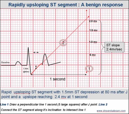
A slow upsloping ST segment( <1.5mv.sec )can be a significant marker of ischemia.This is especially true in established CAD or individuals at high risk . For so slow up sloping a .5mm allowance is given to filter out false positive (ie to improve sensitivity) . So for slow up sloping ST segment , to be reported as positive it should depress atleast 1.5mm or some times 2mm.
Available evidence suggest a rapidly upsloping ST segment (> 1.5mv /Sec) is a non ischemic response irrespective of the quantum of ST depression at 80msec. However , a rapidly upsloping ST is rarely depressed beyond 2mm .( This is because , the geometric hyperbolic curve of ST segment does not allow a situation of 3mm ST depression at 80msec with rapid upsloping )
What is the angiographic correlation of slow upsloping ST segment depression?
Few studies are availbale to address the issue. It is believed slow up sloping of ST depression is often associated with CAD but it is very rare to find a critical and proximally located CAD.Left main disease is almost never manifest with slow upsloping ST depression.
What is the significance of slow upsloping ST in clinical situations like unstable angina ?
It is rare for cardiologist to diagnose or “even look for” slow or rapid up sloping ECGs in coronary care units. But , a patient with stable CAD , sinus tachycardia , angina can exactly mimic a stress test situation .
Some of the low risk UA , mainly secondary UA due to increase demand situations manifest with slow upsloping ST depression , while classical thrombotic occlusions produce the typical horizontal or downsloping ST segment depression.
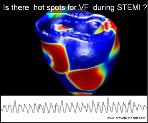
Is there any hot spots in the heart that triggers primary VF following STEMI ?
Posted in Uncategorized, tagged nstemi, stemi, ventricular fibrillation on September 3, 2009| 1 Comment »
Primary ventricular fibrillation is the number one killer in STEMI.It is believed to occur ( Rather it occurs really !) in up to 25 % of all patients with STEMI before they reach the hospital and another 4% after reaching the hospital.
What triggers this primary VF ?
Easily answered : It is the acute ischemia in majority.
Why it triggers in only in some patients? The rest reach the ER safely and some casually walk in to the OPD few days after a STEMI
This can never be answered with our current knowledge base. Some call this as fate !
Scientists should work hard on this issue, if we know the answer we could possibly prevent the number one killer of the mankind at bay.
Many factors are being analysed to find the reasons for primary VF
- Extent of infarct
- Area of infarct
- Intensity of pain
- Adrenergic drive
- Gender
- Myocardial critical mass
- Is it the left main STEMI ?
- Is it a bifurcation STEMI ?
If nothing explains the VF it is always safe to blame it on susceptibility and inherited risk for primary VF , which of course is very much likely as the K+ channel activity and it’s response to ischemia is largely inherited
Is there any hot spots in the heart that are hypersensitive to ischemia ?
Some studies have clearly documented increased incidence of primary VF in infero posterior MI , and RV MI
than anterior MI . J Am Coll Cardiol 2001; 37: 37-43
Why ischemia of a certain location of heart should be more prone for primary VF ?
The answer is any body’s guess.
Some intriguing possibilities are ,
- RV is a anterior chamber , when infero posterior MI occur in association with RV MI the ischemic zone encircles a almost 50% of heart like a band .This could be one explanation for more incidence of VF in infero postero RVMI.
- Any MI which involves a antero -posterior axis of heart is likely to trigger a VF
- Some of our patients who survived a primary VF had a short left main and early bifurcation with a large diagonal branch.The lesion was noted in the bifurcation.This raises a possibility , if a STEMI occur at a bifurcation with two divergent areas of acute ischemia it has a high chance for precipitating a VF.
Related video by the author
Ignorance based cardiology -You tube
Potential research areas
Genetic susceptibility
Environmental Energy flows and primary VF
Some believe a role for astrological forces and VF
What is primary RF ablation for ventricular tachycardia ?
Posted in Uncategorized, tagged primary rf ablation on August 31, 2009| 1 Comment »
Ventricular tachycardias , especially incessant ones not controlled by drugs are very troublesome . Radio frequency ablation is the treatment of choice currently. Principles of electrophysiology would demand acccurate localisation of the tachycardia focus and then ablate it with RF energy .This requires induction of the clinical arrhythmia on the EP table, mapping , identifying the circuits and ablate the optimal points of reentry or slow conduction or P potentials
In reality , some times ( or Is it many times !) , tentative ablation
in the ” V-tach Zone” without mapping is more easier and surprisingly more effective than the much scientific approach of localising the circuit and inducing the arrhythmia.This is referred to as primary ablation
Is it not a crude method to blindly burn cardiac tissues ?
No, we are not worried by the crudeness , as long as it is safe and effective. Experience have made us clever, inducing a VT in the EP lab can be very demanding to our senses and it is a true stress test for the cardiologist’s patience and endurance.Primary ablation has reduced fluroscopic time, procedural time and most unexpectedly increased the success rate!
So even a relatively unscientific blind burn may be better than a scientific burn ?
Yes. It seems to be , at least in idiopathic VTs of fasicular origin and some VTs in RVOT.
Heart is a 400gram organ , it can afford to lose few grams of tissue , especially when it is pathological and behave aberrantly
Reference:
A nice paper from India