Feeds:
Posts
Comments

Archive for the ‘cardiology -Therapeutics’ Category

Left ventricular dissection is a rare complication of STEMI .A case report

Click on the slide to see the video  hosted in  youtube

Slide 1

Slide 2

Reference

http://www.ingentaconnect.com/content/bsc/echo/2009/00000026/00000003/art00006

http://resources.metapress.com/pdf-preview.axd?code=g4kqby7wnkjepetx&size=largest

Read Full Post »

Human body is a collection of trillions of cells.  Life  is nothing but , a bundle of energy flowing across each of these cells  .Every  organ  has a  specailised mode of communication among themselves and others. When a cell is in an excited state , there is a  likelihood of spontaneous electrical activity.This can happen in nerve cells, cardiac cells , GI tract,  or virtually in  any cell  which has a porous cell membrane and ionic fluxes across it .

  • Each cell membrane has a resting membrane potential . It  varies between -60 to – 90mv in most cells. When this potential increases there a propensity for  arrhythmias in heart  and convulsions in the brain , peristalsis in intestines and so on .
  • Drugs  like local anesthetic lignociane acts by blocking the  Na+ channels and there by neural activation .Similarly magesium  acts on these channels to reduce the excitability of these cells.
  • We know,  the sharp ascending stroke of cellular  action potential is mediated by Na + .Blockage of this channel blunts the action potential voltage and thus  the  early and late after depolarisation is prevented
  • Magnesium sulphate’s anticonvulsant action is directly  attributable  to this membrane stabilising action

Thus , membrane stabilising action  can be termed as “membrane sedating”  action

Read Full Post »

Thrombolytic therapy was a  mini revolution  when it was introduced two decades ago .It has since evolved  , not only in the  molecular structure  but also in it’s usage pattern.

The first generation streptokinase is continued to be used even today  . While the latest generation thrombolytic agent TNKTPA(Tenekteplase) is threatening  to push the  old warrior out of  CCU.

(Of course the  American Physician & Pharma  community  never  gave the due respect to  streptokinase  !)

The two common indications  for thrombolytic therapy  are

  • STEMI
  • Acute pulmonary embolism

Uncommon indications

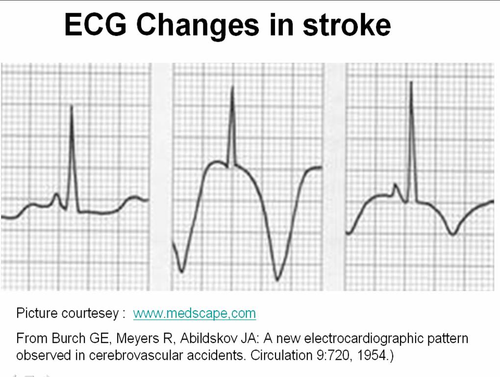
  • Stroke( Can be common in few institutions)
  • Prosthetic valve thrombosis
  • Rarely DVT

From the beginning , there has been a controversy  about the thrombolytic  dosage and  the speed with which it is to be administered .Let us recall , streptokinase was initially  used  in  various regimes ( 5-30lakh units between a 10 -3hr infusion )  Later ,we arrived at a consensus at  15L units  in 1 hr infusion . TPA also experienced the same . Which  settled  for front loaded regimen(35 + 65mg)  . The confusion reappeared when we developed bolus thrombolytic agents( TNKTPA) .

In STEMI thrombus formation  is  often a one time process  while thrombolysis is a continuous process. In pulmonary embolism both  thrombus formation  and lysis  is often continuous process  .

The success of thrombolysis depends on the sustained  drug concentration ,  the pressure at which the drug interacts  the thrombus.

Many times it is prudent to administer  intensive heparin after thrombolysis  to prevent recurrent thrombosis. Further ,  most of the pulmonary embolisms  will require long term anticoagulants.

How to maximize the success of thrombolytic agents ?

  • Local catheter based thrombolysis can be tried  within the coronary ostium (Largely unpopular)
  • Within the pulmonary artery for pulmonary embolism (Still considered an useful option )

It  makes sense , to administer these thrombolytic agents over a prolonged period of time so that the lytic process gets wider recruitment of the natural lytic mechanisms.

When a drug is infused continuously , the drug  reach the thrombus in  a pulsatile manner , which facilitates thrombus dessication  (Like drip irrigation ) . A long acting drug even with a high concentration may not be  very effective , since  the  drug is required to produce a mechanical effect  here . (Unlike say a long acting antibiotics !)

TPA in Pulmonary embolism

The inadequacies  of  2 hour infusion of TPA is  glaring in acute pulmonary embolism .We believe   a 48-72 hour streptokinase infusion   has a definte edge   over a short and brief TPA infusion.

Issues need answer

It is yet , not understood why we can’ t infuse TPA as  a   long term infusion like streptokinase .

Advantage  of bolus TNK TPA  in  pre-hospital phase of STEMI

The argument in favor of bolus dose  thrombolytic agent  is  the ease of administration .

The other the major advantage claimed  is ,  a 10 second  TNK TPA   in STEMI  can  substantially  reduce the time window   and facilitate  early completion of thrombolysis .

Counter point

But , the  later concept is hard to prove  . . .

In fact , there  are  no controlled studies  available for assessing the   efficacy of TNK-TPA   vs  Streptokinase   with reference to various time windows. We presume so many things. An  incomplete   early thrombolysis  may not be better than a  more  successful  but  slightly delayed TIMI3 flow .

As scientists,  when  we try  to answer these  question we  ask for data .  Are we getting it any way ?  Are the existing data reflect  fact ?     We  wonder,  will we may never get   an  hourly  angiographic  data base  about the IRA  patency  in  TPA bolus  vs streptokinase infusion .

It is most unfortunate,   with  many of the critical questions   still to be answered ,  the cardiology community believes ,  they  have  reached the  summit  of  knowledge  about thrombolytic therapy . Current perception is , the research on  existing  thrombolytic drugs  is  deemed to have been complete .

In this hyped  era of interventional coronary  care  ,   it is a remote possibility   to have any  further comparative studies on thrombolytic agents .

The greatest threat faced by  us  today  is the destiny  of  modern medicine is   often  decided in  few corporate board rooms  and   hence   research questions  rarely  emanate from bed side !

In this scenario, where we are not likely to generate   genuine  clinical  data ,  the only way to move   forward is   to go  by  our experience – ” Genuine  experience to be precise . . .”

Final message

Ease of administration should never be the criteria in choosing a thrombolytic agent . It   can severely    compromise the quality of thrombolysis  ! especially in pulmonary embolism and to a certain extent in STEMI.  Success   rarely  comes  with ease  . . .

Many believe , the choice  between  streptokinase   & TPA    goes much beyond it’s academic reasons.  TNK TPA (Tenektepalse) has come in a big way to replace streptokinase  even   in developing countries.  Ofcourse it is backed by a huge study  ! (ASSENT) .

The cost effectiveness and worthiness  of TPA over streptokinase  was  never proved comprehensively.

Note of caution :

The observation made above is   based on personal  opinion  in  about   20 patients  . Readers are  argued to do their own  analysis on this issue and come to a conclusion .

Read Full Post »

It is said every clinical diagnosis needs to be substantiated with  documented objective  evidence .

Probably,  the commonest cardiac emergency , that can be  diagnosed purely by history is UA.

Yes , unstable angina is a symptom not a  disease entity !

By definition UA is

  • Any  new onset angina  of severe grade
  • Progressive crescendo angina
  • Angina with radiation to new site
  • Angina not controlled by nitroglycerine
  • Any angina after a PCI /CABG

If you read the definition again, you will realise ECG or enzymes never come into the  diagnostic picture .UA can be diagnosed even before one has a look at  the ECG ! So, it is too obvious one can diagnose UA irrespective of whatever is recorded in the ECG. Normal ECG is one such possibility.

When a patient is having severe  compromise  in the  blood supply to his / her heart  , how  on earth ,  it  is possible to have a normal ECG ?

It only tells us,  ECG is not a fool proof method to exclude ongoing ischemia . When we know , ECG can miss even a STEMI  it is not a big deal it misses a UA.

Apart from  the electrical blind spots of conventional 12 lead ECG, following are the other  explanations offered for a normal ECG in UA.We know UA occurs with ST depression(Classical ) , T inversion,  rarely ST eelvation

So UA can occur with

  • Pseudonormalised t waves
  • Pseudo normalised ST depression
  • Cancellation effect of two  opposing  subendocadrial ST segment vectors ( As in multiple active plaques PDA   and LAD lesion )
  • Even Ischemic cascade

Final message

Even though  UA  CAN  occur with normal ECG  , we are uncomfortable to   diagnose  UA without   documenting ECG changes . We should realise this fact , as missing a diagnosis of UA , just beause the ECG is normal  could have very costly consequence !

Read Full Post »

STEMI is the commonest cardiac emergency . Many believe , we  are close to  conquering  it .  It is hardly the truth .

  • The  mortality is  up to  30 % out of hospital and another 6-8  % within CCU  and another 2 %   at  30 days due to recurrent ACS   .This  is followed by an   annual attrition rate OF 25  due to progressive LV failure  .
  • The commonest mode of death is electrical,  ie primary VF.
  • Mechanical deaths are also equally important. Free wall rupture carries 100% mortality . Ischemic MR, Ventricular  septal rupture (VSR ) may also result in deaths.

Here is a case history and ECG of a  patient with STEMI .

After thrombolysis , the paradox happened . ST elevation  increased by 4mm and soon the patient became restless with worsening pain and became silent instantaneously ,  with monitor showing EMD and asystole .A diagnosis of free wall rupture was made.

What we used refer  in our CCU (Madras medical college Chennai .One of the oldest CCU in  South Asia )

as   “Action pontentialisation”  of surface ECG . This ECG finding has  great  clinical significance .

Here is a zoomed up view of a qrs complex of  the patient , which is very

closely resembles an action potential

Picture courtesey  http://ocw.tufts.edu/Content/50/lecturenotes/634488/634591

Pathological basis of  “Action potenial”  Like ECG

  • When the ST elevation is huge and wide it mimics  an action potential .
  • Myocyte action potentials are normally recorded epicardially in physiology lab where a  micro electrode with glass pipettes directly enter the myocyte.
  • A giant ST elevation and a sustained dome indicate , the quantum of  electrical injury is  very large and the  ECG electrodes is picking up the myocyte electrical events like that of a intra cellular electrode.
  • It is to be recognised  ,  ST elevation in chest leads is substantially taller than limb leads   because the exploring electrode  is located just above the myocardium . But,    when a  huge  ST elevation  is recorded  over a limb lead (as in this patient )  one can imagine ,   how intense the electrical  charge  of  the myocardium  should  have been  !

This heavy downpour of electrical energy that  emanate from the myocardium   means two things

  • The area of infarct is very substantial
  • The tissue in question is  very unstable .

Clinical correlates of  action potential ECG

  1. Damage is transmural , the   infarcted area is soft, friable and often hemorrhagic .
  2. The pericardium is also  likely to get involved in the injury process .
  3. The myocardium is  rupture prone or already torn .
  4. Even minor hemodynamic stress can be fatal in these patients
  5. An episode of vomiting, a fall in blood pressure,   an episode of  LVF or a short run of VT is suffice  to result in a fatality.

The death happens by a sudden rupture ,  EMD and asystole .

Can a life be saved  by the much fancied Emergency PCI  ?

Not really. The PCI  can not reverse the myocardial damage ,  so it’s role is little . But , any way it should be done and  .  .  . it  will  be done  in most institutions to give the benefit of doubt (Of course , with  a definite the risk of doubting  !)

What is the risk  of  PCI in these situation ?

The infarct related artery * if opened up can convert a bland infarct into a  “angry red”  hemorrhagic  infarct .This   is as good as  giving  the patient ,  a  farewell  party for his journey to heaven !

Note : Primary PCI  definitely  saves life in STMI . The  * is applicable only in persistent ST elevation , late after an acute MI.

How could  have the above death prevented ?

As one of the comments to this article  suggested, we need to have methods to identify impending rupture early and accurately .This should  followed by a prophylactic  surgical intervention (Reinforcing the friable myocardium – with a patch or mesh  )  .This is again not  a easy decision to make .

Final message

When the ECG  assumes  a shape of an  action potential ,  it is often a sign of  imminent  death  . Even though it may sound a pessimistic  view  it is often the truth  . Of course , an  emrgency PCI or  CABG  are  the only options available , we have  to be remember the above truth  ,   as we   play  those sophisticated  games  within their coronary arteries.

Read Full Post »

Human body is  now  approached by many of the physicians as  collection of  multiple  organs . This is  the price we pay for modernity in medical science. The era  of great physicians  in general medicine has gone . Now, a  super specialist  of one organ  is  rarely concerned about what is happening to the patient’s  other organ ,  it is  considered    foreign to him  ! While ,  this is the dominant thinking pattern of   modern-day specialist

Let us  travel intime  and  go to the year 1954 . . .

Three  physicians from Michigan ,USA  published  one of greatest observation in clinical sciences , namely the ECG changes in various forms of stroke .

Now , a shrewd physician  , will  suspect a subarachnoid hemorrhage (SAH) by looking at the ECG when the clinical situation demands . But , what we need is every one should develop that skill . We have seen errors happening  even in big institutions (or is it because it is big ?)  when  an elderly person comes with deep T  inversions with or without  altered sensorium being rushed into  CCUs  & cath labs instead of  neurology units.

We  need to teach  our junior  colleagues  . . .  That ,  ECGs of patients with  acute neurological syndromes  (ANS)  can mimic as acute coronary syndromes (ACS) ( especially in elderly ) .

The following ECG changes * are observed during stroke

  • Deep  T wave inversion –   Sub arachnoid hemorrhage
  • Cerebral thrombosis   –      Prolonged QT interval, U WAVES
  • Cerebral hemorrhage –      ST segment  shifts /T inversion


The ECG changes tend to occur very early after CNS injury.May last up to 1 week. They are not useful to identify the type of stroke. But , deep T wave inversions strongly suggest SAH rather than ICH or thrombotic stroke.

What is the mechanism of these ECG changes ? 

It is a clear proof that heart and brain are interconnected by neural network. All the noted changes occur during myocardial repolarisation . (ie ST segment )  The current thinking is  (Ofcourse , it is same as our thinking  in 1950s !)  it is mediated by adreneergic surge  initiated by CNS insult  transmitted to  myocardium by the sympathetic system.

Why should SAH produce more  ECG changes than others ?

It is possible the net adrenegic drive from the brainstem and spinal cord will be greater in SAH as it  spreads the entire CNS  through the cerbro spinal fluid. While localised ICH and infarct is  likely to generate less adrenergic impulse. 

Reference

Read the link to circulation 1964 .With courtesey to circualtionaha.com

http://circ.ahajournals.org/cgi/reprint/9/5/719.pdf

This came 50  years  ago , we still quote their work and no one has improved their work . 

Final message

If  only  we make the  clinical bed side teaching as a  regualr habit ,  we  do  justice to   our  great  physicians of the past ,   who enriched  our  life  with their  clinical  skills  and  passion for knowledge  sharing .

Read Full Post »

As the name suggests   dilated cardiomyopathy  would imply  cardiac chambers will dilate , at least some time in the course of the disease .It can be minimal, mild or massive. A new entity called  non dilated cardiomyopathy is also gaining wider acceptance . (That will be dealt seperately )

Logic would suggest , the first chamber to dilate in DCM  should be the left ventricle because it is  facing the direct load of systemic blood. But we also know , whenever  LV is stressed , left atrium comes to it’s assistance .

Left atrium does this    by total self sacrifice ( by all  means!)  increases  it’s  force of contraction, elevating it’s  mean pressure or even increasing it’s rate (AF) .

Like most  other critical questions in cardiology  ,  the factors that determine LV dilatation in DCM ,  is  also poorly understood !

  1. Is it the after load ?
  2. Is it the  muscle mass ? or it’s turgid  or flabbiness ?
  3. Is it the interstitial integrity?
  4. Is it the blood volume ?(LVEDV ,  LV residual volume )

When the issue is complex , it is  usual  to  make the   the unknown  genetic defects  ,  the scapegoat !

As of now the most important determinant of LV dilatation  could be  the behavior of the desmins, the gap junctions and myosins the titins etc

If  the LV of a DCM patient  refuses  or  resists  dilatation what  might happen ? Is it good or bad for the patient ?

Here is a catch .  A  LV  that does not dilate  obviously should be  be good for the patient  is in’t ? Medicine is not that simple.

When   LV  fails to  dilate  it means it has become  too  stiff and rigid    and pass on the  burden to  to LA which  faces the music. And in the process it dilates.This is the reason , we  observe  diastolic dysfunction in vast number of DCM patients.( Currently it is estimated > 75% DCM will have significant diastolic dysfunction )

So , now we can imagine how complex the sequence of hemodynamic stress in DCM that determine the chamber enlargement.( RA, RV  dimension in DCM is a separate issue !)

So now answer this question :  Which chamber dilates first in DCM ?

  1. Left ventricle
  2. Left Atrium
  3. Any of the above
  4. Both of the above dilate simultaneously

The answer must be 3 .

Why  recognising this sequence of  chamber enlargement  in DCM   is important ?

  • It gives us an opportunity to assess the dominant mechanism of LV dysfunction.There are reports , where some  DCMs  have more diastolic dysfunction than systolic dysfunction  .This will have important therapeutic implication.Further , many of the infiltrative   disorders of LV can have features of both DCM & RCM .
  • When a RCM begins to dilate it is usually  a harbinger of terminal heart failure. But,  it need not be always true .  A small restrictive LV  , when  dilates ,   may acquire a  slightly improved diastolic properties , as the  LV becomes more placid . And ,  if it happens the LA size may regress.
  • The role of LV restriction devices like, Acron mesh, Dor procedure, plication  in refractory  DCM is not well defined. All these   modalities actually  adds  a small dose of diastolic dysfunction in these patients who have grossly dilated ventricles. This fact is  very important  , as presence of any preexisting  significant diastolic dysfunction in DCM makes  the role of LV restrictive devices and surgery a big question mark !

Read Full Post »

Imaging  coroanry artery is  generally  in the   domain of interventional cardiologists. MDCT has helped us to change that.

The  humble echocardiography can   identify the origin* of   coronary arteries   in  most   persons. The resolution power of modern day echocardiography is  2mm and the left main  ostium is >3.5mm in 99%  of population . If some body says one can’t  visualise the coronary artery by echo ,   it can only reflect their ignorance or lack of patience to get an optimal image. Of course technological limitations are there.

*  To be emphasised again , only the origin can be identified.

Can we identify ostial leftmain or proximal  left main disease  by echocardiography ?

It should be possible in  few .

Can we place  a doppler sample volume  within  the left main and measure coronary flow velocity ?

When obsterticians are able to  assess the  uterine artery flow  in a bulky uterus ,  it should be possible to do the same in  a coronary artery . Motion artifacts is the issue in the heart.  Micro sample voulme (<1mm) are expected in the future  that will make a non invasive coronary flow assesment a distinct possibility.

Read Full Post »

Hypotension is one of the dreaded complication of acute STEMI.

  • It can be due to either a  mechanical complication or hypovolemia.
  • The hypotension in inferoposterior MI is  often related to enhanced vagal tone and easily correctable with atropine  and fluid  administration.
  • RVMI is the classical example of hypotension that may improve with fluid resuscitation
  • Hypotension,  if  not reversible within 12  hours  ,  is more likely to  represent a more sinister mechanism like pump failure, MR or ventricular  septal tear etc .

A new mechanism for persistent  hypotension is increasingly recognised.

This is due to the

1.Loss of LVOT dynamic activity.

2.Excessive  dynamism of LVOT.

LVOT contractile and ejectile falure

Even though LV  outflow tract  contain  less  contractile myocytes  , it has an important mechanical  job to do. We know , it’s  primary job is that of a  conduit  but  it also  has to  eject the blood into aorta with sufficient force.  In fact, it is thought much of the acceleration of blood velocity occur in LVOT . So, LVOT  plays a key role in maintaining the cardiac index.  An excessively dynamic LVOT will impede the forward blood flow as in HCOM.  Similarly  less dynamic contraction  of LVOT  results in  low velocity propulsion , that interferes with   proper delivery of blood from LV cavity into the aorta .

These factors get amplified in  acute MI , as it is a compromised situation with fluctuating HR and contractility. So a properly functioning  LVOT conduit is  absolutely mandatory.

STEMI due to a proximal LAD obstruction   located can involve the septal .If the first septal branch  happens to be a major one,  there will be  definite impact on the LVOT function.

Excessive dynamism  , LVOT   desynchrony  LVOT collapse .

LVOT has a medial border formed  by IVS , an  anterior surface and  a posterior surface .The lateral border is relatively boundary less , except it is guarded by  the anterior mitral leaflet.

But one should recall , the AML comes towards the LVOT only in diastole . When it comes in systole it becomes a pathological event  called  SAM  (Systolic anterior motion )

The LVOT wall desynchrony can occur in both anterior and posterior MI.In a mulivessel CAD  this can happen when there is disproportionate inferior to anterior wall motion defect.

Management.

  • There is no specific management strategies aimed at restoring LVOT function.
  • Emergency revascularisation will attenuate the mechanical dysfunction
  • Dosage of powerful inotropic agents should be moderated in dynamic LVOT obstruction.
  • Spontaneous recovery  may occur in few

http://circ.ahajournals.org/cgi/reprint/116/5/e110.pdf

Haley et all Mayoclinciproceedings 1999

Read Full Post »

Magnesium is a powerful cell membrane stabilizing agent. It is well recognized to act on the cerebral motor cortical cells and  suppress seizure , especially in eclampsia of pregnancy .

Mg SO4 is still the drug of choice for seizures of pregnancy. It  was soon realised  ,  the  molecular basis of  cellular excitability    is    similar  for  every cell  . And  thus , we got this  great antiarrhythmic drug !

  • Magnesium is a  cofactor in the enzyme Na /K ATPase in the myocyte cell membrane
  • Integrity of this enzyme is essential for proper maintenance of the intracellular potassium levels.
  • Many times hypokalemia can not be  fully corrected by administration of K + alone .
  • Co- administration of magnesium  increase the intracellular K +    and hyperpolarize the cells and make  it less excitable.
  • Further , magnesium competes with ca++  ions  to enter the cells and thus   it is a natural calcium blocker. This property also helps in controlling refractory calcium dependent  cardiac arrhythmia.

Indications for magnesium

  • Torsades de pointes . Note:  Magnesium does not shorten the QT interval significantly but still effective in torsades.
  • Any refractory VT especially , post MI.
  • Digoxin induced , hypokalemia dependent atrial tachycardias, MAT

It is administered 1-2mg boluses of 2-3 boluses.

Where we should not use magnesium ?

Routine Use of magnesium in recurrent non sustained VT following MI is not recommended .(Courtesy ISIS -4 trial )

Reference

http://www.ncbi.nlm.nih.gov/pmc/articles/PMC1368485/

Magnesium : Nature’s own calcium blocker

http://www.ncbi.nlm.nih.gov/pmc/articles/PMC1368485/

ISIS 4

Some think ISIS 4 was a delibrate attempt to defame magnesium !

Read Full Post »

« Newer Posts - Older Posts »