Feeds:
Posts
Comments

Archive for the ‘Cardiology -unresolved questions’ Category

Any structure which has a foundation on which it  stands is generally called base or basement. By tradition   base is  synonymous  with bottom .Ironically , for  heart this rule does not apply. This is the core area of confusion. The heart is a complex shaped  3D organ with multiple surfaces with  4 chambers connected  each connected to its own  great vessel .

shape of the heart base and apex of heart

We know  heart is  simply suspended by these vessels inside the middle mediastinum and  anchored  with ligamentous attachment to chest wall, diaphragm  through pericardium .

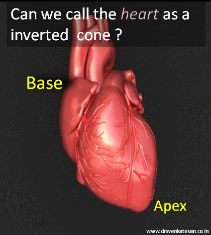
Now, traditionally the heart was thought to  be cone like stricture with  apex located near the left mid clavicular line .

base of heart apex  waht is the shape of the heart

If there is  an apex there must be a base  .So ,  we  reasoned the highest and farthest part of the  heart  must be  the base .  Paradoxically , this base is at the top ,  and  not in the  traditional sense  of bottom .

Like wise ,  during auscultation  the  events  in the base represent the great vessel  and semilunar activity  (Anteriorly )

Baseless  base

Clinical base is not the anatomical base . When a patient lies  on his back  essentially  the atria and posterior aspect of ventricles form the  base . In  erect posture the  diaphragmatic surface becomes  the true anatomical  base  of the heart .

Further confusion during  Echocardiography

It is well known , basal LV function is a critical determinant of LV function .But we should be very clear  what area we are talking about ! Basal  aspect   can  lie   either  superior , inferior , anterior  even  posterior surface  of left ventricle .(It took me years to realise this simple fact !)

Similarly   basal septum  can mean either a LAD zone or even RCA zone one has to specify basal  anterior septum to define LAD zone ischemia .

(Please note , this is contrast to clinical cardiology where  the  base of the heart refers only to anterior aspect of heart ! )

FInal message

Calling  a  particular portion of the heart as base  would be an  anatomical misnomer . This  nomenclature is based ,  more  by  tradition and  our faulty  perception .(As visualized   by anatomists and pathologists )

We have come a long way  from the Da Vinci  days. Modern cardiologists  with  sophisticated imaging techniques would like to  call  the basement of heart  differently  in the future and correct the nomenclature issues.

Read Full Post »

In one of  our patients  who had a recent STEMI , CAG revealed  70% LAD  lesion  with   TIMI 1  flow .The distal run off was slow .He had moderate LV dysfunction with no major symptoms. The angiogram was done routinely .( Yes . . . routine CAG- the term I do not relish ,  while it is becoming a way of  life for all learned cardiologists !)

I  was discussing   this case with my fellows and  about medical management of CAD. I  told them  Aspirin will help prevent ACS, statins will stabilise the plaques , beta blocker would prevent cardiac  events by blunting adrenergic surges and Nitrates is a powerful coronary vasodilator that  will improve the  coronary blood flow

A final year MD fellow  threw a  direct  question at me .

Sir,  do you mean  Nitrates  would increase the  TIMI  flow from 1 to 3  in the long term in this patient ?

I was taken-aback  for a moment . . .   and thanked my student for a valid question .

Nitroglycerine is  a powerful coronary vasodilator  we are taught for nearly  half a century . Oral nitrates are used  liberally in the chronic management of  angina. It is a multi billion  dollar market.

Has it been documented to improve coronary blood flow in the long term ?

No

Why then it is  used long term ?

It is a clear case  of  inappropriate medical therapeutics .

* The confusion is partly due to our mix up the mechanism of  relief of angina from coronary vasodilataion. Realistically , NTG should me known  more as a powerful venodilator reducing the preload . It  dramatically reduces the LV filing pressure  and relieves sub-endocardial stress  .This is the major determinant for angina relief .(Of course after-load reduction also helps)

Final message

Nitrates  should be used only for relief of an episode of angina or just to prevent It .This may surprise you  , Nitrates has no documented  efficacy  in the long term management  of angina.

Read Full Post »

Both  entities   refer  to contraction of vascular smooth muscle.Vascular smooth muscle tone  is under the control of autonomic neuro and humoral system.

Local mechanical , stretch  receptors also  play a role .( local temperature -Cold vasoconstriction  , warmth relieves it ) Abnormal elevated  vascular  tone occur in pathological situations ( Vascular interventions, surgery , drugs, extreme cold ,   )

Both terms are closely related often used interchangeably .There are some subtle differences though , mainly in the usage pattern.

Vasospasm

Is generally a   regional   phenomenon implying central mechanisms  are less  operative.

A local  trigger mechanical or biochemical .( Even a mechanical drag can cause a spasm

It is often transient .

Occurs primarily in small , medium blood vessels .(I  wonder whether  Aorta can go for  spasm !)

Clinical examples

  • Catheter induced vasomotor in cath lab (Radial spasm)
  • Spasm associated with plaque  Prinzmetal angina
  • Cerebral vessel spasm after sub arachnoid hemorrhage ( Need not be transient )

Vasoconstriction

This is also result in  constriction of blood vessels

But the blood vessels involved  are in  micro circulation like capillaries and arterioles .

Often sustained (If transient does it become vasospasm ?)

Vasoconstriction  often involves wide areas and may result in elevated regional or systemic vascular resistance . Hence  it often  result in elevated  blood pressure.

Clinical example

  • Normal Blod pressure  regulation y autonomic mediated vasoconstriction
  • Arteriolar  constriction with Nor adrenaline , dopamine .
  • Capillary Vasoconstriction  due to cold (Raynaud phenomenon )

* Please note other terms like vascular tone , vasomotion   closely competes with above .

Final message

Most of the confusion in medical terminologies are man-made.Both vasospasm and vasoconstriction are  often used  interchangeably .It  is ideal to use  the former for a transient , localised response  in large caliber vessel , and the latter when it occurs as  a general response to altered autonomic  tone and  involve the micro vasculature.

Read Full Post »

This  is the story of a 55 year old  women ,  who was received  in our CCU  with a  dramatic STEMI (ECG looked like an action potential ) ,  LV  S 3  and  hypotension.    It was impending cardiogenic shock.Since we do not have full fledged primary PCI  program  , thrombolysis was planned. She had  cardiac arrest   immediately after  starting streptokinase infusion . She  was  promptly shocked  and  revived .  The ECG changes rapidly  reversed(ECG -3) . Every other  hemodynamic parameter got stabilised as well . To our surprise   ( few hours later ) this patient  was  so comfortable , sat up on her bed ,  demanded a discharge . (Which was refused of course !)  One week  later coronary angiogram was done, a near complete recannalisation of RCA was documented.

ECG 1 on arrival
Inferior MI 2  

ECG -2 Developed cardiac arrest  10 minutes  after  starting the Streptokinase Infusion

primary VF 2

ECG -3 .Taken few minutes following   the VF

inferior MI evolved 2


Acute myocardial infarction (STEMI)  kills more than a million life every year . Majority of death  happens within an hour of onset of symptoms. Ventricular fibrillation  is the arrhythmia of death. Why this occurs  only in  few , while  many are  immune to it ?

God keeps  this secret  close to his chest ,  how and why  he selects   candidates for this arrhythmia !

Scientists are still  far away  in finding the truth . But , one thing  is obvious .The  moment   coronary artery is totally occluded  , the heart begins a fight  and try  to  get rid of this obstruction . In the process ,  it  goes into convulsion (VF)  with a foolish belief  , it  can shrug of the thrombotic insult . Death often   ensues if  not intervened . (Very rarely  VF can be a non sustained one  and patient survives cardiac arrest !)

VF  as  a electrical  response  to  reperfusion injury .

Often times ,  we witness patients  to  go  for  VF  very early following thrombolysis . The  thrombus in situ is an irritant , it  triggers the inherent fibrinolytic system (Natural TPA included) If it is successful  it opens the occlusion ( atleast partially )  and salvages the myocardium .If the fate is against  the patient , very early reperfusion of IRA triggers  VF  .  If this occurs at home   survival  is  low .If  the VF occur at hospital the probability of survival is near 100 % .

               The  intensity of  natural lytic mechanism  is the major determinant  of   early reperfusion . Ironically  the same  factor   determines  occurrence of the deadly  VF .

I would believe  , the STEMI patients  who die early (even before reaching  the hospital ) are (un) blessed with a  fighting  heart  ! Ironically , the lazy hearts  reach the hospital  alive ! (slow &  steady win the race !) .  Of course , reperfusion  injury is not the only mechanism of VF . Other common suspect is  left main STEMI .

Link to related video “Ignorance based  cardiology ”

https://www.youtube.com/watch?v=J9DH6Vr04es

Final message

While , VF  is  referred  to as arrhythmia  of death , it may  in-fact , represent  a common form  of  reperfusion arrhythmia in  the setting of  STEMI !  .  . .  Hence , it can  Initiate  a new lease of life in  many   lucky ones !  I hope the title of this article  makes sense  !

Read Full Post »

  1. The concept of  FFR is based on pressure gradient  alone.In any hydraulic model (Both biological and non biological systems ) pressure difference  is the least   important parameter  that determines flow.
  2. FFR  is unphysiological  as hyperemia   is  artificially induced one .(Adenosine  is not the only parameter that determines it !)
  3. Serial obstructions and branch point hemodynamics are  conveniently ignored.
  4. Reproducibility  remains a big question mark .
  5. On safety  issues  FFR  is a suspect.( Often times , it  requires expertise comparable to  that of a  complex  PCI !) .Beware , the FFR unit has stiff catheter system and is an additional health hazard .  I have witnessed   atleast two cases  where  insignificant lesions were  made significant by  FFR related Injury .
  6. And  now the  knock out punch ,  ! Probably the most vital  issue for which FFR should be banished * , it is not taking into account of vulnerabilty of a plaque .( An FFR > .9 with a hanging , eccentric , mid LAD lesion was left alone by one of the  academically up to date ,  evidence  based interventional cardiologist!  )
           (*If perfomed  in isolation without IVUS/OCT  )
I am still wondering how this concept came into cardiologist domain and into the cath lab .It should have  never been let out of theoretical physics labs !
Final message
The best way to assess physiological significance of an anatomical obstruction is  to  do  exercise  stress test .
If  the lesion is  able to sustain good exercise capacity , it  can be deemed physiological unimportant.
While , this is an explicit  proof  in single vessel disease  ,  even  in   multivessel  CAD ,   EST  is   a  collective  measure of  coronary  reserve flow .( Something like instantaneous equivalent of virtual  multivessel  FFR  )
Moderated After thought
FFR is a highly specialized theoretical  tool , that has very limited role in cath lab .
The two major practical (Non academic)  use of FFR   is to shun away  those   internet fed ,  annoying, pseudo  intellectual patients ,  who constantly ask for  angioplasty  for  obliviously insignificant lesions !
FFR comes very handy  to  bail out  cardiologists at  times of distress   ! (To escape  from the wrath of our patients   after a sub optimal &  technically inferior   PCIs   and   in  the  long term confabulations  in   restenosis  after stenting !  )

//

Read Full Post »

Thrombus laden plaque  is sine qua-non of UA/NSTEMI . That’s what we  have been taught  !  right ?  It may be  true in many  situations , but please remember there is another concept  called  demand ischemia , where in there is  no active thrombus ,  still resting  angina may occur due to  increasing heart rate etc.

I just wanted to test how far this concept is understood ,  by the  fellows in our coronary care unit . Following  is  story of a patient who arrived at CCU with  angina  at rest .  I showed  this   ECG asked them the  management .

positive est and unstable angina

History was  purposefully blinded . 5/6 cardiologists wanted to admit the patient either in CCU or rush to cath lab.  Heparin/ Fondaparuinux was prescribed by all. Tirofiabn was suggested by few.It is a  high risk UA with left main disease some one  mumbled .

I silently listened to them and  revealed the history . This patient  has just finished the  exercise stress test , it was terminated as he had angina at peak exercise. and was  reported as  positive . A date was fixed for elective   coronary angiogram. 10 minutes later ECG totally normalised  , and the patient went home (Boarding a crowded Chennai  city bus )

The fellows realised the importance of history . In fact no body asked for it ?  I felt  bad  as  all my fellows failed in this test That reflects bad teaching on my part !

What is the mechanism of ST depression here ?

  • Fresh thrombus ?
  • Mechanical occlusion ?
  • High  heart rate ?
  • Combination of high rate and probable flow limiting lesion .

(Severe forms of  stable angina can occur at rest . So do not equate all rest angina as true  unstable angina !)

Final message

Do not label an ECG straightaway  as acute coronary syndrome when there  is  baseline  tachycardia and ST depression . Spare few minutes and apply your mind !

If  a combination of ST depression  and angina  can be taken  synonyms with UA  every EST positive fellow should be labeled as UA and admitted in CCU. Please remember any tachycardia with a fixed tight lesion will  mimic UA . Further ,  since there is no thrombus here  and there is absolutely  no role for heparin.

Read Full Post »

ST segment elevation is the key parameter on which the fate of millions of infarct patients are recognised and managed. It is ironical we do not have standardised reference point for measuring the quantum of SR elevation .

This is especially difficult when ST segment blends with forward limb of T waves.

While we have reference point for measuring  ST depression  (Like during EST ). . . why we do not have one for ST elevation ?

Now we have adopted a rough criteria .Read below .

where will you measure st segment

How to measure ST elevation in ECG
How to measure ?
Measure The ST segment 40 ms from J point.
how to measure ST  elevation in STEMI 2
                        I lost track the source of this Image .(STEMI hand book 2012 ?)
Final message
ST segment  elevation  is  the key parameter in ACS.  Quantifying  it becomes  important in  assessing  the efficacy of reperfusion strategies and risk stratification. Fresh ST elevation can represent pericarditis, reinfarction or an early dyskinetic  segment . Unless we have  proper reference point there is  a room  for  error in this simple parameter.

Read Full Post »

This is an  ECG which  I reported  yesterday in my clinic . I thought it was a  near perfect example for sinus node premature beat .

sinus premature beat spb 2

(Of course I need to explain  why the  P morphology  slightly  differs )

A  sudden unexpected  QRS  complex is often called as  ectopic beat . If it occurs prematurely (ie earlier than anticipated )  it is called as premature beat. If it occurs late it is refereed  to as escape beat .Please note the difference is not absolute .

Sinus node is a dramatic bundle of energy with divine powers that  drives rhythm of life !

The pacemaker cells are arranged in a compact fashion with  differential properties from cranial cells firing fast and caudal cells little slower. The neural control is under constant Neuro/electro/humoral  servo control mechanism.It is well known the pacemaker shifts it’s firing location within the SA node in fairly regular fashion .The entire SA node has rich adrenergic and  cholinergic  innervation , with  a dominant control by the later . (This is  why the intrinsic heart rate is  in the tachycardia  range (around 116 )  when SA node is denerved  pharmacologically )

wandering-pacemaker

sinus premature systole spd sinus node ectopics002

SA node ,  being  a complex structure ,  it is not surprising to note  few beats to fire  slightly late  or  prematurely.If it occurs late it is called sinus pause ,  if it occurs early it is sinus premature beat , if  both occurs  interchangeably  we refer it as  sinus arhhytmia. (Read  about sinus pause here)

What is the clinical significance  of   SPD ? (Sinus premature depolarisation )

It is a  very benign entity that it is  merely an  academic fascination . By  stretching my  imagination  I  can  correlate  it  with few possible  clinical issues.

  • May be it has potenital to trigger a  SA nodal reentry tachycardia  or In appropriate sinus tachycardia/bradycardia.
  • It may be imporatnt in sinus node modification process.
  • However ,the main issue is  thee  cardiac physicians  in their enthusiasm should not mistake it for some serious  cardiac arrhythmia !

Related article

https://drsvenkatesan.wordpress.com/2009/04/14/can-premature-ectopic-beats-occur-in-sa-node/

Read Full Post »

The link between brain and the  hand  starts right from fetus .  It is a well known fact  vertebral artery   competes with hand blood flow  . In the right side , there  is one more  vascular issue !  .Bracho cephalic  artery  arises  directly  from aorta and supplies the  right  hand and  right half of brain.

It remains a mystery  why left brain  is   blessed with a  separate  origin ,  while right has to share it with blood meant for hand  .It is beyond science  . . . isn’t

It is possible the left hemisphere  of brain   has more   purpose   to be alive  ,  with bulk of the cognition work to do . Hence   God created a  separate  supply to it !  Of course , he  would   have never  thought ,  the  possibility of  his ” mean” creations   adventuring  within the   arterial tree  !

Click over the Image for animation

right radial artery coronary angiogram  pci  risk of stroke 002

Please remember  whenever  we   play with   catheters and wires  through   radial route , we  are  hugging  and scraping   the artery meant  for cerebral circulation !

Final message

Femoral Interventions  enjoys a proven  track record. Currently ,  radial route has virtually taken over with  few  advantages . However , the  overall stroke risk in the two approaches  remain  low but genuine (.4 %) .It may be true , arch manipulation is more  with  femoral but  the threat to  vertebral and brachiocephalic circulation  is more with radial .  When the available evidence are  not conclusive  and  new ones are not forth coming  . . . it is wiser to rely on common sense !

Reference

I think  this 2011  study  from the  prestigious stroke journal  has convincingly answered the issue

cholesterol and ateromatous emboli following coroanry intervention 2

cholesterol and ateromatous emboli following coroanry intervention  radial vs  femoral 2

It concludes , the right radial approach  is indeed risky  to develop cerebral  micro embolism   when compared to right femoral

A Review article in  Circulation

cholesterol and ateromatous emboli following coroanry intervention 2  radial vs  femoral 2

Other references

1.http://stroke.ahajournals.org/content/38/7/2176.full.pdf+html

2.Transient Cortical Blindness after Coronary Angiography Journal of International Medical Research. 2009;37:12461251,

3. Stroke and Cardiac Catheterization Circulation. 2008;118:678683,

Read Full Post »

In HCM every myocyte is  genetically made  defective . Myofibrils are in disarray every where . Still , can we identify some vulnerable zones that acts as arrhythmic  focus ? If that is possible , we  have a opportunity to abate that focus .

In HOCM  , which is the most stressed area ? LVOT ?  Septum, ? When we say stress , it can mean either mechanical or electrical .

VENTRICULAR TACHYCARDIA 002

Does electrical instability involve the same zone as mechanical stress ?

How often VT originate from LVOT in HCM ?  For this we have good clinical model _, the patients who underwent alcohol septal ablation.

What happens to the incidence of VT  post septal  ablation  ?

“It is reported  post septal ablation the incidence of SCD  becomes  equal to general population” (Read the paper below )

If that is true , it is obvious the  arrhythmic  focus is also ablated along with LVOT myocardium .

Outcome of HOCM after alcohol septal ablation

Though many studies claim  so !  It  fails to convince us  .  HOCM is a diffuse disease of  myocardium.  Even a cluster of myocyte disarray  with fibrosis   can be a future focus  irrespective of it’s location .

However ,  it is always possible relieving the mechanical stress of the LV can definitely reduce the likelihood of an electrical event .(Even if the arrhythmic focus is intact elsewhere !)

* We know RVOT is  developmentally arrhythmia prone zone . We also know HCM involves RVOT (After all ,  IVS  is legally shared by both ventricles !  ) . Some of  the monomorphic  VTs with LBBB morphology may originate from RVOT in HCM .

Management of recurrent VT in HOCM

  • Drugs (Amiodarone/ Calclum blockers/ Beta blockers/Disopyramide)
  • ICD- (Probably mainstay  )
  • Very rarely ablation (If localised focus is well documented )

Reference

1.A case report for successful ablation of  VT in HOCM   http://www.ncbi.nlm.nih.gov/pubmed/9255687

2.http://www.ncbi.nlm.nih.gov/pubmed/23076968

//

Read Full Post »

« Newer Posts - Older Posts »