If science is considered as a journey towards truth,.. knowledge, data, and statistics are the key companions in this infinite voyage to an unknown destination. While hundreds & thousands of scientists do travel in this turbulent road daily, pursuing their mundane work, there are very few researchers worried about the true purpose of their journey, the quality of the road they travel, the dangerous fault lines they create.
It has become a taboo topic to criticize medical science even after realizing the fact that we are compelled to follow and glorify some of the best nonsense.
Dr. Jhon Loannidhis Professor of statistics and public health from Stanford University is of a different genre. He became so popular after his landmark paper
RT-PCR: Real-time polymerase chain reaction, a sophisticated gene sequence-based biochemical test. Thanks to corona, this complex medical investigation has become a household name.
Jones proposed his criteria to diagnose acute rheumatic fever in 1944, we still use it to diagnose with many modifications . Currently, AHA position statement – 2015 by Gewitz et all is being followed. (Circulation 2015)
From Braunwald textbook of cardiology. Apart from this, there is one catch .Even if the child fulfills Jone criteria, there needs to be evidence for preceding streptococcal sore throat, either by culture or antibody. Now, can we include an RT-PCR as a new parameter to diagnose streptococcal infection is the question?
Why we Insist on evidence for preceding GAS?
It is for a the simple reason, many entities other than rheumatic fever may fulfill Jones’s criteria. (Still disease, HS purpura OR even simple viral arthritis etc) Some may even call it an essential criterion in the past. Practically it is not done is a different story.
Tests for preceding streptococcal sore throat are ASO titer and anti-DNAase B. How about the now glamorous RT-pCR for streptococcus ? Though it was suggested as a useful test in the past ,the cost and logistics were prohibitive so it was never considered to be included in the Jones scheme of things.
There could be three roles for RT-PCR testingin Rheumatic fever /RHD
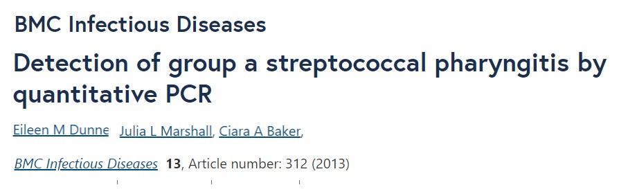
1.RT-PCR as evidence for recent streptococcal sore throat.(GAS organism) Which still not practically used often but has big scope.(Ref 1)
2.To rule out co-viral (influenza-like) infections as a cause for fever and Joint pain (Used in population-based screening in a high endemic area (Ref 2)
3.There could be one more indication (experimental though) Micro-RNA detection by RT-PCR to identify children who are prone for progression to RHD. (Ref 3)
Final message
Now, we must introspect. While billions of dollars are going down the drain on RT-PCR for diagnosing a common cold pandemic which has no specific treatment. Will WHO and other cardiovascular preventive authorities consider to include RT-PCR as screening test for GAS in children. This will enable early start of primary prophylaxis and prevent RF/RHD a century-long scourge of the third world.
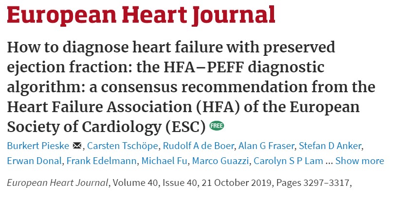
Heart failure has been classified in many ways, with prevailing levels of our knowledge and ignorance. It is based on a variety of factors like rapidity of onset, etiology, chambers involved, hemodynamics, etc.
Forward vs backward failure
Acute vs chronic failure
RV/LV or Biventicular failure
Systolic vs diastolic heart failure
High output vs low out failure
Ischemic vs non-ischemic failure
Reversible vs Refractory HF
None of them have really helped at the bedside though it helped us understand the condition. Now, in the last decade, we have crash-landed on our favorite obsession to classify HF ie based on Ejection fraction. We believe we have found an exciting new classification. (HFrEF/HFpEF/HFmrEF).We embraced it, even after recognizing EF as a battered LV functional parameter due to its high load-dependence with a dubious reproducibility.
If we rely too much on echo, there can be a few more classifications for HF
HF failure with preserved diastolic function(25% of all DCMs with HFrEF )
HF with preserved mitral valve function
HF with preserved Global longitudinal strain(Still normal EF%)
HF with preserved RV function
HF with preserved Torsion and Twist.
Finally, HF with normal Heart (Anemia/CKD etc) In anemia heart never fails in true sense. In fact, it works at peak capacity.(More of a Success than failure). Similarly isn’t odd to put primary CKD/CRF in the CHF basket.
Probably the most important and practical classification could be
Primary vs secondary HF (Primary means all muscle diseases under MOGES system )
Valvular vs non-valvular failure (Surgically correctable MVR/DVR/Mitral valve repair)
Revascularisable or Non-revascularisable HF (STICH study responders)
ICD/CRT eligible HF vs Non-eligible HF ( Rule out DANISH study non-responders)
Dr Thomas Lewis said over 100 years ago, the essence of the practice of cardiology is to recognize HF early. Looking back at the literature, there will be no dearth of classification for HF. It will come and go according to academic and Imaging whims. Of course, that may aid in ruling out primary cardiac conditions. But, we must always emphasize to the next-generation that HF is often due to systemic*(reversible too) conditions in substantial numbers. Here the heart is just a bystander watching helplessly, trying to adapt to a remote systemic comorbid problem. Such hearts don’t require cowboy aggression but gentle care by concerned physicians.(One study reveals weight reduction and systematic exercise program adds more life to HF than drugs and devices. Will link the reference/ or try google)
*Eg: Anemia is the commonest cause of HFpEF on a global scale. .CKD, undiagnosed autoimmune disorders, malignancy, are other classical examples. Let us be first a physician then a cardiologist, that will ensure our we don’t miss important treatable conditions with our short-sighted definition of heart failure based on EF%.
Reference
1.Y. Juilliere, J.N. Trochu, P. de Groote, et al.Heart failure with preserved systolic function: a diagnostic algorithm for a pragmatic definition Arch Mal Coeur Vaiss, 99 (2006), pp. 279-286 View Record in ScopusGoogle Scholar
History is rarely kind to the original heroes in the scientific world.The classical Blalock-Taussig shunt,(BT shunt) the term we heard for the first time in the early clinical years of MBBS .We know, it as a dramatic surgery (Palliative though) connecting subclavian artery to the pulmonary artery for the commonest congenital cyanotic heart disease -Tetralogy of Fallot.
Now, half a century later, came to know, there is a gripping story of an oppressed black hero behind this famous cardiac surgery. This post is all about the fascinating life of Vivien Thomas, a humble carpenter’s son from Nashville. While he dreamed to become a doctor, circumstances and fate had some thing different to offer .He could join only as helper in the wards of John Hopkins, Baltimore . His extraordinary hand skills were recognised by then surgeon Alfred Blalock and made him as an assistant in the Hopkins animal lab.He was working on a project to resuscitate traumatic shock victims then. Dr Helen Taussig who was a pediatric cardiologist was wondering whether Dr Blalock could offer some surgical cure for the sick blue babies under her care.
When Dr Blalock was brainstorming the problem , it was Thomas ,who created dog models of hypoxic circulation and helped create the concept and methodology of diverting blood from subclavian artery to pulmonary artery .He single handedly operated on nearly 200 dogs. He literally taught the chief surgeon Blalock the delicate vascular suture tricks .
Come October 24th 1944 , the first blue baby was operated , with Blalock Insisting Thomas to stand beside. History was created -first heart surgery in USA. Which later on became the most famous concept that gave a fresh lease of life to thousands of children with TOF.
It’s painfully emotional to watch the Vivien Thomas standing right behind Dr Blalock,guiding his boss anxiously,with his hands tied just because he is not a qualified doctor. The others in the team included Dr Denton Cooley and Helen Taussig.
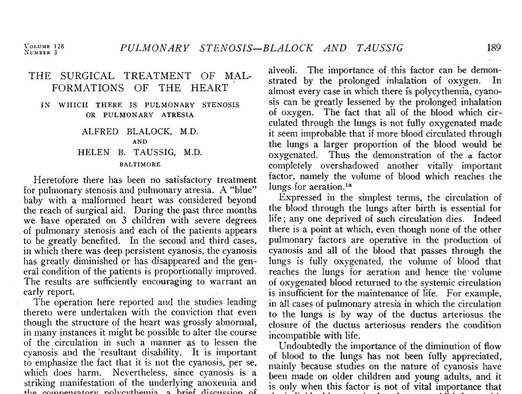
No surprise, when this famous work was reported in the media, the entire cardiology community rejoiced as the news broke out over the globe .It was published in JAMA in 1945 (Blalock1945.pdf ) . Did you guess it , yes, the name Thomas was not to be found anywhere though. How can you expect it ? , after all , he is a black lab supervisor working with dogs !
Thomas’ , work was never recognized for the next 30 years until a grand occasion (Lord made?) that happened in the Baltimore in 1971. His dream of becoming doctor became a moment of truth. Baltimore school,of medicine finally recognised his work and conferred a honorary doctor . Unfortunately Dr Blalock was no more by then to attend to his famous pupil. Its 2020 , 80 years after the monumental surgery , the BT shunt has since been renamed as Blalock, Thomas ,Taussig shunt . A new exclusivecenter for congenital heart surgery in Baltimore has come up in their name. What a great end to this black man’s journey in troubled racial times.
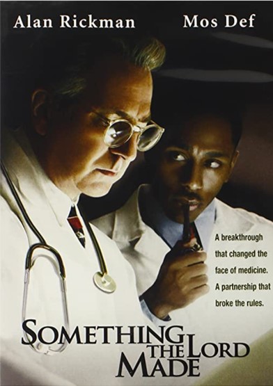
Thanks to Hollywood minds who thought this story deserved to be made as movie. “Something Lord made” directed by Joseph Sargent. It was a gripping scientific roller coaster .No surprise it got so many awards including three Emmys.
Every physician,especially the cardiologists should watch this movie. I can vouch, the one and a half hours you are going to spend will enrich professionally and Intellectually. Lucky to find this movie free on you tube.
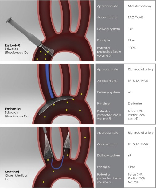
The incidence of stroke during TAVI up to 5 % (minimum ). Stroke risk reduction during TAVI is a critical requirement that can be a deterrent against this wonderful Intervention.
Many devices are being considered
EmbolX (Edwards life science)
Emrella
Sentinel (Claret medical)
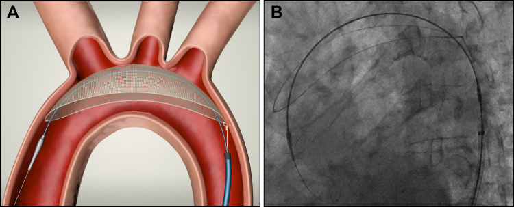
TriGaurd (Keystone)
TriGaurd 3just got the approval from CE and appear promising. (REFLECT trial) It is inserted through the transfemoral route , deflects embolic material to descending aorta since it covers all the three branches of Arch.What happens to these deflected particles? Any bodys guess.
So , in my understanding it converts potential brain embolisation to peripheral microemboli , wh
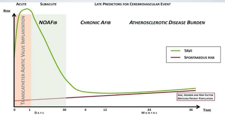
While, these innovative aortic arch filters reduce the risk of periprocedural embolic stroke, please mind, TAVR patients continue to be at significant risk for stroke over a long period. This is due to other late causes like TAVR leaflet thrombosis, atrial fibrillation, arch atheromas, and bleeding due antiplatelet agents.
Can you guess how many drugs a busy physician on an average writes in their prescription in his office ?
Three ? , Five , Six , . . . . Nine,? There is no specific study available for this non-academic query. I have got stunned to see a maximum of 18 drugs in one prescription. So, it should be anything between 1 to 18. May be a mean around 6 or so (Make your own guesstimate)
There is strong evidence to suggest writing a drug prescription has become a (un)conditioned habit-forming act. My professor* used to say generally 2 to 3 drugs are sufficient for most of the common illnesses we encounter (Only in extraordinary situation one may need to go beyond this )
One evidence less estimate though a random observation among the physicians suggested the bottom half of any long list of drug prescription is redundant and it doesn’t really address the specific problem the patient is suffering. Meanwhile ,the concept of poly-pill is making drug compliance easier in many cardiovascular and diabetic diseases.
*William Osler
Final message
Number of drugs human body can handle simultaneously without any harm is often an ignored chapter in the Principles of clinical pharmacology and therapeutics.
Let us mind the length of our prescriptions and ensure less harm to our beloved patients.
Related material
This was my old presentation made about polypharmacy in CHF :Perils and pearls
Its almost like playing a billiards game in absolute blindness.
It is not an unusual scenerio, to see the balloon catheter delicately bending at IAS puncture site , dodging and deflecting with random jerks as it tries to steal a entry in a few diastolic milliseconds time window when the fish mouthed mitral valve opens in sub square cm areas of MVO trembling in fast atrial fibrillation.
Agree ?
Gathered some tips to cross a difficult mitral valve during PTMC.
This is a PPT presentation taken from archive (Made in 2012)
Often times Its noted we tend to struggle more at the mitral valve crossing than at IAS puncture during PTMC. Experience prevails over Image assistance. Assessment of LA size , IAS plane , and sub valvular disease seem to be critical. Probably the secret of success which I found out was , smart guys never hesitate to repeat IAS puncture site for optimal trajectory .Over the wire technique is not forbidden.
Unfortunately, TTE guidance is of little use to cross the mitral valve. Co-registration of fluro/3D TEE is promising , but most cardiologist continue to rely on their experience.
I don’t know whether you have seen this before. Surely , I haven’t seen a presentation such as this one.
Place: Annual scientific meet ASE 2013. Minneapolis
Presentor:Dr.Partho Sengupta, Mount Sinai hospital, New york.
Its a 3D presentation in “space” without a screen by Holography.
The stunning 15 minutes lecture take us into the myocardial architecture, with speckles , flow vortex echocardiography and fluid kinetic energy mapping.
Don’t miss, a dramatic live teleporting of ASE president on to the stage.
Can you Imagine , where does this technology take us to the future ?
Patients may reach doctor’s offices by holographic teleport for a medical examination or vice versa. Yes, it’s all going to happen someday.
Coronary collateral circulation is one of the major determinants of symptoms and outcomes in chronic CAD. But, we generally shrug off the value of coronary collateral circulation in acute coronary syndrome. The fact is, it has a myocardial mitigating effect following sudden total occlusion.
When does it appear? We did a small analysis (PDF version)
We found it is noted in 25% of patients. With reference time of appearance, 6% had it within 12hrs and in few, it was noted as early as 6 hrs. One caveat is, we may not know whether its preexisting collateral due to chronic multivessel CAD. I am sorry to note this study did not address the outcome analysis. We however documented patients with good collaterals had negligible wall motion defect and near-normal function post PCI. Some of you can pursue research in this area.
Potential role of collaterals in ACS
It limits the infarct size
Keep the myocardium alive and give us time to intervene
Can converts a potential Q-MI to non-Q MI
Possibly prevent primary VT/VF and hence dreaded sudden death in early STEMI
Prevent early adverse remodeling of the left ventricle.
When these points appeared just my assumptions, Dr. Ali Aldujeli, (Lithuanian University of Health Sciences, Kaunas) in his presentation, at TCT 2020 confirms many of them are Indeed true
Final message
I agree, in the era of instant gratification with primary PCI, relying on coronary collaterals may appear a lesser professional virtue. Still, we may need to respect nature. Many times it bails us out.
The contents of the this blog is being published as Kindle E book , as per the request of many of the readers. Every article will continue to be open source in this site. Again I shall reiterate the book format is not aimed at any commercial intent. It is only to facilitate learning in a single book format Here is the link to book https://amzn.in/d/euhL5vu
Click below to see who is watching this website live !
This site will never aim for profit. Still ,this donation link is added at the request of few visitors who wanted to contribute and of-course that will help make it sustainable .
Please Note
The author acknowledges all the queries posted by the readers and wishes to answer them .Due to logistic reasons only few could be responded. Inconvenience caused is regretted.