Feeds:
Posts
Comments

Right ventricular function assessment has always been difficult in view of it complex shape and limited imaging planes in echocardiography .

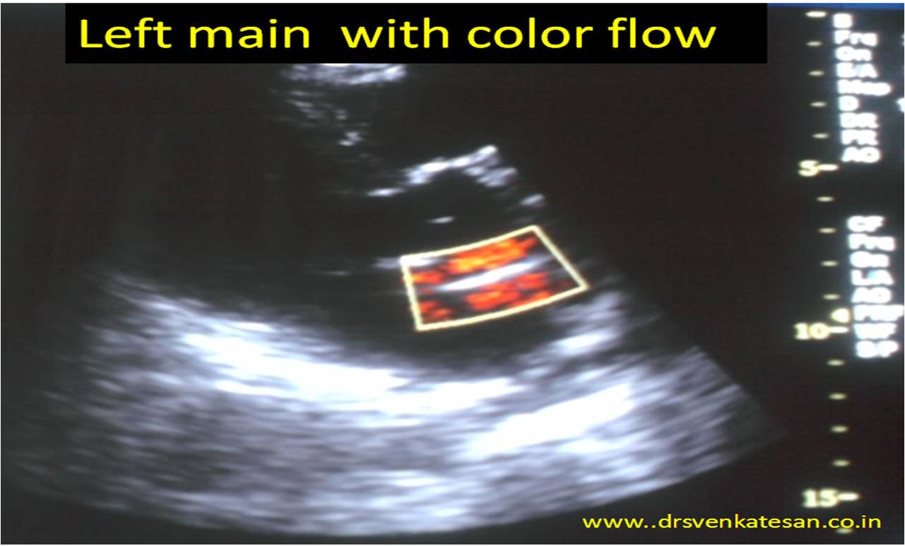
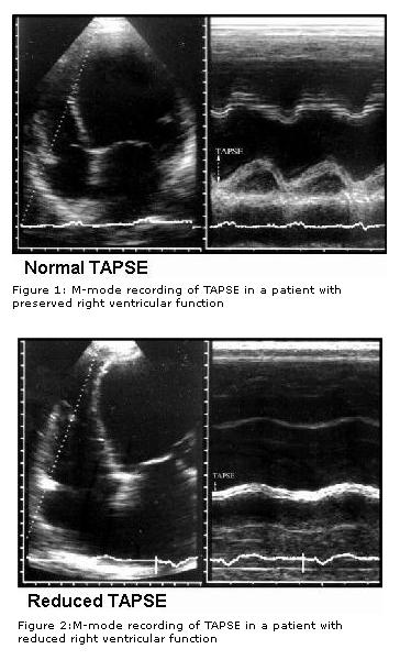
Recently , we learnt tricuspid annular motion can give  a quick assessment of  RV function . This was accomplished by M-mode echo of tricuspid annulus. (TAPSE)  tricuspid annular plane systolic excursion . This simple parameter has  brought the  maligned M mode echocardiography into limelight again.

tapsee rca motion by coroanry angiogram rv systolic function tricuspid annulus motion right av groove atrio ventricular grrove mapsee m mode echo

Currently coronary angiogram is done just like a non invasive echocardiogram across the nooks and corners  of any country

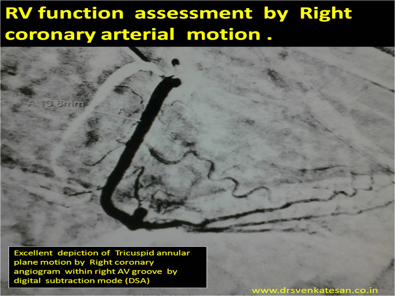
Modern day  cardiologist is expected to  look  beyond the coronary artery  narrowing  when reading coronary angiogram.  If only we give little importance to how the coronary artery moves with  reference to  cardiac cycle we can  get  excellent information about  mechanical properties of heart.

Every cath-lab  work station has a DSA mode . With this one can measure the coronary artery swing and document   it  objectively  .Right coronary artery swing  faithfully reflect  RV longitudinal function . This motion is more accurate than the TAPSE by echo . We have found the normal excursion to be 15-20mm (Slightly  lower than TAPSE) . Similarly LCX motion give us an estimate of longitudinal LV function and LAD motion can tell us how IVS moves .

Final message

Coronary artery swing*  is a   new  method (  rapid  and accurate) to assess  cardiac  function  in cath lab !   We should utilise this more often .I feel it may throw more valuable  than the sophisticated but complex 3D reconstructed and post proceed  imaging modalities  to assess individual  chamber function .

* There is no published reference available for  modality  .It is so simple  concept  i think ,  it does not require any major experiments for a  proof !

Reference

RV  function assessment ASE  guidelines

http://www.echobasics.de/rv-en.html

Normal RV function Indices .

TAPSE (tricuspid annular plane systolic excursion) < 2 cm

TASV (tricuspid annular systolic velocity)< 15 cm/s

Tei-Index (myocardial performance index)> 0,50

TAPSE  can be correlated with coronary swing

Further research potential 

Now we   require  comparing  the  TAPSE   with the quantum of RCA swing by angiography.  I have asked my fellows to look into this  aspect . I guess TAPSE by  Echo over-estimates the true  motion ( normal 2 cm )   seems on higher side. It includes translational motion of echo which is eliminated in angiographic annular movement .

Right ventricular infarction (RVMI ) is a  common  cardiac emergency in coronary care units. It can be termed as a mechanical complication of infero-posterior STEMI .However ,  around 10 % of anterior  MI do develop this complication . Onset of refractory hypo-tension in spite of correcting hypovolemia  suggests  RVMI.RVMI generally comes under class 3  (Cidar Siani /Diamond -Forester classification of STEMI )  , ie   silent lung with systemic hypotension.       (RV shock requires an unique definition , as it can not be included in traditional  definition of cardiogenic shock as the PCWP is likely to be normal.

How to manage a full blown RVMI  who is not showing  signs of improvement ?

Following is an extract  from our coronary care unit experience

(do not ask for evidence for everything !)

  • Consider immediate angiogram  to know the  anatomy of the problem .Try opening the RCA which is most  likely to be  the culprit (Any associated critical  LCX /LAD lesion  must be attended too ! )If  the duration of MI is beyond 36 hours  culprit lesion may be  left untouched or at least not our primary target !
  • Inotropic support (Doubtamine continuous infusion is preferred .Milrinone  for the rich !)
  • There is no specific RV assist devices available.(LV  assist device has no role in RV )
  • Restrict fluid (Opposite  to RVMI guidelines) There have been instances of  overzealous fluid therapy resulting intra-cardiac  hypervolemia. IVS encroaching LV worsening the cardiac index .
  • Pacing is  definitely required in severe bradycardia or CHB .  Dual chamber pacing is the  ideal  choice to maintain AV synchrony as we desperately need  the  atrial booster pumb  for a failing RV . (Please realise , VVI  pacemakers ,  can still save lives as it takes care of extreme bradycardias  effectively )
  • PCWP in the setting  of RVMI is an  unreliable parameter of true cardiac function.(In almost 90 % of RVMi some degree of LVMI is present ) . In RVMI  PCWP is determined by a  delicate balance between LVEDP and the  onward stroke volume from a failing RV .) The alter tend to bring the PCWP down former would keep it high . Which component is  operating at a given point is a  wild guess  . The situation get quiet complex in the setting of multiple vaso-active drugs , pacemaker , ventilator
  • Balloon Atrial septostomy  /dilatation might help ( Hypoxia may worsen as elevated RA mean pressure may shunt right to left  however cardiac out put might improve)
  • Pericardiotomy  or simple splitting  of pericardial  layers has been tried   (Improves RV restriction effect)
  • If the patient is on ventilator keep the PEEP well below the standard recommendations (RV will struggle more ! )
  • Pacing catheters  can irritate the RVMI in their  raw zone and trigger recurrent ventricular arrhythmia .( Often  labelled wrongly  as Ischemic electrical storm !)
  • Call  Nephrologist  consult  if  renal function  deteriorates . Peritoneal  dialysis is preferred  .  It  is worthy to know , deaths have occurred on hemo dialysis  table.

Final message

RV shock  carries a dismal  outcome , almost  reaching  as that of an LV cardiogenic shock. Ironically ,the most important prognosticator  in RVMI  is the quantum of LV involvement !

If only  . . . we get  an  image like this , echo can help rule out most  left main disease with conviction .

Have a close look  at it ! One can get a good image of  coronary ostia in short axis view . But , here it is well visualized  in long axis .

left main

I tried to put color flow within left  main .

left  main color flow

What about pulsed  Doppler across left main ?

After all it needs 2mm sample volume and this left main was near 4.5mm . So keep trying !

In this complex world , simple innovations fail  . . . just because  they are simple !

Here is a new* PTCA  catheter which has a  two balloons  ,  the distal one dilates the lesion and the proximal one has a stent over it  . The stent is just deployed after the dilatation by the proximal balloon . The proximal balloon  not only help us prepare the lesion before stenting it also helps  in crossing difficult lesions  . Further   , it can be even used to post dilate the lesion . It can be a non compliant balloon  as well . It appears a  good  concept .

*Not really  new  I  believe  ,  Accuramed  owns the patent for  this twin balloon catheter over a decade now .(First twin balloon Gemini PTCA was used in 1988 )

gemini balloon catheter twin two ptca pci

I do not know why we haven’t adopted it yet ,  while many  dubious  innovations are making merry around the world !

GEMINI DOUBLE BALLON STENT ACCURAMED 2

GEMINI DOUBLE BALLON STENT ACCURAMED  3

The only downside could be ,   combinations of stent and balloon sizes are limited . But ,  it is not a major issue .The ability to  fine tune the stent apposition  moments  after the procedure , by h a simple pull back  is just amazing !

This catheter is  made by

A C C U R A
Medizintechnik GmbH
Max-Planck-Str. 33 61184 Karben Germany
Tel +49-6039-9201-0 Fax +49-6039-9201-22
E-Mail info@accura.info
http://www.accuramed.de

Final message

Two  balloons  over a  single catheter  is a  new development  .I wonder it can be  the standard of care in all PCIs . Hope somebody takes  this concept to the next level  for the benefit of our patients.

Link to the manufacture

http://www.accuramed.de/fileadmin/daten/en/Gemini_System.pdf

Surprises are hall-marks of medical science . The cardiologists do  get  it ,   in enough doses   from  echo  labs  on a regular basis !   . One such thing is  the total ECG-ECHO myocardial  territorial  mismatch following  a STEMI .  Human myocardial segments are divided by cardiologists  by 17 segments by echocardiogram . Long before  echo came into vogue ,  electro-cardiologists  divided the  heart electrically into three zones to  localise MI . (Anterior , inferior and  the  poorly defined entity  lateral walls* ) .Inferior and posterior  segments are  almost used interchangeably. So , when we have 17  echo  segments to be fit into these three electrical category !   were  bound to have  some overlap . The issues of fitting in septal segments is really complex as septum  is a three dimensionally engulfs all three electrical surface of the heart .

* By the way , anatomists  never agreed about existence of walls in heart.They simply said  , heart has smooth  surfaces that blends with one another.  We cardiologist have  built imaginary walls and struggling to come out it !

We will   try to answer the question that’s been asked here .  “Inferior MI”  by ECG   . . . “Anterior MI”  by  echocardiography . How common is that ?

Possible causes for this wrong call

Technical errors  in  acquiring echo  imaging plane  or  it’s interpretation is the commonest . Many  times  ,  obliquely obtained long axis view  wrongly and strongly  suggests  a septal  MI  instead of   inferior posterior MI. This is  because  in  apical 4  chamber view  bulk of   septum  (Basal and mid third )  lies   in the  infero-posterior region .

wall motion defect

Perhaps ,  misunderstanding this  septal  geography is  the  commonest cause for  erroneously  calling inferior MI as anterior  in echocardiography . (A simple clue is the presence of MR . (It  fixes the infarct in infero-posterior zone with 90% accuracy )

Rotation  and  posture of heart

Alignment of the septum to the rest of the chambers  can influence  , how three inferior leads is going to look  at the septum (There can be  considerable errors  -Electrical myopia ? as these leads are located distantly )  . The plane of the septum is such that  in horizontal hearts  septal electrical activity  will be directed infero posteriorly inscribing a q waves in inferior leads rather than anterior leads . One can expect such ECG /Echo discrepancy in the following subset as well

  • Post CABG patients (Any pericardiotomy will make the septal motion  erratic )
  • Obese persons
  • COPD

There are three  more  situations  ,  which   mystified me   with  definite  ECG/ECHO  mismatch

  1. LVH and STEMI  is always an engima . Counter clockwise rotation when accopany  LVH  that masks anterior MI  electrically . It  however inscribes a   q wave in inferior leads.
  2.  In dominant LCX lesions  ( with at-least  one  major OM    )  and  left main bifurcation  STEMIs  ,  combination of  anterior and inferior  wall motion defects are  quiet common . When a such  a  MI evolves ( with or without  revascularization )   regeneration of R wave can be  time shifted . Septal R wave may appear  much earlier and inferior R may follow or vice versa . .Further,  anterior MI  may  evolve as  Non q MI  making it  ECG blind ,   still  echo may pick up the WMA . So there can be important  ECG-ECHO mismatch in myocardial segmental geography .
  3. Further , WMA  need not  always be an  infarct  .Any new episode of ischemia  can result in WMA . Hence a patient  with inferior Q waves  in ECG may experience anterior wall motion defect meagerly  due to fresh episode of   ischemia (This we should not attribute  to  old anterior  MI. It is also possible intra-myocardial conduction delays can elicit remote wall motion defects.

Final message

By general rule  , ECG  correlates  well  with  ECHO  for localising myocardial segments   . At times ,  it  can  really be tricky , and we  get into above situation  in echo labs.

While ,  it is common to observe  ECGs  to mimic  inferior MI  at the first look  and  subsequently echo  revealing  anterior  infarct ,  the reverse is also very much possible .

The  mechanisms are varied and technical  issues are for more frequent than true clinical discrepancy .The issue has important management implications.

Of course ,  coronary angiogram will pin point the   anatomy , still  it also has  strong limitations in localizing myocardial segments (to which it supplies ) especially with multi-vessel  CAD and  collateral dependent circulation .

The WordPress.com stats helper monkeys prepared a 2012 annual report for this blog.

Here’s an excerpt:

About 55,000 tourists visit Liechtenstein every year. This blog was viewed about 530,000 times in 2012. If it were Liechtenstein, it would take about 10 years for that many people to see it. Your blog had more visits than a small country in Europe!

Click here to see the complete report.


* Obfuscation:  hiding of intended meaning in communication, making communication confusing, wilfully ambiguous, and harder to interpret

This world can not be a perfect place and  it is foolish to expect the same !   What is published in medical literature is at best , an abstract thinking  of an unfinished agenda . Still public think  science is   . . . what doctors say ! They feel doctors can not simply watch a person  dying. They want us  act like  God. This is  how medical men became Demi-Gods  by default.

Here was a big opportunity . Who exploited it ? Obviously the greedy corporates  who embarked  on a dirty journey to en- cash this trust  and fill their coffers .This is the foundation  on which the  basics of medical market economy rides !

It is an un-pardonable on-going deceit among  modern human civilization . It has  spoiled  the trust between the patient and doctor and  probably  irreversibly  contaminated  in recent decades !

There are very few positives  though,  with occasional noble medical  souls (Like  BMJ,Lancet )   trying to keep the sinking ship afloat !

This sounding board article (Now we rarely  get to see )  from NEJM way back  in 1975  exposes a  shocking revelation  politely . Now, 40 years after ,  the importance of such article has grown  many fold . We are witnessing  every day ,  medical scientist break  stories ( Yes  . . . it is story )  in general media  with  absolute academic cowardice !

We expect more such  face bashing articles from NEJM . It would definitely  make   immense  good  for  our profession  which needs it  desperately !

Reference

I’m linking the original NEJM article ; Hope it does not violate copy right !

http://www.bumc.bu.edu/facdev-medicine/files/2011/03/Crichton_M_nejm1975_293_1257_medical-obfuscation_structure-function.pdf

6 minute walk test is the simplest of all functional testing in cardiac evaluation . Though  walking  is  a routine day to day motion ,  it is  essentially  a hemo-dynamic stress for the  heart ,  especially so for an ailing heart . Even though  it appears  simplest  of all investigation  there are strict guidelines  available for performing  this .

It is  surprising  American thoracic society  has come out  with a  specific guideline for this .Many of us  are not aware of  existence of such guideline   ,  hence this post  , with courtesy of ATS I am  giving a link.

    Guyatt G. H.,Sullivan M. J.,Thompson P. J.,(1985)  The six-minute walk: a new measure of exercise capacity in patients with chronic heart failure. Can. Med. Assoc. J. 132:919–923.

Butland R. J. A.,Pang J.,Gross E. R.,(1982) Two, six, and 12 minute walking tests in respiratory disease. B.M.J. 284:1607–1608.

History and genesis of the concept

It was originally used in COPD in 1976 with a 12 minute walk .Cardiologists abbreviated it to 6 mts for their convenience.

McGavin CR, Gupta SP, McHardy GJR. Twelve minute walking test  for assessing disability in chronic bronchitis. Br Med J 1976;i:822-3.

In modern times we have an Android application  for 6 minute walk test

This article   provides  you every thing  you  want to learn  about ASD device clsoure .The anatomy , the art of doing TEE in cath lab etc.Do not ever shy away from lesser known journals .It is simply amazing  to find  hidden treasures .Thanks to  Mr Tim BernersLee  invenor of the Internet !


mexican cardiology journal


Sample this arricle

 http://www.elsevier.pt/en/pdf/90140903/S300/


Even though cardiologists consider themselves master of ischemic heart disease , their collective clinical acumen is  put into  acute stress test   when they  confront  a patient with dilated LV and severe  LV dysfunction.This is not  a  rare situation  in clinical cardiology we stumble upon such instances often .Most of them are conferred a  tag  of DCM .

The differentiation from ischemic  vs idiopathic or primary muscular is not a  wasted academic exercise  , since   ischemic  DCM  may get reversed with revascularisation .We have  various  tests to differentiate  ischemic from idiopathic like CAG,MRI, 3D RTE, etc . Still common sense would tell us   95 % of times we can  differentiate ischemic DCM from non ischemic by asking  two critical questions  in the  bed side  echocardiogram

  1. Is there a regional wall motion defect ?
  2. Does all 4 chambers of the heart is enlarged ?

Idiopathic DCM is primary disease of muscle hence  the cardiac   muscle as a  whole  fails  ( We know they are a single  folded  muscle sheet )

Since  Ischemic DCM  primarily affect left ventricle and left atrium  RV,RA enlargement  are terminal events.

* Please note the traditional dependence on CAG to  diagnose  ischemic DCM is fraught with a risk of missing small vessels  induced  DCM,

*** If atrial fibrillation is present longstanding it can dilate both atrium but still RV will be normal  in sized in  ischemic DCM until very late stages

Here is a  20  second flow  chart  to differentiate ischemic  DCM  from idiopathic

ischemic verses idiopathic dcm