Feeds:
Posts
Comments

Posts Tagged ‘coronary angiogram’

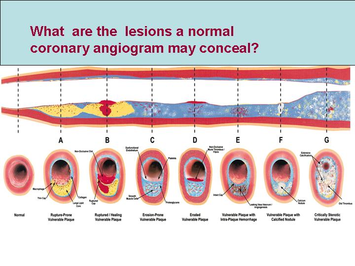
                                         Coronary arteries are the major site for human atherosclerosis .CAD is considered the ultimate determinant of  cardio vascualr health of our global population.Coronary atherosclerosis has a predilection for proximal sites and branching points.Typically it occurs in leftmain, LAD ostium, LCX ostium, proximal LAD, diagonal origins, OMs RCA  and its branches .

4010940_766_gr1

Septal branches , even though divide very early  from the LAD , it  is  uncommon  to get affected by coronary atherosclerosis.  Even for an experienced   interventional cardiologist  , it  would be very rare to have  performed a  PCI for septal disease.

Why septal branches of LAD is rare to suffer from atherosclerosis ?

We don’t know the answer yet.

But , it is thought,septal branches are near perpendicular branches .The branching angle and incidence of atherosclerosis has a peculiar relationship.IAt any bifurcation  point , the atherosclerosis tend to occur ,  if the angle is more acute , and is  less common in abtuse angles .It is  almost rare  ,  if branching happens at   exact  90 degree angle or so !

The other reason for septal branches being immune to atherosclerosis is  , it runs within the muscle in its major course. The constant squeezing action(. . . and possibly bridging also)  makes it difficult for the  process of atherosclerosis to sustain and grow .

Can you still get a  septal CAD ?

Yes,  usually as  a component of bifurcation or trifurcation lesion. Some times a diagonal and septal are very close together and  atherosclerosis involves  both ostia.

What is  the implication for the  cardiologist to perform  a PCI with stenting in a septal branch of LAD  ?

PCI and stenting in the septal branches are more prone for crushing and fracture   as it is constantly exposed to the mechanical effects of muscle contraction.

Any other significance for septal branches of LAD ?

  • Isolated septal myocardial infarction can occur.This could be even a embolic manifestation.
  • Septal branches of LAD are potential target for therapeutic embolisation (By injecting alcohol)  in patients  with hypertrophic obstructive cardiomyopathy(HOCM) .This manover aims to produce a controlled septal myocardial infarction and thus paralysing the left ventricular outflow tract and reduce the dynamic LVOT gradient. This form of treatment, was glorified till recently now considered experimental !

Read Full Post »

coronary-artery1

                                                      Coronary arterial circulation is the life line for  the human heart  and it’s survival.Typically it is supplied by two coronary arteries,  left and right coronary artery.Both, together carry about  250ml of blood every minute.( Approxinately  equal to a  cup of  coke !  ).These coronary arteries  generally divide in a predetermined  fashion , and have multiple branches . It is a  mystery , what  decides this  branching pattern

Is it like a our palmar crease  ? or the cerebral gyri ?

However , it does follow a certain rule,  one major coronary artery  will follow the  four  important grooves of heart. In the left side ,  left main coronary artery (LM) originates in the left coronary sinus (Size varying between 1mm -20mm)  and usually bifurcates into LAD and LCX. The left anterior descending artery (LAD) runs in anterior interventricular  groove while ,  the right atrio ventriculo groove carries the right coronary artery(RCA) .Left  circumflex artery (LCX) traverses the  left atrio ventricular groove.The most inconstant branch is the posterior descending artery (PDA) which runs in the posterior interventricular  groove.PDA  can arise from either RCA, LCX or both or even from LAD.

The major branches of LAD are called diagonal and septal  while the branches of LCX are called obtuse marginal(OM).There can be two to three diagonal and OMs. 

What is ramus intermedius coronary artery ? What is the incidence of Ramus ?

The left main coronary artery  instead of bifurcating into two ,  it trifurcates into three vessels.(LAD, LCX, Ramus)

The real incidence could vary betweenn (10% to 30%) depending upon the series.

ramus

What course it takes ?

It generally goes in the angle between the LAD and the LCX.It may either behave like a large OM or a diagonal branch.It supplies the lateral free wall of the LV many times.The peculiarity of this vessel is it does not run in a anatomical groove .It simply slides over the free surface of LV.Rarely, a  very abnormal course of ramus,  criss cross the aorta and pulmonary artery .

How common is atherosclerosis within  Ramus ?

We don’t know yet. But it is very likely since it is an early branch from left main, it  might  have a  predilection for atherosclerosis  as like LAD or LCX ostium.In fact now we recognise more of  trifurcation lesions involving  three branches of left main .

What would be the ECG finding if a large ramus is the culpirit vessel during STEMI ?

This scenario could be rare.

ACS in ramus could  present as ST elevation in 1/Avl /V5,V6

  • Lateral MI
  • Apical MI
  • High lateral MI

But it is realised , whenever the ECG changes are not fitting with typical ASMI or a lateral MI one should suspect a ramus lesion

 What is the significance of ramus for an interventional cardiologist ?

ramus-2

                                                   PCI in ramus is a rare opportunity for a cardiologist .The issue here  is,  if ramus is involved  adjacent LAD and LCX is also likely to be involved .So it would logically be a multivessel , complex angioplasty.Isolated ramus lesion could be tackled easily.Another issue here could  be ,since this vessel is not within  any anatomical groove  stent deployment would have a poor  support and prone for mobilisation and migration .

Read Full Post »

                                     Hypertension is considered a major cardiovascular risk factor.Hypertension  can have multiple physiological and pathological effects on heart . The common response to  raised arterial pressure is the hypertrophy of the left ventricle ( LVH). This can increase the risk of heart failure in few ( Mainly diastolic failure)  It is a leading cause for stroke  and   less often a  coronary event.

What links Hypertension and  coronary artery disease

                                           Coronary artery disease is almost synonymous with atherosclerosis. There is no separate entity called hypertensive coronary artery disease. But HT can accelerate the process of atherosclerosis. It is widely understood, hypertension can cause  physical endothelial damage and functional impairment of endothelial function.The physical damage ie enothelial disruption , or erosion is a very uncommon phenomenon . So currently  there is sufficient clinical experience  HT is considered dangerous for coronary artery only if it is with the  company of diabetes and hyperlipidemia. (This will seem controversial as it is against the findings of iconic Framingham trial!)

What the medical community refers to hypertension , may not be really so inside  for the coronary arteries.

                                             The relationship between brachial cuff blood pressure and the intra coronary pressure has very little linear relationship. So one should recognise it is the intra coronary hypertension that has a immediate impact on the coronary events. Now only , we are beginning to understand the complexities  of the relationship between HT and CAD. If we analyse a series of individuals HT per se is not a very serious risk factor for CAD* , but it is a number one risk factor for stroke. 

Why HT in isolation  often result in stroke , rather than a MI ?

While HT  is notoriously common to result  intracerebral hemorrhage, the same HT  would not cause  intramyocardial bleeds . Why ?

What is protecting the myocardium against this complication ?

                                      The exact mechanism  is not clear.Acute surges of blood pressure can increase the risk of stroke many times  but  rarely precipitate  a coronary event(  But may cause a LVF) . The reasons could be the coronary endothelial shearing stress is less than the cerebral blood vessels.Both cerebral and coronary circulation has  auto regulatory mechanism . The coronary auto regulation is more robust in that it does not allow  intra coronary pressures to reach critical levels .There is no clinically relevant intra myocardial hemorrhage reported  even during malignant hypertension.

*But a  high intra coronary pressure can sometimes  result in spontaneous coronary dissection and plaque fissure .Lipid mediated injury is vey much facilitated in a high pressure environment.

Has Controlling blood pressure  to optimal levels  , reduced the overall CAD morbidity and mortality ?

                    The answer is yes, ( But not an emphatic yes ! ) Some studies had been equivocal. It is very difficult to say , how much benefit is attributable to BP reduction  per se  and   how much is attributable to indirect effect on atherosclerosis prevention.

Hypertension during ACS

                            High blood pressure during an episode of unstable angina or STEMI can increase the myocardial oxygen demand and worsen the ischemia. It requires optimal control with nitroglycerine ( Preferably ) or beta blocker and ACE inhibitors.Even though HT is commonly associated  with ACS,  one can not be sure the ACS is preciptated by HT. Many times the sympathetic surge during an ACS keeps the blood pressure high.It is a common experience the blood pressure suddenly dropping to normal or hypotensive levels once the pain and anxiety is controlled.

Hypertension during thrombolysis

                           High blood pressure is a relative contraindication for thrombolysis.It need to be emphasised here, It is the  the fear of stroke that make  it contraindicated .The heart can tolerate  thrombolytic agents delivered at high BP .In fact logically ,  hemodynamically and also  practically it is obseved , thrombolytic agents administered at relatively high blood pressure (140-160 systolic) has better thrombolysis than a patient who is lysed at 100mmhg.

                       The coronary pressure head which contain the thrombolytic agent (streptokinase and others ) need to have pressure jet effect on the thrombus.So the  mean coronary perfusion pressure becomes  a critical determinant of success of thrombolysis.

                            It is a paradox of sorts , very high blood pressures are a relative contraindication for thrombolysis and at the same time normal pressure patients fare less well to thrombolysis.

 Final  message

                        Hypertension continues to be a major cardiovascular risk factor.It has direct and indirect effects on the heart.Generally HT is more of a risk factor for stroke than CAD.A slightly high BP ( Just around the  upper limits of normal or just above it ) has a hemodynamic advantage during thrombolysis.(Class C evidence )

Read Full Post »

Answer: Do  coronary angiogram  for all patients  who had suffered from an acute myocardial infarction* ( Forget about all those mulitpage ACC/AHA  guidelines !).

For an  interventional cardiologist ,  it is often  considered a crime to  follow a conservative  approach !

*Caution This one line guideline is not based on scientific fact  but reality based . Ideally one should identify  high risk subsets among the patients who had an AMI .Patients who had complications during the MI get immediate CAG. Others need  a focused LV function asessment ,  pre discharge  sub maximal excercise stress test or perfusion studies .But this concept has been  virtually replaced by pre discharge coronary angiogram for all ,  in many  of the centres in the world.

Read Full Post »

Coronary angiogram is a video graphic  snap shot of coronary arterial lumen which is filled with radio opaque dye. This is some times called as coronary luminogram . It is a paradox , when we say normal coronary angiogram we can only mean  normal coronary lumen. But  generally, this can provide sufficient  information regarding the status of  coronary blood flow.There are three structured layers in coronary artery wall . Coronary angiogram  can not give any information about the status of the intima, media or adventia .

Lesions A to F may be totally missed by conventional coronary angiogram

Lesions A to F may be totally missed by conventional coronary angiogram

A patient with normal coronay angiogram can have diffuse  atheroscelrosis or  localised atherosclerosis within the media of coronary artery .Many times these atherosclerotic plaques grow outward into the adventia and fail to encroach upon the lumen to be detected by coronary angiogram. These plaques , even though has an hemodynamic advantage, in that it doesn’t block blood flow , has a serious risk for sudden rupture and result in an acute coronary syndrome.

So what is the message?

A normal coronary angiogram can never convey a meaning of normal coronary arteries.


A person who has a normal coronary angiogram has no guarantee that he won’t develop a coronary event in the near future.(But the the chances are very low)

If coronary angiogram has serious limitations  what is the next alternative ?

Intra vascular ultra sound imaging(IVUS) can give us an idea of the coronary arterial wall anatomy. This investigation , though available for clinical application is too complex for regular use.So , you  can’t subject every patient with normal CAG  to an IVUS  (Intra vascualar ultra sound) to confirm the normality. The best option is what we follow every day in our practice  .Tell your patients   with normal coronary angiogram , that they are likely to  have  normal coronary arteries  ! don’t add up to their anxiety by saying,  in spite of normal CAG  still they  can carry  gross atherosclerosis in their  arteries. Anxiety can precipitate an coronary event. Too much technical information to the patients  can be counter productive. Instead  advice regular life style modification,  blood pressure ,diabetes, lipid  control  etc .

Read Full Post »

                    Lateral myocardial infarction is not a common site when compared to anterior and inferior MI. But the lateral MI has some unique features, since it involves free wall of the ventricle.The laplace law mediated wall stress is more as the dyskinetic segments bulge with a long radius .Due to this,  lateral MI has a distinctly high risk for free wall rupture .Further pericardial rub is more common in thse patients.Ischemic mitral regurgitation and vulnerability to LVF is also more prevalent if the lateral wall is involved.  Generally lateral MI pateints have a turbulent and complicated course than a simple inferior or anteroseptal MI.

The angiographic correlation of  lateral MI is rarely reported in literature.

The following leisons are commonly observed.

1. Proximal  LAD with large D1  involvement

2. Isolated large D1/D2 disese

3.A left dominat LCX  with large OM1 disease

4.Large ramus disease

5.LAD total and RCA to LAD/D1 collateral pattern

Final message

If we encounter a lateral MI either alone or in combination with inferior/  posterior MI , it is better to manage these patients  aggresively with early triaging for CAG and revascularisation.

Read Full Post »

« Newer Posts