Feeds:
Posts
Comments

Archive for the ‘Cardiology – Clinical’ Category

Right ventricle is a passive venous component of the heart .It simply acts a  transit pump for blood to reach the lungs.

It  is true  , RV is dispensable in many complex congenital heart disease as we  can connect the great veins directly into the pulmonary artery  by  Fontan , Glean and it’s clones  bye passing this chamber . Still , by no means the importance of this chamber is to be underestimated.  RV dysfunction and failure  is the key to survival  many  disorders.RV shock is is cause of sudden cardiac death in acute pulmonary embolism and RV infarction .

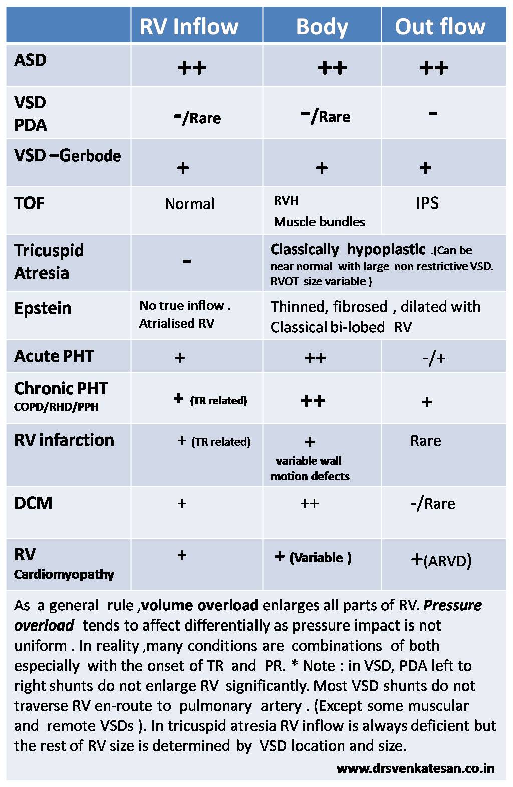
RV is an unique muscular chamber .It is more of a triangular shape. It has  three different parts connected by three different angle .There is no true  apex  for RV , it is   connected  to Inflow and outflow in peculiar fashion .

In the  following table I have  tired to  describe  of how different parts of RV  behave in various disorders.

what is the morphology of  RV enlargement RV inflow outflow body sinus portion of RV

Read Full Post »

While many of us are preoccupied with wires and balloons ,( coronary  myopia ! )  , our radiology  colleagues are making rapid strides . Let us spend some  time  to understand  how  the myocardial segments  are inflicted the  final insult . We need to realize , there is a pattern  to  this myocardial  end game of scarring and fibrosis.

MRI is the  gold standard to assess the myocardial architecture . It has a role in both assessing the anatomy , function  , perfusion and viability .

how to differentiate ischemic dcm from idiopathic dcm myocardial scar epicardial transmural

  • LV function is assessed  by cine MRI
  • Viability  stud by  delayed enhancement MRI (DEMRI , also called as  LGE- Late Gadolinum enhancement  )
  • Myocardial scar best assessed by DEMRI*
* Why do you require DEMRI to identify scar ?
One can detect scars in plain MRI but contrasts make it better .Hence delayed enhancement in by DEMRI is used  to detect scars.
Is it ischemic  DCM or Non ischemic DCM ?  ( That is the question we commonly ask  
We rely too much on CAG anatomy for this. It can be misleading. Cine MRI with DEMRI  gives the answer straightway with high degree of accuracy  .  CAG is required in all  ,  but if it is normal , or  has insignificant lesions  , the dilemma  of ischemic DCM would continue !)
**Note ,there is one   simple algorithm proposed by the author   to  differentiate  Ischemic DCM from Idiopathic DCM  without MRI – Click here to  Link
Following  scar patterns in DEMRI help us to arrive a diagnosis.
Favors Non ischemic  DCM
  • Mid myocardial scar
  • Epicardial scars
  • Global sub-endocardial scars
  • No scar(Ironically if  no delayed  hyper-enhancement is noted it is likely to be non Ischemic DCM )
Favors ischemic DCM
  1. Regional transmural scars
  2. Localised sub-endocardial  scars
* Ischemic DCM will always involve subendocardium as ischemic wave front goes from sub-endo to epicardium.
examples for Non Ischemic DCM
  • Amyloidosis (Can be restrictive as well )
  • Chagas
  • Fabrys

Why is  scar localisation and Quantification important ?

Apart from differentiating various cardiomyopathies  it has  few clinical implication .

  • Since scar indicates irreversible damage , if extensive  it will  argue  against any re-vascularisation .
  • Scar location becomes vital if we plan CRT .It will be futile  to place a CRT lead over a scar.
  • Scars are often  form a macro re-entrant circuits for VT .Help us localize or zeroing in VT focus.
  • Scar quantification is helpful risk stratification of patients  with HOCM .and their family.
Final message
Myocardial scar location and quantification  is the new mantra in a  patient with dilated heart with cardiac failure.
It may be more important than even a coronary angiogram .MRI  will prevail over   any of the available echocardiogram modalities to assess the scar pattern.
Reference
myocardial scarring mri

Read Full Post »

I know ,there is a VSD out there !  but I am  unable to  get the gradient across it.This situation can be quiet  common .The reasons could  be technical, anatomical or  hemodynamic.

As a rule ,  if we hear  a  pan-systolic murmur clinically ,   one must be able to catch  a good  Doppler spectrum somewhere by  echocardiography . However , If  the murmur is restricted to  early or mid to late  systole, VSD  jet is often attenuated in echocardiography .

In the  following situations ,  VSD  jets  may not  record a distinctive Doppler spectra. Invariably the velocity is low , spectrum is short,  less intense ,  lacks good shape and borders are hazy !

  • A closing  VSD
  • A Small muscular VSD
  • VSD with  Severe pulmonary hypertension
  • VSDs with muscle bundle criss crossing
  • Double chambered right ventricle (DCRV, where VSD usually drains to high pressure chamber.)
  • VSD  associated RVOT obsruction  (Note: classical TOF VSD will never generate a murmur)
  • VSD with sinus of valsalva aneurusms (  Doppler  jet  can be really  difficult to record )
  • Inlet VSDs are missed because  convectional  views of echo are perpendicular to these inlet jets.(Short axis better  )
  • Another common situation  is post STEMI VSR.Both a small apical VSD or multi tract  VSD associated with  infero posterior  STEMI   gradients are  difficult to obtain.

What is   inference ?

Doppler spectrum will help detect  small VSDs and color doppler will not miss even a tiny VSD.Doppler spectrum across VSD  is dependent many  factors other than the presence of VSD. However some large VSDs are detected better by 2D echo rather than doppler signals.

Final message

Presence of a Anatomical  VSD does not  imply it should generate a noise.The murmur as well as Doppler signals  are  primarily  determined by the pressure difference on either side of VSD. After all , one of the  largest VSD  that  we encounter

Read Full Post »

The pericarditis  of acute rheumatic fever is not a true infective pericarditis.It is more of inflammation .It is primarily  T cell mediated  reaction . Neutrophils rarely take part  in this inflammation and hence  no significant  exudation . Hence , there is less  sticky and adhesive molecules inside the pericardial space .The most inflamed layer is epicardium which a nothing but visceral  pericardium .This layer lacks the tensile strength to constrict the underlying myocardium.

why rheumatic pericarditis does not go for constrictionFor constriction to occur the fibrinous  ( thick ) layer of pericardium need to be involved . In rheumatic fever  even though it is  pancarditis ,  fibrous layer is not  involved. Further the inflammatory gradient is thought to spread from within  (Unlike tuberculosis )

Note :  In chronic tuberculous pericarditis,  diffuse inflammatory process  invade from the exterior surface . Very often ,  one can not differentiate  layers. In extreme cases even myocardium and pericardium can not be separated .

Summary

The peri-cardial effusion of acute rheumatic fever

  1. Is transient ,non infective and resolving (Unlike valvular inflammation !)
  2. Less of neutrophil activation  (Less adhesion)
  3. It does not involve the thick , tensile  fibrous layer of pericardium hence lacks the contractile force .

Other lingering  questions

1.How common is tamponade  in acute rheumatic fever ?

2.How important is the mass of the effusion (Viz  a Viz  Intra pericardial pressure !) in causing tamponade ?

//

Read Full Post »

Takotsubo cardiomyopathy is an  unusual response  of the left ventricle to extreme emotional stress .The catecholamine  surge  has  a profound stunning  effect of LV apex  and  a paradoxical hypercontractility of basal LV.

The exact mechanism is not clear , Following factors may contribute.

  1. Multi-vessel coronary artery spasm,
  2. Cardiac microvascular dysfunction.
  3. Abnormal myocardial fatty acid metabolism,
  4. Reperfusion injury  after an ACS *

However , the most accepted mechanism is Endogenous catecholamine-induced myocardial stunning and microinfarction

Why is LV apex alone affected  ?

The adrenergic receptor distribution is high in LV apex .They are exposed to high concentration  and gets stunned easily . Basal LV has less adrenergic innervation  , so it shows less of catecholamine toxicity , instead  it exhibits.  hyper-contractile mode. However, this rule is not absolute.

One more suggestion was apical balloons correlated with wrap around LAD.(Báñez B et all 2004)

what is the mechanism of apical ballooning syndrome 2 takotsubo cardiomyopathy

Image courtesy Circulation December 16/23, 2008 vol. 118 no. 25 2754-2762

*Some consider ACS should never be  linked to  Takotsubo.But it is not easy to differentiate.(Carrillo JACC 2009(Kosuge JACC 2010)

Reference from this site

A link to related article -Ischemic Takosubo  from this site .

https://drsvenkatesan.wordpress.com/2012/05/27/in-extreme-mental-stress-left-ventricle-becomes-a-banana/

Reference from other journals
what is the mechanism of apical ballooning syndrome takotsubo cardiomyopathy

1.Báñez B, Navarro F, Farré J et al. (2004). Tako-tsubo syndrome associated with a long course of the left anterior descending coronary artery along the apical diaphragmatic surface of the left ventricle.]”. Revista española de cardiología (in Spanish; Castilian) 57 (3): 209–16

2.Carrillo A, Fiol M, Garcia-Niebla J, Bayes de Luna A. Electrocardiographic differential diagnosis between Takotsubo syndrome and distal occlusion of LAD is not easy. J Am Coll Cardiol. Nov 2 2010;56(19

3.Dorfman TA, Iskandrian AE. Takotsubo cardiomyopathy: State-of-the-art review. J Nucl Cardiol. Jan-Feb 2009;16(1):122-34

4.Kosuge M, Ebina T, Hibi K, Morita S, Okuda J, Iwahashi N. Simple and accurate electrocardiographic criteria to differentiate takotsubo cardiomyopathy from anterior acute myocardial infarction. J Am Coll Cardiol. 2010;55(22):2514–6. doi: 10.1016/j.jacc.2009.12.059.

how to differentiate takotsubo cardiomyopathy from anterior stemi

//

Read Full Post »

In one of  our patients  who had a recent STEMI , CAG revealed  70% LAD  lesion  with   TIMI 1  flow .The distal run off was slow .He had moderate LV dysfunction with no major symptoms. The angiogram was done routinely .( Yes . . . routine CAG- the term I do not relish ,  while it is becoming a way of  life for all learned cardiologists !)

I  was discussing   this case with my fellows and  about medical management of CAD. I  told them  Aspirin will help prevent ACS, statins will stabilise the plaques , beta blocker would prevent cardiac  events by blunting adrenergic surges and Nitrates is a powerful coronary vasodilator that  will improve the  coronary blood flow

A final year MD fellow  threw a  direct  question at me .

Sir,  do you mean  Nitrates  would increase the  TIMI  flow from 1 to 3  in the long term in this patient ?

I was taken-aback  for a moment . . .   and thanked my student for a valid question .

Nitroglycerine is  a powerful coronary vasodilator  we are taught for nearly  half a century . Oral nitrates are used  liberally in the chronic management of  angina. It is a multi billion  dollar market.

Has it been documented to improve coronary blood flow in the long term ?

No

Why then it is  used long term ?

It is a clear case  of  inappropriate medical therapeutics .

* The confusion is partly due to our mix up the mechanism of  relief of angina from coronary vasodilataion. Realistically , NTG should me known  more as a powerful venodilator reducing the preload . It  dramatically reduces the LV filing pressure  and relieves sub-endocardial stress  .This is the major determinant for angina relief .(Of course after-load reduction also helps)

Final message

Nitrates  should be used only for relief of an episode of angina or just to prevent It .This may surprise you  , Nitrates has no documented  efficacy  in the long term management  of angina.

Read Full Post »

One of my otherwise  well behaving  patient ,  suddenly asked me this question ,  before leaving my  clinic  after a 15 minute consult .

Doctor  . . . I am taking the clopidgrel and aspirin  for 5 years like a vitamin tablet  . . . is that all right  doctor ?

I just got curious, I checked  the prescription again . Yeh ,  he was right !

I have  been mechanically writing  Tablet Clopitab  A  since 2008 !

Clopidogrel abuse long term

For what ?

Some sort of CAD !   Was it for ACS ?   No , it was for chronic stable angina . No PCI,  . . . no DES !

Why the hell  he is taking dual anti-platelet  therapy for 5 years ?

Some  body  , some  where  , has  prescribed it . This man  is taking it  for years together with absolute sanctity.

I was amused  . . . it is also  my mistake . Why it never struck me  to scrutinize the prescription ?

I thanked him . I  removed clopidogrel  from the list , and asked him to continue tablet Ecosprin 150mg  for some time .

( And  now I  had a genuine doubt  ! Does he have CAD at all ! I browsed his file , I couldn’t  find a true documentation for CAD  as I feared  !)

I asked him to get back with an  exercise test ,  . . .  if it comes negative i  can even stop the aspirin as well  I  explained  him  ! (Now he got amused !)

Final message

Who wants Knowledge ?

It is  dumped every  where , free of cost  . . .  both in real and cyber world .Applying it requires more sense  .  and my patient  taught me that  !

Patients  not only  help us  earn  our  bread  and butter , they  do  enrich  our brain  as well ! Never get humiliated when a patient teaches  you a lesson in medicine !

Read Full Post »

The other day my fellow got a call  from surgical ward for emergency ECG opinion for a  suspected Inferior MI .It later turned out to be an acute cholecystitis.

One of the important  anatomical mis-perception  among physicians ,  is to consider  inferior, posterior  and diaphragmatic surface  of heart  as separate entities .They are all  closely linked.In fact, they  more often  mean  the same  anatomical zones !

Heart is a dynamic suspended organ within the middle mediastinum .It  can assume a vertical or horizontal position due to number of surrounding anatomical  and physiological factors. (Diaphragm, Lung , being  important ).The ratio of intra thoracic vs Intra abdominal  volume  &  pressure determine whether the posterior surface of the heart is going to face the back of chest  or simply sit and  rest on the diaphragm .We know a horizontal heart is likely to inscribe q waves  in inferior leads .

acute abdomen diaphragm inferior wall mi cholecystitis pacreatitis

Courtesy : Basic image source from digitallab3d

The  diaphragm can be termed as an  anatomical causeway , that isolates   thorax  from the  abdominal  cavity .Close encounters between the organs separated by this delicate biological  membrane is  always possible .This is especially true for electrical signals  which show little  respect for anatomical barriers .

This is the reason there are too  many abdominal conditions that mimic  inferior MI during a painful  emergency (and vice versa  when inferior  MI mimics  acute abdomen .) In  our  department , we   have witnessed  the following conditions mimicking Infero-posterior ACS.

  1. Acute ascites with polyserositis
  2. Gross obesity with APD
  3. Posterior fat pad ( Necrosis ?)
  4. Thickened pericardium
  5. Minimal posterior pericardial effusion
  6. Diaphragmatic pleurits
  7. Esophageal spasm
  8. Fundal air  trapping and ballooning after a heavy meal !
  9. Acute duodenal ulcer perforation ( With gas under diapharam causing q waves)
  10. Acute cholecystits
  11. Diphragmatic hernia
  12. Achalasia cardia
  13. Pancreatitis

Final message

Do not rush to make a diagnosis of inferior wall MI when  you encounter inferior q waves  with  or without ST /T changes , especially  when the symptoms are atypical .

Read Full Post »

cardiac auscultation murmurWe know clinical auscultation is an art . It is more of a  special sens rather ! It is a combination of natural and acquired ability of your brain to phase out a sound or series of sounds . Sound perception also has  two point discrimination  like touch .(Auditory cortex -Temporal lobe maturity)

It involves selective blanking  and noise cancellation techniques. Ambient noise  contamination is more in youngsters . Elderly men  often have otosclerois so they are benefited by  this handicap .Your chief maybe one of them .So simply do not bother.

Finally ,  clinical acoustics   require lot of imagination . Seniors professors  know what they are expected to hear in a given patient . They look for it rather than  they hear  it  .This is the secret of their  magic ears .

The famous quote  “What the mind do not  know  . . . the eyes do not see”   is very much  applicable  for the ears as well ! 

What your  temporal lobe  do not expect  . . . the ears do not hear!

Beware , even experienced cardiologists  mistake   systolic events   with that of  diastolic and vice versa !

Final message

With due courtesies  to great men like  Potain ,  Leatham , Austin flint,   and other pioneers of cardiac auscultation , I would modify  the  title  of this article .

The science of  cardiac auscultation  may appear more of an  auditory illusion to many  youngsters today  . Still , dedicated  auscultation , with a sound clinical knowledge in a quiet  room  with  a good stethoscope  would  make this illusion  into a  reality !

Read Full Post »

Spinal cord is a busy  neurological  highway to brain .It  runs  24/7  non stop  with unlimited  horizontal and vertical lanes .It is such a compact  structure , it can  easily  get confounded   when multiple signals converge,  diverge, summate , deduct , reflect back,   or cancel out .
A 64 year old women came to me for  second opinion  regarding   chest pain . A  cardiologist  had  just adviced her  an  emergency   coronary  angiogram and also suggested she may require an  urgent  PCI  as well .
I listened to her history in my office  . . .  In  her own words .
Doctor , I am  getting  sudden   compressing  type of  pain which  starts in the centre of the chest and soon transmits to the left shoulder and  gradually reach the inner aspect of the hand up to the little finger . And occasionally it is very severe and some times i feel like sweating as well ! I am unable to predict when it comes doctor !
It was  so convincing  but one  feature was  not fitting In . She said , she used to walk  daily   and do all house hold work with no pain . She also  recalled about the  acid peptic disease , and neck pain periodically due to cervical spine problem.
Her resting ECG was normal .She was  afraid to do a stress test . After thinking  for a minute , I had no  other option  but  to endorse  my colleague’s view and asked her  to go for coronary angiogram .
One  thing I  suggested differently was , I told her it was not an  emergency , I also  conveyed my gut feeling  that it is unlikely to cardiac  pain . One week  later  CAG through radial route  was done . Both of  us were  happy  to find a  normal  coronary  angiogram !

Final message

Pain is a  feeling . It can be  perceived  at  multiple levels  . The site of origin , spill over on transit and at the level of brain .  A patient with multiple  potential source for pain can either summate , deduct , reflect  or cancel out .This can confuse the clinician in a dramatic fashion as it did to us ! . To complicate the matters  further , gastric pain can trigger a cervical  pain and vice versa . (Spill over effect)

Read Full Post »

« Newer Posts - Older Posts »