Feeds:
Posts
Comments

Today is one of the most auspicious days in Indian traditional festive time. Saraswathi pooja, a celebration of the Goddess of knowledge and education. I would like to share one of the all-time great quotes on learning from Thiruvalluvar a sage poet who lived in the southern Indian state of (mine), Tamil Nadu in 4th -5th century BC  2500 years ago.

 This Thirukural number 391 in the chapter of education goes on like this. (In the Tamil Language)

In English

Karka, Kasadara, Karpavai , Katrapin,

Nirka , Atharkku Thaga !

It says

Karka : Learn

Kasadara: Here comes the punch. Kasadara means pure.  He says simple learning is not at all-sufficient. One has to learn from good sources, learn deep that should be devoid of errors, contaminations, and falsehoods.

Karpavai  : Thus you learn all lessons in life meticulously.

Katrapin:  So, after this hard and enlightened learning, what we should do?  He answers next.

Nirka Atharkku Thaga: This means , don’t just stop with learning, follow it with action in a righteous way. Unless we do that he warns to conclude ( in another poem in the same chapter) there is no purpose of learning itself and we are again at risk of becoming illiterates.

So, what does this Thirukural teach the Nobel professionals who follow cutting edge medical research?

I think I need not elaborate . . . Acquiring knowledge and true learning has become two different processes.

It’s just a sample of one kural (Quote) among 1330 poetic quotes written in 133 chapters by this great philosopher of Tamil Nadu who shared the same timeline with Aristotle and Socrates of ancient Greece 5000 miles west of India. For those ,If you are interested in his monumental work on literature which can be referred to as the manual for effective living  (I wish to call it as “Standard operating protocol”  for human life)  please follow the link.

It appears,antiplatelet agents are waging a turf war on the CAD battlefield. It is no secret either, the fight often goes beyond academic reasons. Though NSTEMI connotes a true cardiac emergency, it consists of a highly heterogeneous population. A patient with UA can be treated even at home (Low-grade angina with little ECG changes, when it’s due to Increase demand situation). While, in the other extreme of NSTEMI, a patient with a GRACE score >200, in Ischemic  LVF, might need an emergency multivessel angioplasty along with Mitra clip ±  ECMO support. 

Antiplatelet agents along with heparin will remain the cornerstone* in the management of NSTEMI/NSTEACS, irrespective of our fine catheter skills within index lesion. They are administered right from the pre-hospital phase/ In ER, CCU/ or on way to the cath lab(upstream)/or within the cath lab/or after CAG /PCI.  It is the right balance between the prevention of stent-related coronary thrombus vs systemic bleed we are worried about. Definitely, DAPT is warranted. (See the chart below) Prasugrel has been reinvented as the most powerful P2/Y12 blocking antiplatelet agent. It squarely beats its other colleague drugs like Aspirin, Clopidogrel, and Ticagrelor in terms of potency as well as its risk of a bleed. This is the current antiplatelet protocol in NSTEMI in a patient planned for PCI after visualizing the coronary anatomy. Note, Aspirin plus Prasugrel combination occupies the top slot among various options. The principle of DAPT strategy is all about Initial escalation to match the heightened risk of thrombosis/ cardiac events and later de-escalate once the risk period is over (Which can vary between 1 month to 12 months or even 2 years)* The popular concept of attributing NSTEMI to platelet clot and STEMI to fibrin clot is no longer valid. The contribution of the individual component(white vs red)  in a given load of coronary thrombus was never quantified accurately. That’s why antiplatelet agents alone are grossly inadequate in NSTEMI. This will be vouched by this NSTEMI algorithm, which begins with red clot busters heparin. 

So, how to handle sharp-edged drug-like Prasugrel?

A powerful drug-like Prasugrel is at high risk of being misused. It has taught us some harsh lessons in stroke. So, we have to be wiser to extract the maximum out of this drug in the presence of a high thrombotic milieu (or at risk of developing it after a PCI.)

Since ECG and clinical features are not sufficient to predict the coronary thrombus. It is suggested to have a look at coronary anatomy and decide only if a PCI is contemplated.Some of the situations where Prasugrel is likely to be Indicated  

  • Any PCI with a stent in the culprit artery.
  • High  thrombus load
  • Prolonged procedure time


    When to Avoid Prasugrel?

    Just looking at coronary anatomy is not sufficient.  Estimating the risk of bleeding is required. Attempting to use various scoring systems during a cardiac emergency is a self-inflicted mathematical burden. In my opinion, none of these scoring systems(CRUSADE , ACUITY,  ARC-HBR) truly discriminate patients in a useful way. Mindfulness with an eye on co-morbid conditions is required.This has to be matched with coronary lesion /PCI complexity. Realistically, the confidence in our technical adequacy of stent deployment shall decide the need for aggressive post PCI  DAPT or anticoagulation

Final message

Just because we know the coronary anatomy, don’t expect prasugrel to be kind enough to lower the risk of stroke. The risk is the same whether we know anatomy or not. It is the funny evidence base we have created that makes us believe it so. Routine DAPT for all patients with ACS is not warranted without assessing the bleeding risk. Meanwhile, there can be an important subset of patients who can really benefit from prasugrel even within coronary care units who are unplanned for PCI. (Which the current guidelines seem to forbid without any valid reason)

Postamble

We know, stents love to befriend thrombus instantly, that demands aggressive antiplatelet/anticoagulants) which beget bleeding. So, should we stent all lesions in a given patient with NSTEMI ? is a very valid question (rarely asked though) needs to be answered by the custodians of patients’ heart. When dealing with a complex PCI case scenario, simple mindfulness with an eye on comorbid conditions and downgrading ourselves to a good general physician mindset is welcome.

Reference

1.The DUBIUS trial downstream vs upstream use of antiplatelet agents in NSTEACS- No difference

https://www.acc.org/latest-in-cardiology/clinical-trials/2020/08/31/21/44/dubius


 2.

Is sudden, unaccustomed, physical exertion a trigger for plaque rupture and an ACS ?

Yes, it is, but don’t get apprehensive. The underlying risk factors, plaque burden, and its morphology matter much to result in a coronary event.

What is the mechanism?

Plaque morphology,  the lipid core, the shoulder region’s eccentricity, the crystallization of cholesterol lay the foundation. The Isometric component of stress surges Intra-coronary pressures and facilitate vascular injury.  Endothelial dysfunction leading to erosion and subsequent acute total thrombotic occlusion is a well-known response to stress. Currently, spontaneous coronary dissection secondary to unaccustomed stress is increasingly recognized to be a culprit.


plaque fissure and exertion physical and mental exertion and plaque

Which is more dangerous? Mental or physical stress* ?

No one can answer this query with certainty. The combination of both can prove deadly in vulnerable patients. The final common pathway for both physical and mental stress seems to be the same. Adrenergic toxicity at the cellular level.

* Mental stress-induced primary electrical events (CPVT/ Inherited channelopathy ) are unrelated to plaque destabilization that is often confused with ACS in many SCDs.

What are the natural protective factors to stress?

Coronary autoregulation,stress-busting hormones like endorphins , natural anti-fibrinolytic systems do play a role. Human beings experience infinite episodes of mental stress in their lifetime. Only a fraction (of a fraction ) result in ACS. It is obvious , there must be some major invisible protective factors. One may call this as metaphysical force ( scientific equivalent to fate ?) operating on a particular plaque to destabilize it.


It was April 15th 1912, Titanic, the Invincible, had just sunk into the dark waters of the Atlantic coast off Newfoundland. Exactly same time around, Dr. James Herrick, In Chicago, Illinois was busy documenting the first diagnosed case of acute coronary thrombosis. A new disease was born ie Myocardial Infarction. This was also the era of the Noble Prize-winning  Invention of the ECG machine by Waller, Einthoven, and Thomas Lewis & co that sow the seeds for the specialty of electro-cardiology.

Though much was studied about MI with pathological specimens in the subsequent decades, there was a lull in the efforts to define the entity of myocardial Infarction till WHO  defined in the early 1970s. It was dogmatic, still fair enough. (Clinical, Enzymes, ECG criteria, with  any two feature, must be present to diagnose )

Since then, the field of cardiology has seen unprecedented development in both the diagnosis and treatment of ACS. We now have a universal definition( EHJ 2019 Thygesen K ) that asks us to triage based on high sensitive troponin followed by clinical and other parameters. STEMI usually doesn’t have much diagnostic confusion.

Nomenclature Issues in NSTEMI/UA

The definition of NSTEMI  refuses to settle, though we have come a long way since the times  UA/NSTEMI were clubbed together as siblings. The term unstable angina was coined by one of the most revered cardiologists of our times  Dr. Noble O Fowler in 1978. They are the same one hitherto referred to as Intermediate coronary syndrome/Pre Infarction angina. Later, if enzymes were raised it was labeled as non-transmural/Non-Q  MI. This became the classical NSTEMI later changed to NSTEACS (Still it is valid)

The semantics surrounding the NSTEMI  is unlikely to end as long as we depend largely on ECG to diagnose and treat complex coronary obstructive syndromes. This, by no means, undermine the importance of ECG in this setting. It will remain the gold standard as far as, I can look into the future.

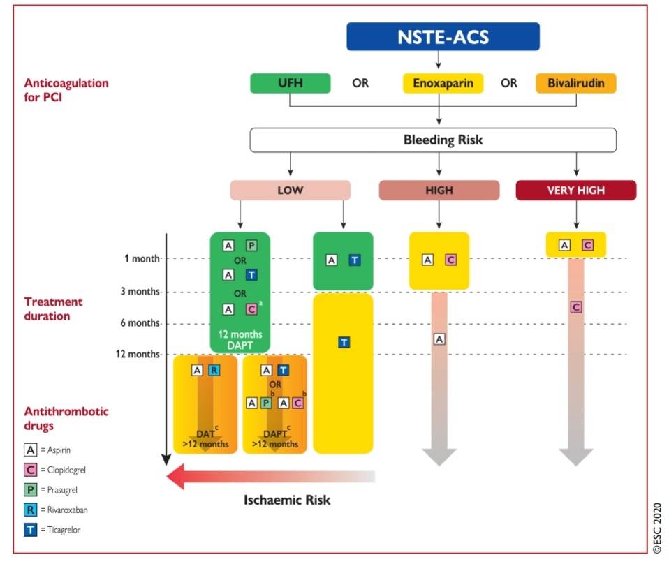
Some observation about the new ESC 2020 NSTEMI guidelines

Anyway, ESC 2020 has addressed this issue. It suggests a new term “ACS without persistent ST elevation” for NSTEMI (Ideally they should have used this abbreviation  NP-STEACS)

(*I guess, the current ESC 2020 guidelines really wanted to get rid of both NSTEMI/NSTEACS for a very valid reason but still it was worried about the confusion it might create so retained the old term NSTEMI/NSTEACS  )

The categories included in the current NSTEMI scheme are

1.Transient ST elevation (How transient ? Prinzmetal/ Non Prinzmetal ?)

2.Persistent ST depression

3.T inversion

4.Flat (Absent ) T wave

5.Pseudo normalization of T

It may include the following as well (Not in official ESC 20220 guidelines)

6*.Hyperacute T (Very early STEMI ? or NSTEMI?

7*.Wellen/Dewinter or its variants

I think ESC is to be appreciated for recognizing an off ignored observation that UA may have a transient ST elevation and end up later as NSTEMI/NSTACS. This group of ACS still poses a challenge for us to understand the overlap between total and subtotal coronary occlusion (Non-Prinzmetal ST elevation)

Final message 

Does this nomenclature issue create problems in management? 

  • Yes, it does. The major implication is in the diagnosis ACS with dynamic ST segments ( ST-elevation / /depression or any combination)
  • If a probable STEMI after spontaneous lysis presents as NSTEMI, Is it the baby STEMI or neo NSTEMI ? One may not rush such NSTEMI patients to cath labs.
  • Of course, many of us are conditioned to follow a “single point agenda “ that dictates all ACS shall reach the cath lab and managed thereafter based on coronary anatomy. If that is the case, I am sure the bulk of this 79-page new NSTEMI guideline appears redundant.(Ref 1)

Reference

1.Jean-Philippe Collet,  ESC Scientific Document Group, 2020 ESC Guidelines for the management of acute coronary syndromes in patients presenting without persistent ST-segment elevation: The Task Force for the management of acute coronary syndromes in patients presenting without persistent ST-segment elevation of the European Society of Cardiology (ESC), European Heart Journal, , ehaa575https://doi.org/10.1093/eurheartj/ehaa575

2 Fourth Universal Definition of Myocardial Infarction (2018). Eur Heart J 2019;40:237-269. 

           Practice of cardiology is simple as long we don’t dwell deep into coronary physiology.

One of my patients asked, why he was told his total occlusion in LAD appears safer now, which was subtotal a few months ago.I told him, it is indeed true. It is the fear of subtotal disease that’s prone to a fresh coronary event. In total occlusion, chances of that happening are less or nil.


How can you say 100% block is safe?  Is that always true?

No, it’s not always true. He was surprised when I said it is not 100 %, even 90% lesion can be safe if it’s not causing significant angina and responding to OMT. Of course, It is the morphology and stability of the lesion that will dictate* the outcome in the subtotal occlusion. If the lesion is stable, FFR is good >.8 (TMT is poor man’s FFR equivalent )  you can leave it as it is. Doing OCT /Virtual histology /NIR spectroscopy to define the vulnerability of plaque is neither practical nor desirable (Extreme academics is injurious to health) 

So it is not the degree of the block that’s going to matter, but the effects of that block on distal circulation that will decide the rules of the myocardial revascularisation game. But unfortunately, both you, (the patients) we (the cardiologist) are finding it so difficult to come to terms with this basic truth in spite of multiple guidelines. 


Meanwhile, CTO however makes it much easier to make a decision. One need not bother the content of CTO unless you plan an Intervention. I guess there is no FFR for CTO. Are we aware of any studies that have quantified antegrade flow across a 10% patent LAD and compare it with the Collateral flow in LAD in 100% CTO?

We have long glorified a concept of the open artery hypothesis. (Mainly in Post STEMI though) No one has dared to test and compare a hypothesis that a closed artery might still score over the open in at least some of the subsets of stable CAD. Such a study can never be ethically forbidden after all its a well-observed truth in the real world. 

Reference 

Trials on CTO  revascularisation DECISION CT (Not useful )   EURO-CTO  (May be useful) 


EURO CTO https://academic.oup.com/eurheartj/advance-article-abstract/doi/10.1093/eurheartj/ehy220/4990878?redirectedFrom=fulltext


Psychological factors both depression and anxiety do confer a significant risk for CAD. However, a distinction must be made between risk factors and triggers. It is highly likely, depression has more consistent correlation with chronic CAD than anxiety. 

Primary anxiety per-se is of less of a risk factor for chronic CAD, while it can still be a trigger for cardiac events. (when it occurs over heightened baseline risk) Primary depression increases the CAD risk many fold by slowing the system making it sedentary and promote endothelial dysfunction, which is the key promotor of atherogenic CAD.

It is also noted, anxiety is less associated with obesity (when compared to depression). Further,catecholamine fluctuations that are so common in anxiety states may act like an exercise equivalent ( It’s my quirky hypothesis to be tested by future generations)

Emotions have a complex equation with neuro cardiac axis .Sudden emotional deaths due to possible arrhythmias or stress cardio-mypathies are important areas for research.

Sharing my presentation in one of the Annual physiologists conference held at Chennai in 2016.

Topic: Emotional Triggers in ACS 


Click here fro PDF version of the presentation


No one would have Imagined a generally Innocuous entity called Diabetes will emerge into a  “single disease sub-speciality” in medicine”. Thanks to the global authorities  & pharma Industry for making this speciality a formidable one. The link between diabetes and cardiology is so strong, now with pharmacological strategies looking for overlapping Indications.


Let me share a presentation in one of the cardiology meet in 2017 at Thiruvananthpuram.TAN CSI meet , India.


Click here for a  PDF version 

The days are gone when anti-diabetic drugs were alleged to increase CVD mortality. New generation anti-diabetic drugs (SGLT-2 Inhibitors) are coming up that actively dictate and demand us to use it for reducing CVD risk.

(Am I crazy, to look ahead for stand-alone Indication for SGLT Inhibitors for cardiac failure in non-diabetic as well, as a powerful osmotic diuretic !)

Superficially, tissue hypoxia might look similar to Ischemia but differs in one important aspect. Though the hypoxic myocardium is short of oxygen, the respiratory excreta from cells ie Co2, lactic acid, and free radicles are promptly cleared and flushed as blood flow is normal. Hence, generally acute  Ischemia of tissues is more cell threatening than regional hypoxia at any organ level. 

How do you classify hypoxia? we need to go to physiology classes again.

There are 4 types. Ischemic -Hypoxia, systemic hypoxia, Anemic hypoxia, Histo-toxic hypoxia.A good reference to read (Ošt’ádal B., Kolář F. (1999) Myocardial Hypoxia and Ischemia. In: Cardiac Ischemia: From Injury to Protection. Basic Science for the Cardiologist, vol 4. Springer, Boston, MA.)

The question we want to address here is the effect of systemic hypoxia on LV function.

We encounter this in different settings.

Chronic hypoxia  :In COPD, there can be slow adaptation, still there will be some definite impairment of myocardial function.(Which may not be important in normal times but will tell at times of other stress ) Many studies have documented COPD to compromise LV function. In fact, DCM has a link with some severe forms of COPD (Personal observation, will try to get the evidence )

In acute hypoxia(Non-Ischemic) it causes organ dysfunction.(Acute pulmonary embolism, and sepsis.) We see this often in IMCU with ventilated patients with  multisytem defects a poorly contractile ventricles. Some of us used to report this entity with a empirical term global hypokinesia due to hypoxia. The effect of systemic hypoxia on cardiac metabolism is complex as the heart has a unique ability to survive with ketones in anaerobic metabolism at least for a few hours to days.

It is truly fascinating to note, how the human donor heart makes a stunning statement (to the coronary blood flow obsessed cardiologists) when it is shipped in ice bags with exclusive support of metabolic juices without a single drop of blood and come alive in the recipients without any damage. Is it not a wholesome proof? , for temporary survival, the heart just require ATPs and high energy bonds, not blood in the real sense. 

Here is a 1962 paper that analyzed how the heart is able to survive hypoxia. Walter F. BallingerII and Heinz Vollenweider Anaerobic  Metabolism of Heart   Circulation Research. 1962;11:681–685

How to study the effect of chronic hypoxia on LV function? 

We may lack animal models but Ironically we have perfect human hearts to study the effect of hypoxia on the myocardium. They are cyanotic congenital heart disease.  Here is one of the rare reports based on only two patients that address this issue. The authors (Neeraj Awasthy et al) to be appreciated for this pertinent observation.I think we need to look further. Even in TGA, the urgency to perform an arterial switch within a month or so is not only hemodynamic regression in LV  mass facing pulmonary circuit but also early hypoxic injury to LV  myocardium.

One more area of research
 
The corona pandemic gives us an opportunity to study the behavior of heart with extreme hypoxia all around.The presumed 5 % Incidence of global LV dysfunction with COVID pneumonia is hypoxia-related or viral myocarditis we are not clear yet.

Reference 


Caution: Please don’t expect much scientific content in this post. I Hope, you can spare a minute to answer this hypothetical question.

What will be the shape of the curve,  If you plot BMI in the X-axis and LDL/ total cholesterol in Y-axis from a thousand normal adult populations?

  1. It will be linear for sure.
  2. Maybe a little curvilinear.
  3. Its likely J shaped with age 
  4. I think it is U shaped
  5. No, it is Inverted U
  6. Sorry, I don’t know.
  7. No one knows.

Why BMI refuse to go along linearly with LDL (Cholesterol)?

Only an ultra-fraction of total body fat is represented as cholesterol within the vascular system. It’s worthwhile to note, total body fat store is about 8-10 kg,  the amount of cholesterol in circulating blood is hardly 10 grams. It is presumed another 30 grams is present in cells. Body synthesise 1 gram /day.I don’t know whether I can make this statement. It may appear total body fat and cholesterol are almost unrelated things in spite of the dynamism of Intermediary metabolism exogenous and endogenous cholesterol levels are modulated to keep the intravascular cholesterol within an amazingly narrow range

How justified are we to expect a good correlation between BMI and Cholesterol? 

Apart from the presumed logic, there are other dynamic factors that dictate how any person deals with excess lipids/cholesterol. 

  • Genetics profile(Hereditary dyslipidemia)
  • Dietary habits 
  • Various hormone sensitivity to cells.
  • Physical activity 
  • Age /Gender 
  • Ethnicity

Most excess fat gets deposited within adipocytes. So it is scientifically impossible to guess the serum cholesterol level by just looking at overweight people. 

Final message

Even after 50 years of vigorous research, we are clearly ignorant about this fundamental question in lipidology and clinical cardiology. This is directly reflected in myriad dietary guidelines that flooding in the academic and public domain. Let us be transparent to our patients about our knowledge or lack of it. At the minimum, we should stop confusing all those healthy, active people with borderline obesity.

Further, we need to come out of our villainous portrait of fat in general. Let us respect the fat as an essential building block of every cell and hence the whole body. We still need to pursue a long journey to identify &  target only the high-risk population who have atherogenic dyslipidemia that impacts the cardiovascular outcome.

Counterpoint

There is indeed a correlation between body weight and serum lipid. Don’t be dogmatic with limited research and knowledge and confuse the academics. Go through the literature right from Framingham /MRFIT/Monica to Interheart study. There is scattered evidence to show BMI do have a reasonable correlation with blood lipids. 

Yes, scattered is the right word, it can mean anything.

Reference

This well-conducted study suggests, within the normal BMI range there could be a correlation with LDL. I don’t know how useful is this data in clinical practice. 


Coming next 

There is one more question, which has not clearly answered. What is the relationship between seum cholesterol and Intra plaque cholesterol?

For lighter reading , Ruben Meerman is able to kindle the hidden science.

When you lose weight , Where does the fat go ?

We can consider Jugular veins of the neck as a naturally present right heart catheter. It faithfully reflects the live pressure and waveform data  from right atrium and ventricle .

Can JVP tell us anything about left heart pressures? Is there any relationship between JVP and PCWP or LVEDP ?

If you tell JVP reflects LV filling pressure in any graduate medical exams, you will be admonished. However in DM or post-doctoral exam, if you say there is no link between the two, you are likely to be chided.(It is unfortunate the answers vary depending upon the level of training , which I feel is not academically correct )

Though the JVP-PCWP link, apparently appears Illogical, it does  have a scientific basis. It is true, there is a huge (& multiple) anatomical barriers between the left heart and Jugular vein in the form of pulmonary arterial & venous circuits, the right ventricle and right atrium.Still ,the hemodynamic principles demand, whenever left heart filling pressure increases, the right heart pressure should increase correspondingly to drive the blood from RV across the pulmonary circuit.This raise should be in the mean pressure. (or diastolic pressure,) it’s rarely related to systolic pressures as RV systole normally generate more than twice or thrice the LVEDP.


This driving pressure across the lungs  is called the transpulmonary gradient. (PA mean minus LA mean) The normal being < 7mmHg. So if there is a sudden increase in LV filling pressure to 20mmhg, there has to be elevated right heart pressures.(20 +7) This will be reflected in JVP as well. So patients with acute diastolic heart failure as in HFpEF must show elevated JVP. This can be documented elegantly In patients with positive responses during diastolic stress testing.  (JACC: Cardiovascular Imaging 

There are important caveats in JVP-PCWP link

  • If the PH is long-standing and precapillary (Reactive PAH) has set in the right heart pressure will no longer reflect the PCWP.
  • If there is any organic Tricuspid valve disease (Both TR/TS) JVP can reflect PCWP.
  • Finally, any cause of RV dysfunction will immediately elevate the JVP so biventricular dysfunction makes correlation of JVP with PCWP meaningless.(Acute pulmonary embolism, and RV infarction)
  • Further confounding can occur if we contemplate RV diastolic dysfunction as seperate entity. (At what level of RV systolic dysfunction, does the RVEDP begin to raise ? I think we don’t have an answer for this . Researchers please note.)

Some more mechanisms of elevated JVP with left heart disease

  • Bernheim’s effect and ventricular interdependence can make JVP elevated spuriously without elevating PCWP.
  • Acute mitral regurgitation left atrial V waves can “tide-back” all the way to PA and the right heart to elevate the JVP
  • In ASD and Lutembachers syndrome  the RA pressure waveforms may reflect the LV filling pressure, though inconsistently.
  • Finally, and importantly in Fontan circuit JVP may exactly reflect the left heart pressure for the obvious reason, as SVC is connected to the pulmonary artery directly.

Final message

JVP will always tell what is happening to the right heart chambers only . It can, no way be taken as a direct marker of PCWP/LVEDP. However, there can be a correlation between JVP and PCWP/LVEDP in a certain subset of cardiac failure. (As in exclusive isolated left heart failure (typically HFpEF) the elevated JVP might just reflect the elevated LEDP provided there is normal RV function )

Reference

1.This study elegantly shows a correlation (or lack of it) in different subsets of heart failure. It tells us very clearly If JVP(RAP) is not correlating or disproportionate to PCWP, it implies RV dysfunction.

2. This paper suggests a really useful scheme to classify heart failure as concordant and discordant with reference to right and left heart.

It throws some interesting facts. I guess it will help us guide diuretic management and prognosticate chronic heart failure.