Feeds:
Posts
Comments

His bundle pacing is the new kid in EP lab. It involves exploration of few 3-dimensional cubic areas of His bundle (4-6mm³ ? ) in the crest of the IVS looking for optimal His pacing site. It aims to provide better recruitment of His Purkinje and hence more synchronous pacing. Still, the modality is in the early stages and has few key limitations. They are, requirement of very specialised leads,(Select Secure™ 3830 , Medtronic) lead instability (Susceptible to RV ejectile forces*), high threshold, and lower battery life and finally uncertainty of distal bundle disease. A need for temporary RV back up the leads in some centers will tell us how confident we are, about the concept of his bundle pacing.

The success rate of HPB pacing has not reached the desired levels.Meanwhile, the area His bundle lead explores is so thin and delicate that requires working around membranous septum. No surprise, a new adverse event is reported.  Yes, the first case of acquired VSD reported following his bundle pacing. (Ref 1)

Now, experts are moving down in the septum to capture LBBB to avoid the above-said limitations of HBP. I will not be surprised we ultimately reach the RV apex , the good old destination again, for absolute safety and stability.

*RV leads are naturally isolated and not much affected by RV contractile hemodynamics

Final message

Wish the concerns about His pacing are exaggerated , best of luck for this new mode of pacing. Medtronic is a pioneer and has a long passionate history. After all, cardiac pacing is one of their top Innovation in cardiology in the last century that made a huge impact in the management of electrical ailments of the heart.

Reference


*The post is not meant for those who understood GLS (Global longitudinal  strain) in a proper perspective. I am writing this, after a surprising answer from many students of Echocardiography, when I asked them what is GLS?  Most answered, GLS currently is the best global LV functional index available. What a misunderstanding? The fact is, EF % will always be the best global parameter*, while GLS remains a regional function index.(*The limitation of EF% is in the methods of measuring it and not in EF itself.)

                                   We are trying hard to ditch LV EF%  by Teichholz’s / even 2D Simpson method, as they are considered a crude way to measure global LV function. Unfortunately, we are doing this without a credible alternative. GLS is being promoted as the next best. The normal GLS is around(-20 ± 2) . Nothing is perfect. Best global LV function probably can be achieved by 3D Voxel Echo/MRI)

Normal GLS with various machines

Please note, the bullseye 17 segment model though brings an illusion of a radial perspective of cardiac contraction, its purely longitudinal stain represented in short axis format.

The much popular GLS (Global longitudinal stain ) is a poor surrogate for global function. The word global is apparently misguiding and conveys a false message. When we refer to GLS, it is an adjective for longitudinal function and nothing to do with overall global LV function. (Though we have many studies to show it has good correlation with global LV function).

The longitudinal function is presumed to contribute 60 % of LV function.  This means GLS is at best 60 % accurate in determining global LV function. Mind you, the heart doesn’t work in a longitudinal plane alone. The muscle fibers of heart are arranged in three distinct fashion (LOC) subendocardial- longitudinal,  Mid-oblique, & Sub epicardial -concentric (Remember LOC ) Each fiber either lengthens or shortens.

The left ventricle not only shortens longitudinally, It also contracts radially, shrinks circumferentially, rotates clockwise at the base (5-10º) , counter rotates at apex (Up to 60º) twist,  & un-twists.  It’s worth reminding ourselves, we are ignoring all these components and happy to fall for GLS.

What can be done to improve the accuracy of true global strain?

The simplest way is, to look LV in  short-axis by 2D and confirm everything is okay with radial contractile forces and deformation. Mind you, the most accurate tool to measure stain is the good old M mode with undisputed temporal (time) resolution ad frame rate the M mode thickening best deformation parameter to measure radial stain ( at a particular plane though).

Is the measurement of true global strain possible?  (GLS+ GRS)

Probably yes. What about GLS plus GRS (Global radial strain)  GLS measured by speckle + RS (Radial thickening by 2D/aided by M Mode)  We are working on a project where the radial strain component is added to GLS. Roughly, it should pull the negative GLS  beyond + 20  (If we assume GRS is + 30 to 60 ) This should be correlated with 3D voxel Echo  /MRI .

Final message

Beware,The “G in GLS” is a perfect miscommunicator. * GLS  can never reflect global LV function. If EF% by M-mode was criticized, for measuring only one aspect of cardiac function ie radial, the same would apply for GLS, in that it measures only longitudinal function. Never discard M mode/2D. It still, pours unadulterated ultrasonic data from myocardial contractile units in the highest resolution. We should continue to use it. In the name of modernity, we make it look outdated.

Reference

M S Amzulescu, M De Craene, H Langet, A Pasquet, D Vancraeynest, A C Pouleur, J L Vanoverschelde, B L Gerber, Myocardial strain imaging: review of general principles, validation, and sources of discrepancies, European Heart Journal – Cardiovascular Imaging, Volume 20, Issue 6, June 2019, Pages 605–619, https://doi.org/10.1093/ehjci/jez041

*


Anti-platelet drugs find a place virtually in every prescription written by a cardiologist for CAD.No doubt, it sits right on top among the highest prescribed medication in the world. They are used in all forms of CAD/ ACS. It becomes  mandatory in  post PCI as a stent maintenance protocol. 

Cardiologists (at least me) are exhausted with so many studies with these drugs. When we thought we are relaxing for a while, the current issue of circulation release a big meta-analysis with 50,000 patient data.It tries to draw fresh battle lines between the three friendly  P2Y12 inhibitors.


  • The findings, from the meta-analysis, directly confront the famed study ISAR React 5 (NEJM 2019)which apparently crowned Ticagrelor the superiority cap over prasugrel  
  • It says Ticagrelor is as good as Prasugrel in any ACS patients. 
  • I guess this meta-analysis is meant to remove the huge faith cardiologists show towards Prasugrel (Still as on date, Prasu is probably  best for stent thrombosis prevention in complex PCIs)
  • While the humbled and knocked out clopidogrel still manages to woo, with its low bleeding risk and cost .(Comorbid patients) 
  • As expected Aspirin, is not even in the fighting ring, just chucked out by the referee for being too smart and threatening the famed heavyweights.(THEMIS brings Ticagrelor even for primary prevention 2020 FDA approved)

What should you believe in? 

This meta-analysis or the ISAR React 5? Don’t believe either,  Then what shall I do? Maybe, go with your Intuition. (Considering the fact, P2Y12 receptors are more attracted to unidentified wall street ligands, than Adenosine diphosphates)

Final message 

Let us hope true breakthroughs happen in antiplatelet drugs so that we no longer need to see these boring  fights between the same old drugs.


This post was originally written in 2013.

A middle-aged man with STEMI  came to our CCU.  It is just another case of STEMI and asked my fellow to lyse.

Anterior STEM ecg

But it was not the case . He, told me, Sir, the patient had a syncope following chest pain and he has injured his face and Jaw. He was actively bleeding. When I saw this face, it was indeed  frightening.Strptokinase induced bleeding

What shall we do ? When a patient  with STEMI presents with bleeding facial Injury

  1. Rush for Immediate PCI (Which was  of course not possible in our place as it happened out of office hours! )
  2. Take that ultimate risk and thrombolysis
  3. Give only heparin ( Many times it is as good as  lysis )

We took a (bold ? ) decision to thrombolyse with streptokinase.(After  a CT scan which ruled out any Intracranial bleed like hematoma etc) Clopidogrel was also given.

absolute contrindication for thrombolysis facial trauma

Patient continued to bleed in the initial 3 hours and was oozing in the next 12 hours. Blood transfusion was contemplated, but it was not required. Dental surgeon opinion was sought, his teeth were pulled and a compressive bandage was applied.It arrested the bleeding.The ECG settled down.LV function was almost normal with minimal wall motion defect. He is posted for a coronary angiogram later.

Final message

 There may not be anything called “Absolute contraindication” everything appears relative

I presented this in the weekly clinical meet,  with a tag line of  How to save a patient, apparently by violating a standard guideline. Not surprisingly, It evoked laughter amusement from learned physicians. I wasn’t. Guidelines are meant to guide us agreed.They can not command us. They are not legally binding documents as well! Many lives can be saved if only we have the courage to overrule when it’s required.

Afterthought

Had this patient has bled to death during lysis what would have happened to the treating doctor? (or )If the patient has died due to MI, because of deferred thrombolysis, what would be the line of argument?

2020 update.

This case scenario is a non-issue as of today. With so much experience, we straight away do PCI . Just manage the oral bleeding if any.


What is the incidence of Isolated systolic pulmonary arterial hypertension (ISPAH) and its Implication? We attempted to answer this question and found some interesting answers. It was published in the Indian heart journal  December 2007 Abstract issue. More than a decade gone. I think this issue is still largely misunderstood. Fellows may pursue this. One more parameter that can be explored is pulmonary artery pulse pressure and effect on progressive pulmonary vascular disease and PVR. Mean while  PAH definition and classification has changed many times, ISPAH definitely requires a place in the new scheme of things.

The abstract

ISOLATED SYSTOLIC PULMONARY ARTERIAL HYPERTENSION
S.Venkatesan ,G.Gnanavelu,V.Jaganathan , Madras Medical College. Chennai

Pulmonary circulation is a classical example of a low-pressure low impedance circulation. It is generally presumed high output states generally do not increase the systolic blood pressure in the pulmonary circulation. In systemic circulation, there can be divergence of systolic and diastolic blood pressure depending upon the cardiac output and peripheral vascular resistance. This has resulted in separate clinical entity -Isolated systolic hypertension.(ISH). It has been our observation many of the patients with PAH during echocardiographic and cath study were found to have an elevation of systolic pulmonary artery pressure(PAP) with normal diastolic PAP . In this context, this study was undertaken to specifically identify whether there is an entity of Isolated systolic PAH
( ISPAH ) and it’s the incidence in various clinical situations.
We analyzed the echocardiographic data of patients who were referred to our echo lab retrospectively. A total of 4000 echocardiograms over a period of 6 months were reviewed. Majority of these patients were referred for routine screening echo from our OPD. Data from patients who were assessed to have PAH were thoroughly scrutinised. They constituted shunt lesions, RHD,PPH, COPD, pregnancy, and patients with unexplained dyspnea for evaluation Those Patients who had both TR and PR jet were only considered for analysis .The Systolic PAP was estimated with TR jet and diastolic PAP with End diastolic PR Jet. ISPAH was diagnosed when the calculated systolic PAP was more than 30mmhg. And the diastolic PAP was less than 16mmhg  Antenatal women formed 2 % of the study population. A total 72 patients fulfilled the criteria of ISPAH Among the shunt lesions it was most common in large VSD( 4/10), followed by ASD(14/35) and PDA( 1/3) . In patients with RHD it was observed in 12%(15/110) , COPD 10%(15/150), in pregnancy and general population it was 5%(23/450). None of the patient with PPH had ISPAH.The mean Systolic PAP was 38mmhg(R 32- 74) The mean diastolic PAP was 14mmhg(R 8-15).The highest systolic PAP was 74mmhg recorded in patient with large VSD.
It is often presumed hyperkinetic states elevate systolic PAP and reactive elevates diastolic PAP .But it is clear from our study the rule is not that simple. Surprisingly many of the RHD patients had only the systolic PAP raised.It is important to recognize systolic PAP was very high in some of the shunt lesions. Taking this alone as an index of severe PAH is fraught with the risk of declining corrective surgeries in these patients.
Perhaps the most important observation from the study is the incidence of PAH in apparently healthy individuals, which is very significant as it could be the marker of continuously increasing chronic lung disorders due to the worsening environment of the 21st century.


A PowerPoint presentation of the paper is available with the author and may be requested.

Aortic dissection is a unique cardiac emergency that tests our collective understanding of vascular anatomy and pathology .It poses the ultimate challenge to the expertise and wisdom of both cardiologists, and surgeons.

It’s all about freezing the Time 

The philosophy of management swings between near-total Inaction* in some (As in most Type B & few Type A as well ) to “No holds barred” approach in others. (In most Type A and few Type B).

*Read it (also) as medical management that includes powerful Aortic pulse attenuation therapy with beta-blockers ( Unfortunately medical management is considered as Inaction by many current generation cardiologists)

Advanced Aortic Imaging is the key

A rapidly focussed TTE usually confirms the diagnosis.TEE might be used but carries a small risk of directly aggravating dissection when performed in an unstable patient. Conventional CT provides good (but limited) information, spiral MDCT delineates the vascular anatomy in a more clear way. MRI probably scores over and adds flow dynamics.

(4D Phase-contrast MRI showing slow helical flow in the false lumen and high-velocity flow in the true lumen. Computational flow dynamics will help assess entry point, plane of dissection, calculate false lumen Index, pressure and wall stress in true lumen and Aortic branch compromise etc . Image courtesy. The Lancet Volume 385, Issue 9970, 28 February–6 March 2015, Pages 800-811)

What is Non-A Non-B dissection?

This is a newly recognized subgroup. It’s not a surprise as the imaging modality improved we found the existence of this subset. In Non-A -Non-B Dissection initial tear involves the Arch and threaten to go retrograde A or antegrade B. We also realized there could be an apparently illogical transformation of type B becoming Type A, later on, due to late retrograde dissection.

The incidence of Non A , Non B dissection is about 10% (Ref 1). Here the outcome between surgery and medical is confusingly sitting at equipoise.

The traditional Debaky and Stanford classification didn’t address this subset.Though some other classification Like (DISSECT (Ref 2) /PENN (Ref 3) tried to clarify .

A new classification based on Type ,entry and malperfusion appear perfect.

TEM classification of Aortic dissection

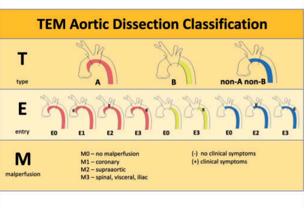
This is a practical classification that uses Stanford as a base model but adds entry point and branch vessel compromise. This is analogous to TNM classification of tumours.(Hans Hinrich Sievers et al)

Where does EVAR stand ?

The management strategy of dissection of Aorta got an important makeover in the last decade (for good mostly ) as interventional cardiologists and radiologists landed in the hitherto to surgical domain with endovascular reconstruction (EVAR) .It is handy mainly in the complicated type B and complicated Non A and Non B . One more viable possibility is the hybrid approach of combining EVAR and surgery in delayed presentation of Type-A.

Single point Principle in Aortic dissection management

Rapid sealing of the entry point and arresting the false lumen progression wherever it is and by whatever means (especially in Type A dissection ) reduces mortality significantly.

Though exit points are important for comprehensive management, one need not waste time to locate and search for exit points. In a specific group of patients, it may not be visible or even absent altogether.

Another critical determinant

Detection and tackling the retrograde dissection and involvement of coronary ostium distortion and damage to Aortic valve , and pericardial invasion is the key to reducing early mortality in Type A dissection

The final outcome in God’s domain

The IRAD and other global registries in Aortic dissection has taught us important lessons. We are continuously learning and the patient outcome is improving.

Still, one unresolved statistical ( metaphysical ) mystery is how to identify? that small subset of patients who are lost because of inappropriately early aggressive Intervention who would have otherwise been pushed into natural survivors of Inaction or less action.

Reference

1.Sievers H-H, Rylski B, Czerny M, Baier ALM, Kreibich M, Siepe M et al. Aortic dissection reconsidered: type, entry site, malperfusion classification adding clarity and enabling outcome prediction. Interact CardioVasc Thorac Surg 2020;30:451–7.

2.Dake MD, Thompson M, van Sambeek M, Vermassen F, Morales JP;DEFINE Investigators. DISSECT: a new mnemonic-based approach to the
categorization of aortic dissection. Eur J Vasc Endovasc Surg 2013;46:175–90

3.Augoustides JG, Szeto WY, Desai ND, Pochettino A, Cheung AT, Savino JS et al. Classification of acute type A dissection: focus on clinical presentation and extent. Eur J Cardiothorac Surg 2011;39:519–22

Metanalysisfor Non A Non B dissection

Pericardial effusion is often detected in patients with Infective endocarditis. Incidence can be as high as 25% . Most often it is mild, can be moderate in few.

Mechanism

  1. Sympathetic effusion in response to endocardial infection. It’s never more than minimal. (Evidence ? it’s only an assumption)
  2. IE related cardiac failure (Raised systemic venous pressure to which pericardial veins drain)
  3. Local sepsis, Abcess formation tracks to pericardial space through transmural lymphatics
  4. Fungal , granulomatous , Tuberculous IE (Rare) Here IE and PE  share the same pathology
  5. Part of systemic sepsis activated Immune mechanism (Polyseroists)
  6. Renal Involvement of IE-Renal failure
  7. Postoperative pericardial effusion in Prosthetic valve IE (Common, often loculated)

Clinical Implication

  • If the pericardial effusion is more than mild, it often denotes worse outcome. This implies more extensive infection or a marker of extracardiac causes of effusion like renal dysfunction.
  • Effusion may predispose to local dissemination of infection and ends up as peri-annular abscess is whether it is a cause or effect of effusion remains to be understood.It is often exudate as one would expect, but transudative  effusions also occur and would indicate more benign course.
  • The sterility of pericardial fluid has not been proven. Culture studies are rarely done from effusions associated with IE.
  • Pericardial effusions appear more often seen in IE of right heart valves. They turn out to be  IV drug abusers.
  • Contained rupture of an abscess needs to be differentiated from effusion

Can we give steroids for PE associated with IE?

Steroids can rapidly plug the inflammatory pores in the from the pericardial surface.It may also prevent future constriction. Currently, routine steroid therapy is not advised in infective pathology . If the infection is confirmed and is being taken care of by antimicrobial therapy there could be a role for steroids with user discretion.

Final message

During the echocardiographic evaluation of IE, the presence of pericardial effusion should be specifically looked for. These patients should be flagged and will require monitoring as the prognosis of PE complicating IE is a concern unless proved benign.

Reference

Two studies one from Spain and other from Egypt looked into this issue specifically.


Severe aortic stenosis will cut off the systolic BP and hence classical pulsus parvus et tardus occurs. This is what , we have been taught all along.

How for it is true?  

One thing is clear from clinical observation. Systolic BP need not be low, often its normal even in severe Aortic stenosis. The issue becomes curious when  high BP is associated with severe Aortic stenosis. This can happen by a variety of mechanisms.(Aging/Loss of Aortic elasticity /Pressure recovery/Hypertension)  I think, there have been little correlative studies of pulsus parvus with central aortic pressure.

Can Aortic stenosis be a cause for systolic Hypertension?   (This academically murky question rose after I stumbled upon this paper )

This paper from the journal of Human Hypertension which was published many decades ago.It sincerely documented high BP in spite of severe AS . The most crucial aspect of this study , however, was the fact that hypertension was completely corrected after Aortic valve replacement. The authors attributed to this high systolic BP as the transmission of LV chamber pressure. This is a frontal attack on the traditional concept of pulsus parvus and systolic decapitation in LVOT obstruction.

I am not sure, whether knowledge always breeds knowledge. Medical science is equally affected by new-onset Ignorance or not recognizing past knowledge.( Like this paper of 1996.) I think this study is done with a good scientific basis and unable to find any serious flaws. Hats of to the authors. This could lead to a further breakthrough in our understanding of transvalvular gradients in Aortic stenosis and the poorly understood vascular- valvular Interactions. With, catheter-based TAVRs become so common, we can exactly measure the pressure dynamics in the Aortic root pre and post valve replacement. (* My take is , systolic BP in severe Aortic stenosis  is preserved until the onset of LV dysfunction)

Reference

Catheter based interventions in TOF  has caught the imagination of  Interventional cardiologists.decades ago. (Quereshi reported first in 1988 Royal Liverpool hospital ) .Somehow it could not develop into a full-fledged modality. The key issue in TOF  is,  RVOT obstruction is infundibular with some degree of valvular involvement. While the valvular component is amenable for easy correction by balloon, the infundibular stenosis requires some form of cutting or splitting. Embryologically,  the malalignment of IVS is the primary mechanism of obstruction. The balloon catheter is will find it difficult to tackle the alignment defect. .Obviously, surgeons can do a comprehensive RVOT reconstruction.

Things are beginning to change. Cutting balloons are available. Various dedicated VSD devices are being developed. Closure of large sub-aortic VSD  followed by  RVOT dilatation appears challenging task but distinctly possible in the near future.

Few cases of palliative RVOT dilatation with a balloon  in critical TOF  is been attempted We hope, in the coming decades at least simple forms of TOF are conquered by the interventional cardiologists!

Hardware: A small profile  coronary  cutting balloon  from Boston scientific .

What is in store for the future ?

3D printing of live heart and designer device or deployable patches for the malaligned VSD is possible. Currently, intracardiac ultrasound would assist the procedure.

RVOT reconstruction with RVOT stenting and percutaneous valves (Melody or Right sided TAVR equivalents) is already been done in post-ICR residual obstructions or late RVOT failure

Coronary cutting balloon flextome tof pulmonary valvuloplasty coronary hard ware

Flextome -Coronary cutting balloon

Balloon pulmonary valvotomy for tof tetrology of fallot

balloon angioplasty for TOF cutting balloon

pulmonary valvotomy in tof tetrology

pulmonary valvotomy in tof tetrology 3

 Other References

1.Boucek MM, Webster HE, Orsmond GS, Ruttenberg HD. Balloon pulmonary valvotomy: palliation for cyanotic heart disease. Am Heart J. 1988;115:318-322.

2.Qureschi SA, Kirk CR, Lamb RK, Arnold R, Wilkinson JL. Balloon dilatation of the pulmonary valve in the first year of life in patients with tetralogy of Fallot: a preliminary study. Br Heart J. 1988; 60:232-235.

 3.Parsons JM, Ladusans EJ, Qureshi SA. Growth of the pulmonary artery after neonatal balloon dilatation of the right ventricular outflow tract in an infant with tetralogy of Fallot and atrioventricular septal defect. Br Heart J. 1989;62:65-68.

4.De Geeter P, Weisburd P, Dillenseger P, Willard D. Valvuloplastie pulmonaire percutanée palliative dans les formes néonatales de tétralogie de Fallot. Arch Fr Pediatr. 1989;46:117-119.