Feeds:
Posts
Comments

Few individual’s works mattered more than others in the field of cardiology. Here was a man born 1914 in Utah, studied at Rush university trained in Mayo, settled in Seattle as a pediatrician. But his passion drove him to become a specialist cardiac physiologist with an urge to find the answers to all those lingering queries that arise as a practicing clinical cardiologist.  He built an exclusive animal lab to study the mechanics and physics of circulation and cardiac pumps in the 1950s 
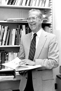
 

                          1914-2001

He can be called the new age, Harvey of the 20th century. He seemed to always bother, how is it that the 6 liters of blood traverse from heart to the periphery and comes back going through vast lengthy circulation with variable pressure and little energy loss.? He also made the very pertinent discovery in neural control, the effect of gravity on circulation. His interest in how venous return would have to match cardiac output was phenomenal. 

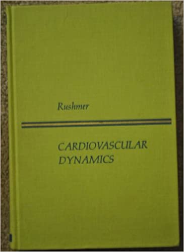
His grasp of cardiovascular physiologic concepts was so powerful and his book on cardiovascular dynamics was so popular. probably the first scientific textbook on circulation. I am sure he had shaped the thought process of so many physicians (I will vouch for myself) and helped create hundreds of cardiologists all over the globe. Dr.Rushmer also did pioneering work on diagnostic ultrasound and doppler. I can recall a video on cardiac embryology edited by him in the 1960s in pre-computer era that probably can not be beaten even today in terms of clarity of content and production value.

Through his thoughts like an engineer and mathematician still, he was able to blend the knowledge together and pass it on to the generation next clinician. No wonder, he was the founder and headed the department of biomedical engineering in the UW. The University of Washington holds an annual Rushmer lecture. 

If one person deserves an award for excellence in cardiovascular science for the 20th century, Dr.Rushmer’s name should definitely, come on top. Though he won several accolades, I feel scientific societies have missed an opportunity to felicitate him with the more worthy award. If the Noble prize in medicine is given for a lifetime contribution to cardiovascular physiology wonder why he can’t be considered for it posthumously.  

It is heartening to note, at the fag end of his career he moved from core science to philosophical and ethical truths of science and technology. He once said, “We’re confronted with the ethical, political, and technological consequences of our medical triumphs. We have to learn quickly how to deal with these profound problems by looking ahead to recognize and avoid complications of our technical breakthroughs’ How true his observation has turned out to be!


Reference

https://www.washington.edu/news/2001/07/16/dr-robert-rushmer-diagnostic-ultrasound-pioneer-dies-at-age-86/

The commonest cause* for repeated entry of right radial catheter to descending aorta is not due to any anomaly. Most times,it is just a skewed angle between right brachio-cephalic artery with Aortic arch, that deflects the catheter to the descending aorta . Just make sure, aortic root is entered with a deeply held inspiration.

*Anomalies of the aortic arch, aberrant right subclavian, Kommerell’s diverticulum, vascular ring must be kept in mind.


Postamble: A true abnormal course

Though, It might appear prudent to avoid the radial route when encountering anomalous subclavian arteries, the reality is different and adventurous. We have acquired great expertise and successful PTCAs have been done through these tortuous vascular highways.

This is a case report from Dr H.S. Isser, Gunjan Garg, from Safdarjung hospital New Delhi. 

A successful PTCA through arteria lusoria : The right subclavian connect to the descending aorta, distal to the left subclavian at the level of ductus arteriosus. and pass retrotracheal and retroesophageal before reaching right arm. Image source and courtesy: H.S. Isser, Gunjan Garg, Arteria lusoria: A challenge for transradial coronary interventionist, IHJ Cardiovascular Case Reports (CVCR), Volume 4, Issue 1, 2020,


One of the topics rarely discussed in heart failure is  CAD as a contributory factor in HFpEF.

This is a copy of the presentation done at the ECHO India Annual scientific meet  2019 at Kolkatta. India.


Will try to find out the recorded version with audio. Here is a GIF run through.

PDF version Download here

Mitra clip is a small metal device that is delivered percutaneously, to clip the incompletely coapting (closing) mitral valve. It was first introduced to treat degenerative mitral regurgitation. It is an interventional imitation of the famous edge to edge Alfieri stitch repair.This procedure in fact converts the single mitral valve orifice into two. In the process, curtails the regurgitation jet orifice significantly. Though the technique looks nice and simple to hear, lots of per and post-procedure issues need refinement. Conceptually it is ideal in primary disorders of the mitral valve. (Read EVEREST 2 criteria for optimal patient selection)  There have been more than 60000 Mitra clips implanted worldwide wide. Thanks to Abbot.

In secondary MR (due to LV dysfunction) Mitra clip has shown mixed results( MITRA-FR not much benefit, COAPT -Did show benefits)

Now, what about Mitra clip as a remedy for rheumatic mitral regurgitation?

This is the question everyone likes to ask. Now we have some interesting breakthroughs. Dr. Ningyan Wong from the National University of Singapore reports probably the first case (Ref 1) . The videos are reproduced with the creative commons license.


Note the classical thickened AML in rheumatic mitral regurgitation.


TEE showing severe MR


Post Mitra clip : A real surprise to note near-total abolition of regurgitation. (This really is good news for the rheumatic mitral valves )

Technical issues

  • Should be isolated MR
  • P2/A2 scallop clipping is the key to success. 
  • The thickness of the leaflet limits the success (Grasping the leaflet will be difficult)
  • Clip Induced mitral stenosis is a distinct risk.

Potential role and future

RHD forms 90 % of valvular heart disease in a country like India. The incidence of Isolated MR in both acute rheumatic fever and chronic  RHD are substantial. If only we refine the hardware and technique to suit these thickened rheumatic valves, Mitra clip is expected to make an impact in this unique group of patients where surgery can be avoided or at least postponed

Though we would very much like to do such a trial in our place, logistics has effectively precluded it. I wish some large centers like AIIMS New Delhi or PGIMER Chandigarh and others can take this concept to the next level.  


Reference

1.Ningyan Wong, Peilin Cheryl Marise Tan, Zee Pin Ding, Khung Keong Yeo, Successful MitraClip for severe rheumatic mitral regurgitation: a case report, European Heart Journal – Case Reports, Volume 3, Issue 3, September 2019

Just roll over the virtual marker along the coronary lesion to get the underlying flow ratio. Blue is an absolute normal segment. Green is ok, orange and red slow-moving coronary traffic jam zones. it’s just like drawing a google map showing life traffic. No wire, no adenosine FFR comes inbuilt in every angio shot. Looks great Isn’t it? This is called QFR. Quantitative flow ratio derived from routine coronary angiograms. It can also guide us to find the optimal sites of both proximal and distal stent landing zone in the best physiological manner.

Which company makes this ?

Any studies done with QFR ?

FAVOR 2 study was reported in TCT. This modality is expected to evolve.

Final message

Whenever possible every anatomical lesion in the coronary should be substantiated by physiological parameter and possibly coronary Imaging to know plaque morphology and vulnerability. Though it is wishful thinking, still for all logistic reasons, most of the real world stenting will be based only on the blind anatomical luminogram.

At this point, please let me utter a non-academic hyperbole. Even a casual query to your beloved patients about their true symptoms and exercise capacity shall make these ultra-modern coronary physiology studies redundant in many. A well-performed and well-interpreted stress test is a good, objective, non-invasive indicator of coronary flow across lesions. It is wise to keep this as a basic clinical foundation in the evaluation of CAD, even as we continue to learn and forget half evolved modalities with rapid expiry dates like FFR, IFR, CT-FFR. QFR shows some promise though. Please watch for next in line coronary physiology – OFR, Optical flow ratio from OCT run through.

Reference

Cardiac myxoma is the most common primary tumor of the heart that presents as mitral inflow obstruction/ regurgitation often with a systemic presentation. It can be either familial, syndromic, or sporadic. Excellent imaging is possible and diagnosis has become straightforward. Surgery is the specific treatment,

What information do the Surgeons need? 

Size, attachment to surrounding structures is the key. The myxoma origins most often in IAS and defining its attachment is crucial. Mitral leaflet distortion, Injury ( and even attachment) is possible. It is helpful for the surgeons if we let them know the mechanism of mitral regurgitation prior to surgery. Echocardiogram including  TEE is sufficient in most. MRI may add some more info. The aim of surgery is to remove the tumor mass completely.

Is myxoma a completely benign tumor?

Another issue is our poor understanding of the recurrence of myxoma. Why should a benign tumor be recurrent? If recurrence is a feature to be counted as a sign of malignancy, myxoma can be definitely a suspect. There seems to be a catch. It is invasive, locally recurrent,  still not malignant. (Whether sarcomatoid degeneration happens is not known. Most pathologists deny this) The problem is, still we are not clear about the cell of origin of this tumor. All that we know is its origin mesenchymal stem cell.


Note 50 % tumor mass enters the left ventricle with diastole. No wonder, as the tumor plops with diastolic cardiac cycle a high-pitched sound simulating opening snap followed by an MDM perfectly mimics rheumatic mitral stenosis. An MR murmur is equally common.

Common sites of recurrence

  • Interatrial septum
  • The atrial surface of anterior mitral leaflet 
  • From contra-lateral atrium rarely

The mechanism of recurrence is either due to incomplete resection or due to its multifocal origin.

4 chamber view showing what appears to be a small narrow pedicle attaching to IAS. Please note echo imaging can be deceiving. Surgeons must inspect the mass in toto before taking the decision to excise IAS or not

What the surgeon needs to do?

The aim of surgery is to remove the tumor completely. It’s painful to diagnose recurrence and subject the patient to another surgery. (We encountered a sorry situation recently) So, when we remove the tumor we should ensure sufficient clearance with normal tissue.Biatrial approach is preferred by some surgeons.( Ahmet Yüksel  Braz. J. Cardiovasc. Surg. vol.31 no.4  July/Sept. 2016)

If the tumor is not well delineated, it is better to remove a significant area of IAS along with tumor mass and subsequent patch closure. Recurrences in AML and contralateral atria is unfortunate and can’t be predicted.

Further, the mitral valve is to be inspected and functionally tested. Minimal repair work or even rarely replacement might be necessary.


TEE imaging of LA myxoma. Note how fragile the edges of the tumor looks. It explains the high incidence of tumor embolus in this condition. Also, to be noted is the forceful impact of the tumor mass on AML that predisposes chronic mitral valve damage.

Final message

Referring a patient to a cardiac surgeon is not a bland ritual. (Have seen many single line referrals such as triple vessel disease referred for CABG) A well-informed interaction by the cardiologist with the surgical team and a possible per-operative echo consult, especially in rare surgeries will bring the best for the patient.   

Reference

1.Reber D, Birnbaum DE. Recurrent cardiac myxoma: why it occurs. A case report with literature review. J Cardiovasc Surg (Torino). 2001 Jun;42(3):345-8. PMID: 11398030


Up to 25 % of LV filling is done by atrial contraction. Atrial booster function is important in LV outflow lesions. This can be critical in patients who have diastolic deformities of LV. ( an audible or even palpable S4 confirms the atrial kick in these situations )  This is how we were taught for decades right. Still, it may hold good in many left-sided condtions, but in HCM it definitely seems to be not true. 

A succinct review of this topic makes a good read.

Incidence if AF in HCM is about 20% (Mostly paroxysmal 70 % , Persistent /Permanent 30 %)

Mechanism of AF IN HCM

  • Increased atrial wall strain(Proven by strain echo studies)
  • Atrial dilatation
  • Atrial pathology (Atrial myocyte  disarray,  myosin is present in atria too. )
  • Unrelated to HCM (SHT etc)

We can confirm with large observatory data, left ventricle handles AF so well. (Ref 1)The onset of AF, (at the least), is, expected to cause some new worsening dyspnea. Even that is not universal (very surprising isn’t it ?)

Does AF correlate with syncope?  again no is the answer. So it’s the LV outflow behavior that determines the hemodynamics not what is happening at the inflow. Even hard outcomes like heart failure, sudden death, net mortality was not found to be altered much by the lesser chamber fibrillation. But, the only issue relevant here is thromboembolism that has to be taken care of.

How is that AF make little hemodynamic Impact in HCM ?

It is difficult to comprehend this scenario. For this to happen the mean LA pressure should remain within the physiological range even when the atria goes to fibrillation. But it seems distinctly possible as many patients with HCM are not aware of this arrhythmia. The LA pressure-volume loop is an enigma. It is likely LA “v” wave loop can adjust to “a” wave deficiency in an exemplary manner.

Further, the hyper-contractile left ventricle can assist itself by sucking blood in very late diastole (to be precise with the onset of systole )and so it need not really depend on the atrial kick.  A similar phenomenon explains the persistence of presystolic accentuation in the murmur of mitral stenosis. The fact that rate control in AF is able to compete with rhythm control in  AFFIRM/RACE study vouch for the negligible hemodynamic impact between SR/AF.

Clinical implication

  • A well-tolerated AF doesn’t preclude the need for thromboprophylaxis. We must ensure  NOAC/Warfarin in all those with persistent AF.
  • Attempts to convert AF to sinus rhythm with all those Invasive LA mapping and Pulmonary vein is unwarranted if not contraindicated.
  • When ICD is indicated additional  Atrial leads to reduce AF is again becomes reductant. 

Final message

Many of the hemodynamic concepts we have learned over the years could be based on logical perceptions that may not manifest at the bedside. Constant flux in our understanding of cardiovascular physiology is required. 

Reference

1.Rowin EJ, Hausvater A, Link MS, Abt P, Gionfriddo W, Wang W, Rastegar H, Estes NAM, Maron MS, Maron BJ. Clinical profile and consequences of atrial fibrillation in hypertrophic cardiomyopathy.Circulation2017136:2420–2436. 


How many times you have treated cardiac arrhythmia in both emergency & non-emergency situations?

Infinite times.

How many times did you really bother to know the mechanism of a given arrhythmia before ordering medication or shocking?

Hmm,.. let me think. (Except for AVNRT/ AVRT, and few VTs, very rarely I have worried about the mechanism  !)

Why is it so? because treatment takes priority and we are able to tame the arrhythmia even without knowing the real mechanism.

The following slide is a gross summary of the cardiac arrhythmia mechanism

Understanding cardiac arrhythmia is vitally important for a few reasons in a few settings.

  • In acute settings, we need to know automatic tachycardias will not respond to shocks. Reentry tachycardias will respond more promptly. (Of course, we may not know it till we shock ) Calcium blockers like verapamil might block triggered activity in MAT. Overdrive pacing is the answer for many automatic tachycardias and some refractory reentrant tachycardias (ATP protocols in ICD has taught us this ) 
  • In the chronic setting when you contemplate mapping, locating, and ablating arrhythmias, mechanisms are important. The task here is locating slow conduction paths and decoding the diastolic circuit around the scar  (If you plan ICD, knowledge about mechanism  becomes redundant again)

  • Finally, knowing the mechanism of arrhythmia is a fascination by itself to help understand the great subject called cardiac electrophysiology, where 100s of ion channels work nonstop drawing the action potential on a moment to moment basis sustaining our life.

A challenge

Can you localize a VT and find the mechanism in a patient who is Ischemic /hypoxic and acidotic? You can never do it. Please note, most polymorphic VTs can’t be localized. The mechanism is either automaticity, trigger activity, or even micro-reentry. You need to shock and look for the causes.(Link to How does the treatment of monomorphic VT differ from Polymorphic VT? )

Final message

Should we need to know about the mechanism of arrhythmia we treat?  Definitely yes, if you have that passion to know the truth, or else just order Amiodarone or shock and check out of CCU. (Of course, we have a very good option of calling EP consult the next day.)

 A review article on mechannism of cardiac arrhymias

Rev Esp Cardiol. 2012;65(2):174–185

 Aorta probably is the most critical structure in the entire circulatory system. (apart from the heart of course !) It is a 1.5 to 2.5 mm thick tube, with a diameter of 2.5 cm/length of 30 -35 cm from the aortic valve to the iliac bifurcation.(Eric Borsero 2011) It handles about 7500 liters of blood every day. Understanding the Aortic pathology has vastly improved at the molecular level with deep gene sequencing that defines fibrillin phenotypes.  Meanwhile, CT ,  4D MRI and 3D prototyping have landed us in a new era where we can feel the exact models of a patient’s virtual aAorta for monitoring and treatment purposes.


While acute Aortic syndromes is the one that bothers us most, even chronic aortic enlargements are equally risky as at any time it can become acute. Though the risk of aneurysm and rupture is related to the histology and molecular disruption at the level of aortic media, we can only rely easily on is its dimension. Traditionally we bother about the diameter since it is the aortic radius that Influences the stress through to Laplace law.(Wall stress equals twice the radius /Thickness of wall)

Click over the Image for ESC Aortic disease guidelines

The annulus is the narrowest part, it gains about 50% width (10mm) at the level of the sinus of Valsalva to reach about a maximum of 35 mm and again narrows at ST to a junction (almost to the same diameter of the annulus) It continues further as ascending, arch and descending aortas with gradual tapering. 

Risk of rupture

Are you aware aorta keeps growing with age?

Unlike many other organs, growth of which gets arrested by adulthood, aorta appears to grow well into middle and elderly age.The aorta grows by 6cm in total length between age 20 to 80 (Ref 1)   The average growth of the ascending aorta is .18mm /year

This fact was reported 70 years ago Dotters famous study Circulation 1950  https://www.ahajournals.org/doi/pdf/10.1161/01.CIR.2.6.915

I used to wonder in many elderly why chest x-ray shows wide superior mediastinum still echocardiogram didn’t show any dilatation.This we call it as aortic unfolding. The term unfolding of aorta may represent the elongation of both ascending and descending aorta. The mechanism is due to multiple factors, like lax ligamentous of aortic attachment, dilatation, and longitudinal elongation.

Unfolding of the aorta . (How to measure unfolding Lee JW, (2014) Aortic Unfolding Determined Using Non-Contrast Cardiac Computed Tomography: Correlations with Age and Coronary Artery Calcium Score. PLoS ONE 9(4): e95887. )

How Important is the length of the aorta?

Risk of Aortic aneurysm is usually attributable to the diameter (Accepted normal 2.1cm/m2) . However, in IRAD registry, 50 % of aortic dissection happened in aortas less than 5 cm. So there is something more than the diameter that confers the risk of an aortic event. Is it the length and elongation?  After years of observation, we now realize it is indeed true. It is a surprise we didn’t realize  elongation of aorta also confers the same aortic wall weakness like that of Increased diameter (Longitudinal deformation/Stretch )

How to measure the length of Aorta?

It is best measured by CT scan or MRI. There is a new parameter called  Wu Index, which is based on both length and width of the aorta , which predicts the risk of aortic event. 


Jinlin Wu, Mohammad  Zafar, Yupeng Li,  J Am Coll Cardiol. 2019 Oct, 74 (15) 1883-1894.

Final message 

Aneurysm of the aorta is traditionally defined based on the degree of dilatation. It’s time, Aortic length should also be included in defining aortic aneurysm. We need to monitor it periodically as well in the population at risk. 

Reference 

1.Adriaans BP, Heuts S, Gerretsen S, et al. Aortic elongation part I: the normal aortic aging process. Heart 2018;104:1772–7.

 
The comprehensive reference