Feeds:
Posts
Comments

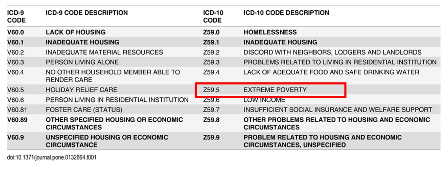
Poverty is a cruelest disease of mankind , the infective vector is not any deadly HINI or retro virus , but mostly the fellow humans themselves ! This is why WHO has included poverty in the ICD code (Z59.5 ) as a disease .

I used to wonder , as a member of Noble profession , should we fight against this disease or be happy to spend my entire life time cleaning the coronary arteries of affluent human-beings and earn few bucks !

Can growth of money eliminate poverty ? 

We may think so  . . .  but it doesn’t most times .Of course ,  affluence bring more jobs to the poor , logically it should alleviate poverty. But , we  know the reality. Its not that easy concept to understand . However we have robust evidence for the opposite ie affluence can aggregate poverty .

If poverty is a disease &  if  mindless  affluence is an Indirect cause for it  , then affluence also becomes a potential disease , Is in’t ? Will WHO include it in ICD listing ?

May I propose Z59.5A to be called  Uncontrolled &  manic desire for affluence ?  Since it is  made at the cost of  fellow humans , it should be clubbed along with poverty as a worrisome disease. Once its included in the ICD manual , I guess it will be unethical to ignore this disease. 

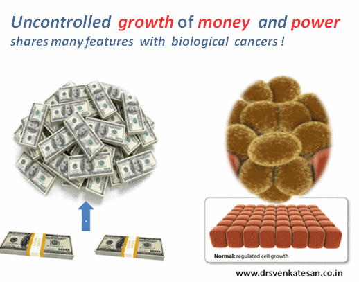
We all aim for growth in life .Nothing wrong in that .There are many facets of life that requires growth. Unfortunately , for most  homo-sapiens ,  growth is synonymous with multiplication of money . . . nothing else seems to matter ! Money when it grows unregulated ,  begins to control you and hijack your  body and mind .

One more issue to  comprehend is , rapid growth of money is possible only at the cost of something (or someone) else – Akin to cancer cachexia ,it depletes the body (Earth ) of it’s resources ( Nature abuse ,  extreme poverty , inequality ,  Third world exploitation , wars, etc ) For the  medical professionals  it is all the more important that growth shouldn’t mean money , as it has a direct and conflicting  impact on some one else’s life  !

mitosis of money 003

Just Imagine ,  if the all the car companies combine together , aim for a  dramatic growth from the current  4 %  to 25 % by 2030  and manage to  achieve it by any means ! . . . This planet will sink in the combined weight of automobile Junk !

It is obvious , uncontrolled growth in any form requires vigorous regulation and  Intervention and will eventually require a radical surgery if the growth goes unabated  !  

Counter point

It is foolish to link growing money and wealth  equivalent to cancer. Unlike cancer cells , money multiplied can be put into use for those in need .It is the principle of charity .But the reality is , human beings who are rabidly after money rarely have this mind set.In contrary the haven’ts have it in plenty. In my opinion, excess money has a dubious  capacity  to  contaminate  human values  ! (Why should some rich and elite opposes affordable health care  to poor ?)

Let us amass wealth and help others . Microsoft  is able to do it. Apple may do it later. What is the need for big companies social responsibility and philanthropy ? If the business worlds  motive and end product  promotes equality and goodness , sans exploitation , the question of charity at a later date never  arises.

Final message

If extreme poverty is a disease ,  forces that Initiates or sustains  poverty cycle can not be a bliss. In this context , the  manic affluence (& the urge for it)  should be included as a communicable disease since it seems to be most contagious as well.

In health care delivery “affluent and modern care” may also connote a sinister meaning. Poor people might think they are deprived of good care because they cant afford it. But ,truth hides deep. True sustainable caring is little to do with affordability+ , since most of the modern health care expenditure is jacked up with junk.

I know in my country people sell their life time assets for what they think as  crucial health care .(Of course universal health care insurance is just beginning to come in. Here again insurance based health care has inflated the actual costs and threatening to impose inappropriate therapies .

As a medical professional we should aim for the cheapest  and best form* of treatment to our patients . Artificially inflating the cost of therapy by worthless drugs, devices, procedures and disseminating them invariably leads to pathological  growth of science.

*If any one thinks “cheap and quality” doesn’t go together in medical care it is ignorance ! Most problems has simple and effective solution.

Post-amble 

 I object this statement -Modern health is nothing to do with affordability.

We need to go to the basics then .What is true health ? Forget about transplants, Organ Assists, Five star critical care .They seem to work in a minority , but drain the world economy. Its Impact on global health is at best minuscule. One Important analysis say 90% health care cost is wasted in prolonging the last 30 days of life of homo-sapiens.(Will get the reference for it )

Please note 

Diseases that occur to  affluent population  is entirely different topic . Can be found elsewhereDisease of affluence


One popular definition of Intelligence goes something like this  “It’s a global capacity  of a living organism  to deal effectively with environment and live peacefully”

When myocytes are confronted with acute ischemia , they  don’t always  jitter . It expresses many behavioral pattern.The damage inflicted is variable as  the molecular mechanism of ischemic tolerance appears to be a virtue ! This might make  much revered time window of myocyte ischemia irrelevant .Each cell has got a unique capacity to survive or die . In chronic ischemia this myocyte intelligence and intention to survive is glaringly evident. How about this phenomenon in ACS ?

myocardial intelligence myocyte memory

Art of  survival 

Why some cells die instantly , some fully recover and few go for hibernation  and others  are just stunned . While apoptosis is programmed cell death with intact cell membrane , hibernation can be termed as programmed cell survival .We don’t know how many intermediate forms of cell surviving mechanisms exists.

One of the famous questions is does the myocyte need blood or fuel for survival* ?

It is a fine balance of various cell  surviving (Anti ) mechanism.How energy is utilised with available ATP molecules is a different  science altogether .It’s  possible like brain,  heart too has  myocardial intelligence  , which does  some independent thinking aided by  fuzzy logic and problem solving algorithms  built within.

Ignorance based ACS care 

While we slog with metals inside the large coronary arteries , the response of the micro vascular territory is at the mercy of God . mRNA instructed DNA codes determine acute mitochondrial  respiratory sensitization.Finally ,the successful reversal of ischemic injury depends upon the cell repair molecule’s crisis management skills !

How else one can explain , some broken hearts recover so well from a major ischemic injury others sink with first insult !

The concepts of pre and post ischemic conditioning  are related phenomenon which can be pharmacologically  mimicked .They  are the major areas of research in myocyte  revascularisation.

Final message 

What is written above  is pure non academic fantasy . Now, read the following article by Dr Kloner which describe the molecular mechanism of  ischemia modification and its impact on clinical cardiology.

Reference 

1.Robert A. Kloner, Shereif H. Rezkalla; Preconditioning, postconditioning and their application to clinical cardiology, Cardiovascular Research, Volume 70, Issue 2, 1 May 2006, Pages 297–307,

Not every one feels the palpitation during tachycardia / Bradycardia /VPDs , Why ?

Palpitation is awareness of one’s own heart beat. It is a complex perception of sensation at cortical level (like dyspnea) . It can occur during physical and mental exertion.However , if it occurs without any physiological reasons , it becomes abnormal. It can mean an abnormality  in heart rate , rhythm or  raise in stroke volume. The first rule of palpitation is both tachycardia and bradycardia can cause it. Tachycardic palpitation is due to valve motion and bradycardic palpitation is due to both motion and increased stroke volume.

The most common mechanism proposed for palpitation is hyperactive anterior mitral leaflet

How and where  does the sensation of  palpitation felt ?

Does  it originate in  the chest wall ?  or Is it the vibrations spreading along  the flow of blood in great vessels ?  or Simply  represent  the vigorous valve motion  sensed by Intra cardiac receptors ? How it is  transmitted to spinal cord where it’s felt at cortical level  ? We are not clear yet. Paccinian corpuscles is thought to sense these vibrations and hand over as electrical signals  to spinal cord either directly from cardiac valves /walls or indirectly from chest wall.

Paccinian corpuscles are predominantly present in sensory nerve fibres located in the dermis of skin. It is also observed in nerve ending to joints, Chestwall, blood vessel and also heart .They act like pressure as well as vibratory receptors * The exact reference for Paccinian corpuscle to be present within the heart is not available to me. Readers may contribute,

Importance of age and gender and IQ

Palpitation is primarily a symptom of young age where the heart is supple and more dynamic. Women tend to perceive more for some unknown reason. Elderly people rarely complaint about palpitation .It could imply aging  with or with out autonomic dysfunction which suppresses transmission of palpitation signals to brain.Chest wall thickness also matters. My guess would be, Chest wall thickness, epicardial fat pad could absorb the vibratory  energy  and chest wall receptors fail to recognise it. One curious observation is,  palpitation is described in a succinct manner by certain patients only. Since , it essentially involves  higher cortical senses , we believe spatial intelligence of the patient  may also be important.

Why Irregularity in heart beat is well recognized?

For the given heart rate , irregular rhythms are felt  more often as palpitation than sinus tachycardia. This is the reason single ectopic beat is easily felt than  sustained tachycardia. A common sequence of  palpitation due to ectopic beat is , a suddenly  missed beat, subsequent pause and forceful post ectopic beat.

Valve morphology and impact on palpitation

Mitral stenosis patients can feel their loud first heat sound (S 1)  or varying  S1 during atrial fibrillation as palpitation; Mitral valve prolapse with redundant , hyper kinetic motion is probably most common cause of benign palpitation.

Sclerosed  and calcific  valves attenuates palpitation. Calcific mitral valve in mitral stenosis make both S 1 intensity and opening snap feeble .These patients are less likely to feel palpitation .

Individual valve pathology can generate palpitation as in Ebstein anomaly , which has a the large sail like septal leaflet that flutters to create palpitation(Apart form pre-excitation syndrome common in this condition) As a general rule ,It is possible semi lunar valves are less likely to cause palpitation than AV valves as the latter only exposed to direct contractile pressure of ventricle.

Right vs left heart origin and localised palpitation

I am not sure one can differentiate left heart from right palpitation. But.palpitation arising from right ventricular  volume overload and increased pulmonary flow like in ASD  are associated with direct local sensation over pericardium . Mitral valve motion can not be localized by  patients .However apical impulse can be felt.Neck pulsations invariably mean high flow states. Venous cannon waves due to high pressure tricuspid regurgitation can be felt with each heart beat (RV systole)

Exertional vs Non exertional palpitation 

Palpitation occurring during exertion often imply its due to excessive handling stroke volume or (Pathological regurgitant volumes) Stenotic lesions are less likely to cause palpitation during exertion it’s never an absolute rule. Exercise Induced arrhythmia always happen in any valve lesions.

Relation with LV function

A dysfunctional ventricle cannot  generate forceful contraction and hence palpitation is uncommon symptom. Cardiomyopathy presents with more of dyspnea rather than palpitation .Even,  an episode of AF do not cause palpitation in such patients .They simply feel breathless (Dyspnea ? Or is it a palpitation equivalent ?)

New age palpitation

With so many foreign bodies and accessories entering the heart  it’s not surprising for patients to feel amusing sounds and vibrations hitherto unknown in human body.

  • Prosthetic valve clicks (Sounds from mechanical valves can be  annoying .Tissue valves, TAVR are more quiet)
  • Abnormal electrical activity  from pacemakers and ICD coils.(Apart form pacemaker mediated muscle twitches)
  • Now, we have entire mechanical LV assist devices  working inside the heart with a 24/7 motors .(LVAD hum its called) Very soon heart is going to become a noisy place and patients would learn to ignore these abnormal sounds

Pleasant physiological palpitation

What brings the unpleasantness during palpitation? (applies to dyspnea as well). It is purely state of mind. While, palpitation due to extreme fear is unpleasant , palpitation due to pleasant emotional arousal (Often referred to as flying butterflies ! (Is it the wings of AML ? ) within the chest  as we hear from some of young  women & men ).Since they know the reason why they get it, cortical input welcomes it ,converts them to pleasant  beats .The Non-academic stuff  is intentionally made to understand how the limbic system and Hipocampus  areas of brain can modify the incoming signals of palpitation that comes from down under.

Thoughts to ponder 

Does post heart transplantation (De-nerved heart) patients experience palpitation ? Again, I am not sure .If palpitation is carried by cardiac nerves it should disappear. Of-course , 30 % of transplanted heart do get re-innervated. When you get a chance to meet a heart transplant patient you ask yourself and find the answer.

*Please be reminded Anginal pain almost vanishes  post transplant.In fact ,there have been instances of cardiac auto-transplant for refractory angina in the past.

Final message

Though all of us can list causes of palpitation without any difficulty , we rarely dwell into exact  the mechanism of genesis of this symptom and  its perception. As we enjoy flying in an exotic world of cardiac  interventions  . . .  the principles of  practice of medicine also expect us to take adequate efforts to understand fully the cardinal  symptoms of our patients . After all , they are the true teachers of Medicine. It is because of their pursuit for explanation for their symptom (Often vague though)  we make our professional progress.

Further  reading

John T.Shepard  The Heart as a Sensory Organ JACC Vol. 5, No.6  June 1985:83B-878

(The heart has variety of sensory nerve endings , still to be explored)

Cardiologists are grappling with at least  half a dozen time windows  in the management of STEMI. (It can be combinations of any of the following :Symptom – DAPT Loading – Door – Needle /Balloon-Sheath, wire crossing etc ) Time windows are Important in choosing the right (or no)modality of re-perfusion . Though superiority of  primary PCI  is thought to be established in academic community , it  may not be in real world. Published studies that suggest pPCI is superior to lysis at any time window  still lack good evidence.

Why is this long drawn confusion  ? 

One of the important determinant of outcome in STEMI , is the thrombus organisation (hardening )time . Some how we have assumed PCI can tackle hardened thrombus  much better than lysis (In fact the outcome in late PCI is as bad or good as lysis in terms of true myocardial reperfusion in this population.This fact will not be visible in scientific data that’s read superficially .One has  to  mine deep for the truth) (Claeys MJ,. . Arch Intern Med. 2011;171(6):544–549)

Two more virtual pathological Time windows.

While we are preoccupied with certain time windows in STEMI  ,may I suggest  two more  Invisible pathological windows. I don’t know , whether these are presumptive theoretical stuff ,  but understanding these time windows will sharpen our decision-making skills in STEMI.

1.Symptom to  ATO time (Acute total occlusion) 

ery gets occluded(ATO ).This is truly Invisible time window .( Pre-Infarction angina  to Infarct time ) Taking the last episode of most Intense pain need not refer to beginning of ATO / Infarct pain. (ACS being as dynamic process in a 24 hour time span an angina  can even be post Infarct angina!)

2. ATO to thrombus organising (hardening) time

It is obvious time is primary factor that correlates with thrombus organisation. But there is much more to it. It’s not the fibrin organisation alone that makes a thrombus hard. ATO gets reinforced by plaque and tissue material ( like steel rods  inside cement) In other words no one really knows  when does the thrombotic process begin or end  and  hardens thereafter. But we know for certain is  tackling a hard thrombus is difficult for both modalities currently we have lysis and PCI*

.(Almost forgot the third modality,  yes its humble drug heparin(.It can do wonders little slow though , Slowness doesn’t matter beyond 24 hrs is it not ?) Now there can be a role for Warfarin also to get rid of chronic IRA thrombus (Moon JY, N The role of oral anticoagulant therapy in patients with acute coronary syndrome. Ther Adv Hematol. 2017;8(12):353-366.)

There are excellent studies that correlated time window to thrombus hardness.At least in  50%  IRAs with time  window less than 12 hrs have thrombus age more than 24 hours Some of the thrombus material aspirated has been shown to be many days old (Kramer et al PLoS One. 2009;4(6):e5817)

Image source : Miranda C.A. Kramer Relationship of Thrombus Healing to Underlying Plaque Morphology in Sudden Coronary Death Volume 55, Issue 2, January 2010

How to arrive at the age of the thrombus  ?

It’s a difficult task to guess the age of thrombus with help  symptom onset and ECG .  There  can be 50 %  error as discussed earlier.

Is coronary angiogram helpful ?

There is no good clue to differentiate fresh from old thrombus by just looking at angio shot. Some experts are able do it (Guess it ?)

Poke and feel with guidewire  : This is probably the best way to tell whether thrombus is fresh or old (Still not fool proof ) Most of us do this in STEMI . All is well if guide wire cuts through  smoothly and nice flow is established.(What we call guide wire angioplasty) Procedure is completed with or without a stent ( &residual lesion) .This is the most gratifying and desirable outcome of primary PCI. (Note : Hardness of thrombus can be overcome stiff wires and force.That doesn’t make it a fresh clot ! This is where we may end up with No-reflow)

nrcardio.2016.38-f4

Image courtesy : Karim D. Mahmoud & Felix Zijlstra Nature Reviews Cardiology volume 13, pages 418–428 (2016) Various forms of thrombus aspirated during primary PCI.

When poke test fails  . . . be ready for a long haul or quit

Thrombus is not a single aged mass of blood. It has lawyers of clot with different maturity  ( like shells over earth ).Hence poking has its own side effects too.Some of it can be violent.When  deeper layers of old thrombus is exposed to fresh blood it can create fresh  cycle of clot activation.( Ofcourse we fight it out with DAPT and heparin) Winner of this fight can never be predicted. To conquer the thrombus or quit is directly linked to the cardiologist wisdom.

What about OCT/IVUS ?

They could help us to assess the morphology of thrombus and give  us Indirect clues about the age of thrombus. Some of the experts say they use it efficiently . My opinion is it adds more glamour than true enlightenment .(Mind you , we need to  cross , clear and flush the vessel to complete OCT. The fact that we are able to complete OCT in STEMI settimg would mean  thrombus is  fresh .In that way it may be useful but without a true purpose.)

Thrombus aspirate analysis : Its more scientific way of arriving at the age of thrombus (Any one want to do carbon dating on this ?) , This again lacks practical use as we need to assess  the thrombus age before poking to avoid subsequent complications. It is also not clear whether thrombus in STEMI is more of RBC and fibrin and net platelet content can’t be quantified.This especially true in stuttering ACS where NSTEMI is threatening to become STEMI or vice versa. (Platelets love to hug each other at high shear force , RBCs do the opposite )

Is the Consistency of the thrombus uniform ?

Here comes the importance of the length of the thrombotic segment. It’s estimated the length of the thrombus segment can be anywhere between 1 cm to entire length of the coronary artery distal to the site of occlusion .The initial proximal part may be soft as its directly exposed to DAPT and heparin.The distal thrombus is flushed only with collateral or a trickle of flow from anti-grade .So ,very likely the distal thrombus is harder than proximal.

How does DAPT loading and subsequent heparin interfere with thrombus organisation ?

Loading DAPT has a definite impact and prevents hardening.(But, one issue is it shouldn’t have been happened before administration)

What is the natural history of organised hard thrombus in IRA ?

  • It transforms  into a CTO.(Many of us believe this is dominant theme)
  • Late total recannalisation – 20% by 30 days
  • Partial recannalisation  (More than 20 % ?)
  • Since wide-spread use of predischarge PCI , true natural history is masked.

Final message

Taming STEMI with pPCI  is not always  a time sensitive emergency procedure . It’s important to recall STEMI patients can harbour thrombus with different maturity .We know STEMI can occur even in  patient with chronic thrombotic process also (even a CTO) . This is proven by a simple fact people walk in 3 days after MI casually. Further, during pPCI both early and late arrivals have equal difficulty though they carry different set of problems tackling the IRA.

If we really  believe principles of coronary care is aimed at tackling coronary thrombosis , wisdom  lies in  judicious use of both CCU and Cath lab facilities .Never hesitate to rush back the patient to CCU for a quick lysis (Or Intra coronary) and avoid the potentially prolonged  battle against huge mass of hard thrombus.

Reference 


Post-ample : A quote 

Importance of  early arrival of STEMI patient to nearest hospital is huge , not because of the possibility getting an emergent PCI . Rather, it is  due to fact that simply reaching the nearest coronary care center dramatically reduce the mortality.(My guess is , this mortality benefit should be more than Lysis/pPCI put together)

Human body is intertwined collection of lives of Individual organs.We believe death occurs when brain dies , respiration stops and circulation ceases . Curiously ,when life ends , these organs  don’t die as a single unit . These three events can happen in any of the six possible permutations.Each organ takes different times to die after loss of life.It is like a crashed computer , where the mother board /RAM memory may be transferred to another and be functional . Out of these three , heart function appears to be supreme as it can function without the need of brain (Science of brain-death) and keep the body alive with intact circulation. Though the concept  called heart dead organ donors is catching up as well. (John Gill et all Use and Outcomes of Kidneys from Donation after Circulatory Death Donors in the United States    

The science of organ transplant has rapidly  evolved .We can transfer an organ from one who is dying or dead to another person who is also dying due to failure of a different organ. Though most organs can be transported and transplanted , we need to maintain life in a dead donor organ . A new and curious specialty  in medicine is emerging which could be named a  Critical Care of Dead or Critical Cadaveric Care Units !) till harvesting happens. Paradoxically ,after the harvest , the organ gains independent  life that has to be sustained.This is where the science of organ transport is waiting in to explode!

How to transport a dead man’s heart ?

There are two Ischemic times in organ donation.

  • Warm Ischemic time
  • Cold Ischemic time

Warm ischemic time refers to the amount of time that an organ remains at body temperature after its blood supply has been stopped or reduced. Cold ischemic time refers to the amount of time that an organ is chilled or cold and not receiving a blood supply. These times quiet vary with respect to different organs.

So far, we have been carrying organs in cold boxes with custodial solutions. (HTK*)

runtranslplant265x125_1234539

This makes the transport time a big limitation of success of the procedure. *HTK -histidine, tryptophan ,a-ketoglutarate, solution 

Whats new in organ transplant science ?

Trans medics is an exclusive company , that has pioneered  in this unique science of organ transport .They have developed separate organ care system for heart ,lungs and liver.

donor heart

It is the first portable heart perfusion system , which increase the time between the harvest to transplant . It avoids cold ischemic time ,replaced by warm perfusion with metabolic activity monitored till  it is transplanted.Ideal far long distance transplant .Soon one can expect trans-continental transplant as well !

This technology could make the difference between warm and cold Ischemic times blurred and in future one may expect to prolong the warm ischemic  period and there is near zero cold Ischemic time.

A heart is perfused by transmedics system while being transported


                                              A  lung breathing in transit

A link to a video

Final message

Modern medical technology can be seen in action , in so many ways and brings both accolades and agonies in equal proportion. While, even flimsy ones tend to get due attention , Transmedics , probably deserves a  grandest reward  for uplifting the science of organ donation . I would think this is the most unique service (playing God )  . . . shipping dead man’s organ  with assured life on delivery  !


Link to Transmedics website

ocs heart transmedics heart transplantation

Click to access 04_yeter.pdf


A middle aged women , with acute onset left sided chest pain and ECG changes was seen by a general physician. He had little hesitation in labeling the patient as  ACS( To be precious he reported the ECG as lateral wall Ischemia) and asked for an echocardiogram to rule out a heart attack  (This is how cardiology is practiced in many areas) .

This patient came to my lab  for the Echocardiogram .The echo window was poor , It showed a structurally normal heart and there was no pericardial effusion. I suspected something systemically wrong in this patient and asked for a X ray chest .

Subsequent scrutiny of this  patient revealed she had moderate left tuberculous effusion. ECG changes are attributed to this. We know pericardial disease can cause ECG changes that mimic ACS. While pericarditis can elevate the ST segment. Can pleuritis without effusion cause ECG changes.  What is the demarcation point between pleural from pericardial surface ?

What are the ECG features of pleural effusion ?

  • Low voltage qrs.
  • Poor R wave or even q waves
  • QRS axis shifts are due to true anatomical / electrical shifts
  • T wave inversion as in this patient

Mechanism of T wave inversion in plural effusion.

T wave represents ventricular repolarisation. To be frank I am not able to give an exact mechanism of such  defects in pleural effusion.

The following mechanisms are  suggested

  1. Left sided pleural effusion can closely mimic pericardial effusion .One can get low voltage QRS in lateral chest leads .
  2. Anatomically I would guess the plural fluid also hugs the heart and the inflamed pleuro-pericardial Interface (Is there fibrous continuity ?) result in some degree of epicardial interference or reversal in electrical polarity.
  3. The true effects of mediastinal shifts with large effusions  on ECG is not clear(Lead V 3 and V4 may pick V5 / V6 signals in left sided effusions )
  4. Fluid altering the electrical conduction property
  5. Associated minimal pericardial effusion and  effectively causing epi-pericarditis as a part of poly-serositis .
  6. After ruling out all plausibility one may think primary ischemic changes as well.

Teaching points and potential error

To label  a left-sided pleuritic pain with ECG changes as ACS can never be considered as a serious error.However , rushing such patients to cath lab or   lytic therapy along with heparin leads to more trouble. ER physicians should always keep in mind T wave inversion in isolation is indeed a rare cause* of ischemia. Still,  as a physician first , we need to have a check list to rule out common non cardiac conditions. Pneumothorax is one another entity that can exactly mimic a STEMI with ST segment shifts and q waves.It’s also possible left sided pleural effusions produce q waves and mimic an old MI as this case report reveals.(Constatine A Manthous Chest 1993)

I think  X-ray chest is least used modality in a coronary care unit for various reasons . Still ,the utility of which can never be undermined and should be used diligently . 

*Of course we shouldn’t  forget a sinister form of ACS  referred to as Wellen’s LAD  syndrome which may present with dynamic T inversion.

Reference

We learn from basic physiology  lessons that human body is made up of 60 % water. What about heart ? There is no reason for the heart should behave differently from rest of the body . If my  assumptions are correct when the normal heart weighs 300g  , 180g of which should be  be water. The same thing could be applicable for LV mass( * Reference requested)

Is there myocardial congestion in cardiac failure ?

Genesis of edema in any tissue depends on local hydrostatic pressures, tissue resistive forces, osmotic balance, and cell membrane permeability. In the myocardium individual contribution of above factors are not known. Apart from total myocardial water content  , myocardial water logging depends upon the trans myocardial venous gradient and the coronary sinus exit pressure in right atrium.Technically ,any severe right heart failure should lead to myocardial congestion at least to some degree.Unlike the lungs , the myocardial edema fluid doesn’t produce crackle , (May cause S -3 gallop instead) . However ,we have modern technology to image water inside the myocardium. Yes, it is called proton / hydrogen imaging or simply called MRI .

This is especially evident In chronic kidney disease , where in the fractional water content within the myocardium is expected to increase further as the whole body is water logged.


myocardial edema by mri in ckd 2

We have seen time and again patients with CKD  improve in LV function immediately  after dialysis . It happens like a magic . The mechanism  is simple .The over-hydrated cardiac Interstitium  threatening to drown the myocytes  is promptly dehydrated by dialysis. This was my wild guess  until I came across this paper which proved the exact points.

myocardial edema in ckd chronic renal failure recovery of lv function after dialysis


Other situations  where myocardial edema may play a significant clinical Impact  (*Includes increased permeability of myocardial capillaries)

  1. Myocarditis
  2. Transplant rejection
  3. Stress cardiomyopathy (Takotsubo)
  4. Congestive heart failure
  5. Acute ischemic injury
  6. No Reflow situation after PCI

Final message

Myocardial interstitial edema in cardiac failure is a grossly under diagnosed  entity.  A water-logged myocardium is classical at least in CKD. We know it can severely compromise the LV function especially, the diastolic function that explains the all too common flash pulmonary edema in CKD.

The number of studies in this  topic (Myocardial Hydrology !)  is minuscule compared to other areas of research in cardiology literature.There is a need to involve both  Nephrologist and cardiologists to explore this curious concept of  dialysable  left ventricular mass in CKD/Cardio renal syndromes !

medical quotes new idea

One more area of research 

It is reasonable to believe,  cardiomegaly in cardiac failure  is primarily related to the increased end diastolic volumes .Still , we are not clear whether there is net increase in cardiac mass as the surface area of the heart increases with dilatation. (Even in DCM ? ) Whenever myocardial mass increases relative increase water is likely. Does the beneficial effect of diuretics in cardiac failure , and the restoration of  LV dimension is due to myocardial interstitial diuresis as well ?

Reference

myocardail edema water content congestion mri n myocardial diuresis interstitial

2.Andrés-Villarreal, M., Barba, I., Poncelas, M., Inserte, J., Rodriguez-Palomares, J., Pineda, V., & Garcia-Dorado, D. (2016). Measuring Water Distribution in the Heart: Preventing Edema Reduces Ischemia-Reperfusion Injury. Journal of the American Heart Association5(12), e003843. doi:10.1161/JAHA.116.003843

3.Cardiovascular magnetic resonance of myocardial edema using a short inversion time inversion recovery (STIR) black-blood technique: Diagnostic accuracy of visual and semi-quantitative assessment Darach O h-Ici, John P Ridgway, Titus Kuehne , Journal of Cardiovascular Magnetic Resonance 2012, 14:22  


Evening cardiac clinic is generally a relaxing place until an occasional patient  with vague chest discomfort present with this sort of an ECG .

He was a 68 y old hypertensive and was on Tablet Amlodipine .After a cursory look at his ECG , eyes wandered to look for some specifics. Suddenly ,my  ECG reading skill was stretched . Is it really LVH ? or Ischemia ? I asked for any old ECG which he couldn’t provide. I had to blink  more than a moment , before asking him to describe his chest pain in detail one more time . I got almost convinced it was not ACS  since he was having only localized pain over left side of chest. Still , I didn’t have the courage to send him home. An Echo was done.

Curious to know what the Echo showed ?


Yes , you also probably guessed right . It is LVH and there is no wall motion defect either.

How to differentiate between Ischemia and LVH ?

This is a common question asked in the board exams.There are number of ECG clues to differentiate the two .Mostly it will help  fellows  to pass exams. Academics rarely comes to your rescue when patients land with  chest pain  especially at odd hours.

Find the answer in this link

How to differentiate LV strain pattern from primary LV ischemia ?


Now comes the real twister , Does presence of LVH  exclude Ischemia in any way ?

Why can’t be ACS in a patient with LVH ?

When I posed these questions , some one suggested global longitudinal strain  with speckle  tracking to rule out ischemic wall motion defect, my resident suggested  high sensitivity Troponin and Ischemia modified albumin.

Whatever is the technological assistance , one thing  is certain,  we need to finally fall back  on patient’s symptoms . Unstable angina is neither an ECG diagnosis nor biochemical  or Echo diagnosis . (Its all about patient description about his angina , that clinches the  diagnosis !)

It remains a fact normal spot Troponin can never rule out  ACS on time ,( Even patients with unstable angina who  harbor  tight  LAD lesion can be both ECG /Enzyme negative )

When we are not sure ,the traditional coronary care dictum  shall operate .It demands admit, observe, with serial ECGs and enzymes. This protocol cant’t be followed strictly for a variety of reasons . We may have to rely entirely on our clinical Intuition accrued over the years.

A modern-day cardiologist might have a different dictum .The simplest solution in such situations is a diagnostic radial snap shot coronary angiogram .It is an easy way out  . . . , and avoids the trouble of spending wasteful minutes of personal conversation with the patient .The ethics of outsourcing history taking , patient record review or even clinical examination has pushed the definition of professional competence in coronary care into murky grey zone.

One more reason we should hesitate to rush these patient to cath lab is the detection of  incidental insignificant CAD (Which will loom larger than life when they are wheeled in from  ER)  that will  lead on to  further inappropriate chain of events.

How relevant is clinical acumen in modern era ?

Clinical acumen in medicine can never be taught in class rooms or read in text books. It is the innate ability to  combine  knowledge, experience ,skill  and  lastly (and most importantly) courage to ignore conflicting and pervasive data from new generation Investigations. Instead of helping us ,they often directly affect our increasingly vulnerable native medical cognition.

Even if some one is blessed with a good clinical acumen it seems to have little value many times as power of Investigations and fear of missing a event will prevail over it ! I have been victim of this phenomenon many times and not able to follow what my mind preach me !

What happened to this patient ?

I had to admit him against my  wish (Of course I was safe!)  He too got admitted reluctantly and was observed till morning , spent Rs 5000 for pack of investigations and stay , was discharged without any issues with a diagnosis of simple Hypertension and LVH.

The non academic bug  didn’t stop there  . . . again  contrary to my conscience  I had to suggest  optional coronary angiogram to rule out true CAD as a precautionary statutory advisory !


Counterpoint 

How do you know this is really not CAD  ? I won’t believe unless and until I see the CAG and its normal.

Ok, Let me post his angiogram if he decides to  undergo it.

We know pleural effusion (hydrothorax) is disproportionately more common on right side in cardiac failure.Though its a well observed phenomenon, the mechanism of which  has not been clear to us. It could be due to multiple  anatomical , physiological factors.


*The are  right and left lymphatic (Thoracic) ducts that drain the corresponding lungs and pleural space . There can be overlap and contribute to the differential occurrence of pleural effusion


Reference 

A meticulous paper written some 75 years ago (1946) from Harvard medical school teach us some important points in this phenomenon.

There is still lot, to be understood about pleural effusion in cardiac failure. We need to know why some pleural effusions tend to occur independent of hydrostatic forces.  It is also noted long-standing transudative effusions can become true exudates. Role of local pleural capillary hypoxia resulting increasing permeability is underestimated.Hepatic congestion and trans-abdominal seepage of fluid is a distinct possibility.

One more area we are not clear is  the relationship  between the  genesis of  pericardial effusion in cardiac failure and concomitant pleural effusion. Post operatively , after univentricular repair (as in Fontan ), pleural effusions can be much problematic with high venous pressure interfering with  pleural drainage.

Impact on symptoms

Finally, even mild pleural effusion can increase the work of breathing and result in dyspnea which is out of proportion to cardiac dysfunction.While we expect the diurteics to clear the effusion of cardiac failure, it doesn’t happen always arguing for a non transudative mechanism in at least some of them.

Further reading

Discerned readers are advised to study the pleural space dynamics in detail.

Link to the original Article of Edgar Mcpeak and Levine 1946


Though heart is known primarily as a pump, it is the four cardiac valves that ensures the critical , non-stop unidirectional flow that sustain the circulation . It is to be recalled these valves originate in the very early days of cardiac development when the primitive heart tube loops , even as the chambers expand from primitive ventricle. Mesenchymal differentiation controlled by various genes that cleaves the valve from myocardial tissue.

While heart by itself is the supreme vascular organ ,  its surprising  few structures inside the heart , like the valves are quiet avascular ( or is it really so ?)

The valves that beat average of 30 billion time in life time ,seem  to get its nourishment from Nowhere ?

Layers of heart valve 


Image source : Huk D., Lincoln J. (2017) Oxidative Stress in Cardiac Valve Development. In: Rodriguez-Porcel M., Chade A., Miller J. (eds) Studies on Atherosclerosis. Oxidative Stress in Applied Basic Research and Clinical Practice. Humana Press, Boston, MA

It’s primarily made up of  complex stratified connective tissue covered by endothelial cells with intervening interstitial cells. The extracellular matrix (ECM) layers are rich in elastin proteoglycan (spongiosa) and collagen (fibrosa). It has been found myocytes and dormant fibroblasts are scattered in the valve leaflets. This forms the basis of focal origins of  Cuspal Ventricular tachycardia J Cardiovasc Electrophysiol. 2002 Jun;13(6):551-6.  from electricaly activated myocytes.

Is there a dynamic cell turnover within the valve tissue ? Where does it get the nourishment ? What is the effect of  aging and CAD on valve tissue ? 

There is growing evidence for valvular extra cellular  matrix are indeed dynamic. This explains valvular lesions in connective tissue disorders like Rheumatoid arthritis. Progressive degenerative changes of aging  involves  remodelling , tissue engineering . Calcification of cardiac valves in endocrine disorders like  hyperparathyroid states implies  cardiac valvular interstitium is in continuum of systemic metabolic pool. There has been very little published evidence  correlating  CAD and Ischemic degeneration of valve.

Evidence for vascularity of valves

It is surprising , this simple question of  vascular supply to cardiac valves has  confronted the scientists for too long.

1.Histopathological and  autopsy studies have revealed vascular channels.(Harper 1938 BMJ)

2.X-ray projection microscopy was used to image the valves after injecting radio opaque microparticles in the ascending aorta. This is probably the one of the few original studies done in UK in 1968 (Clarke et al ) it revealed significant  vascularity of the valve .It was found 16 % the tricuspid valve, in 10% of the mitral valve, and was maximum in Aortic valve (24%).

3.The fact that statins reach the Aortic valve tissue in lipomatous Aortic stenosis indicate significant vascular port of entry to valves.

Direct evidence : Why don’t we ask the surgeon ?

When I asked my surgical colleagues  Does the valve  bleed  as they cut it  ? Most of them were amused with my query , still Iam not sure I got the answer right. At least one of them said since the heart is on pump ,  it won’t bleed any way !


Conclusive evidence : From a 1968 study 

It is academically humiliating to note we have to go back 50 years in time to get a proof for vascularity in cardiac valve.(Or anything new I am missing ? readers may share !) I think , this is strongest proof  for valve cusps are supplied by end coronary arteries. However the penetrance is not deep into all layers of the valve and distally

Is hematogenous spread of Infective endocarditis(IE) occur ?

How does systemic infection reach the valves ? We still believe the IE is due to direct colonisation from circulating blood .Hematogenous spread of IE do occur but difficult to prove. It seems infection of valves primarily happen from outside initiated by  endothelial injury over the valves .

*There seems to be a contribution from  systemic bacteremia  reaching  from within through the pre-existing capillary twigs as well.(Evidence elusive !)


Final message

Is cardiac valves vascular ?  It is curious even in this era , we struggle to answer this query with certainity.

Yes, they are vascular structures , but at best it has partial vascular capillary network. It is also possible  the valves can live a durable life even without these vascular Innervation.(I haven’t heard of a condition called mitral valve infarct or necrosis in RCA/LCX STEMI which supply the base of the heart)

Still, this partial  vasculaity can become Important at times of pathology like infection or degeneration. Expecting more research in this unique area of valvular heart disease

Reference

1.JOHN A. CLARKE AN X-RAY MICROSCOPIC STUDY OF THE BLOOD SUPPLY TO
THE VALVES OF THE HUMAN HEART BY   From the Department of Anatomy, University of Glasgow, Glasgow Brit. Heart J., 1965, 27, 420

2.https://europepmc.org/backend/ptpmcrender.fcgi?accid=PMC1252531&blobtype=pdf

3.Dow, D.R. and Harper, W.F. The vascularity of the valves of the human heart. J Anat193266610–617

4.Ritter, S.A., Gross, L., and Kugel, M.A. Blood vessels in the valves of normal human hearts: from a study of 700 human hearts. Am Heart J19283433–446

5. Saini N, Saikia UN, Sahni D, Singh RS.  Vascularity of human atrioventricular valves: a myth or fact? J Thorac Cardiovasc Surg. 2014 Jan;147(1):517-21.