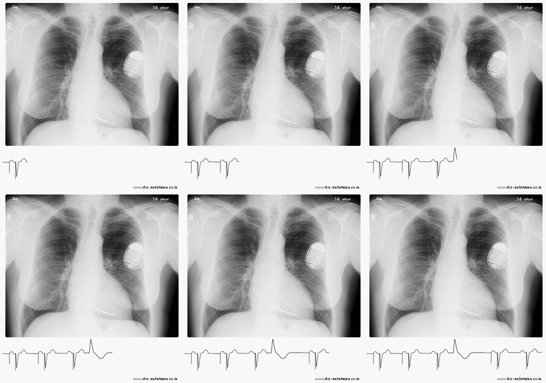
The gradient across coarctation is not simply (& solely ) determined by degree of obstruction , as one would believe.Understanding the hemodynamics and various factors that can influence the gradient is essential Relieving the obstruction /gradient by stent or surgery may not be synonymous with successful treatment as we understand now the entire aorta right from the root to abdomen can influence the gradient ,along with systemic factors.We also know , some of these patients harbor histological abnormalities in the entire stretch of Aorta , what is being referred to as pan aortopathy , that may influence the long-term outcome.
Posted in Aortic diseases, coarctation of aorta, Infrequently asked questions in cardiology (iFAQs) | Tagged coarctation of aorta, collaterals in coarctation, cp stent in coarctation, effect of bicuspid valve on coarctation gradient, effect of ht on gradient in coarctation, gradient across coarctation of aorta, mechansim of hypertension in coarctation, renal hypertension in coarctation | Leave a Comment »
A cardiologist is a physician who has trained himself in a special way to deal with any problem of heart.Ironically , it exists only on paper.The field has developed so vast no one can master everything .There is no such “Pan or global cardiology expert” .In fact it would be shortly become unethical to try to become one !
Pediatric cardiology has developed into such a big field , doing a echo in newborn or infant has become a comprehensive job and requires special talent .This unique and excellent study from Narayana Institute , Bangalore published in the prestigious Annals of pediatric cardiology throws up interesting realities about the quality of echo report done by adult cardiologists in children .The error rate appears huge and stands at prohibitive 38%. While many errors were minor , major were also not insignificant (23%)
With bulk of the pediatric echo involves in the critical decision making process of device closures and interventions the data required becomes vital .The commonest cause for error is probably not due lack of knowledge and but to due to lack of commitment and continuous exposure in doing echocardiograms in those age group.
While this paper decently skirts the issue of quality of pediatric echo done in medium sized hospitals without pediatric cardiology service ,I can say the error rates or inadequate reportage could be significant in such hospitals with apparently good ranking .
Final.message
Of course ,we have many adult cardiologist who do excellent pediatric work , It looks like , as a general rule performing pediatric echocardiograms by non -institutionalized adult cardiologist may not be appropriate ! It may be wise for them to avoid doing echocardiogram in small infants with truly complex disorders (even perceived complex) till they gain the required expertise and confidence.
I recall an adverse issue happened years ago , when I had missed an associated PAPVC in ASD that made my surgeon anxious on table .In a country like ours there is no one to audit our work , “our conscience remains the only option” to deliver the best for our patients especially so, when they are tiny lives in distress.
After thought
Who am I to suggest who should do echocardiogram ? , after all every cardiologist is licensed to do that . One simple suggestion would be , if not confident they can at least mention in their report it is only preliminary evaluation and need to be followed up with an expert . I do that whenever its required and gives me peace of mind as well !
More controversies* to come
Can adult cardiologist do pediatric intervention ?
* Controversy : One of the meaning for this word is “It is a thought process set into motion , that aids digging up hidden truths ”
Reference
Posted in bio ethics, Cardiology -Therapeutic dilemma, cardiology-ethics | Tagged adult vs pediatric echo report, echocardiography expertise required, errors in echo report, errors in echocardiography, ethical issues in cardiology, inadequate echocardiogram, incomplete echocardiogram, mistakes in echo report, pediatric echocardiography who should do ? | Leave a Comment »
We know aortic regurgitation causes a deluge of hugely popular peripheral signs of aortic run off , which are taught right from 2nd year medical school.
When the aorta leaks it reflects in the entire vascular tree .How is that a leak in the remote aortic valve cause a quincke’s to and fro pulsations in the finger pulp ?
Is the blood in the finger trying to follow the regurgitant jet that go back into left ventricle ? Does the to and fro murmur of Duroziez over the femoral artery imply there is reversal of blood flow in femoral artery ?
Things are little complex than it appears
It is true the initiating event of collapsing pulse is the regurgitant jet , however the mechanism that amplifies and sustains it , lies in the altered peripheral hemodynamics.
The systemic arteriolar resistance is dramatically low in chronic severe AR by a reflex phenomenon , as cardiac out put is increased and vascular tree adopt to it. So, with each beat when blood is ejected two things happen in diastole .While a small fraction runs back into LV , the rest of blood runs off , as if it goes in a free way making all peripheral pulses dynamic , bounding and collapsible.
Hence as the name suggest all the peripheral signs of AR are due to the peripheral mechanisms rather than primary event of aortic run off into left ventricle.
Why carotid pulse does not show the collapsible nature of pulse in AR ?
If aortic leak into LV is the dominant mechanism , carotid artery should obviously manifest a collapse ,but it doesn’t ,as carotid has no direct continuity with the peripheral low resistance circuit
What is the hemo-dynamic correlates of descending aortic flow reversal in severe AR ?
The central vascular tree manifest some reversal till the regurgitant velocity fades off . This can occur in severe AR, extending into certain length of aorta. This can be picked up by Doppler probe. Please realise it is only the wave form that get reversed not the actual blood stream.( The momentum gained in systole continues to push forward in-spite of the pulling back forces of regurgitation)
Why peripheral signs are absent in acute AR ?
Acute AR even if it’s significant does not cause a collapsing pulse because it takes time for the peripheral vascular tree to go for vasodilatory mode.Further ,LV is also less compliant keeping the LVEDP high and regurgitant fraction low.
Summary
Answering the title question ,the mechanism of Aortic run off in AR is both central and peripheral. However clinical signs are largely due to high cardiac out put and the resultant adaptive response of the vascular tree due to low systemic vascular resistance triggered by reflex dilatation of small arterioles of the peripheral vascular bed.
Posted in Aortic diseases, Aortic regurgitation, Cardiology - Clinical | Tagged arterial pulse in aortic regurgitation, diastolic run off in aortic regurgitation, mechanism of collapsing pulse in aortic regurgitation, peripheral run off, peripheral systemic arteriolar dilatation in aortic regurgitation | Leave a Comment »
In the last few decades we have understood a major concept in the genesis of cardiac arrhythmia.Slowing in the propagation of cardiac impulse is a key trigger to precipitate a reentry circuit and initiate a tachy- arrhythmia.Still , many conditions like first degree AV block, chronic RBBB or even LBBB are benign entities as along as the heart is structurally normal .They seem never increase the incidence or life time risk of cardiac arrhythmia . Longevity is unaffected.( Or do we assume many things ?)
How is this possible ? or is the theory of slow conduction triggering reentry is flawed ?
Think again . . . if these patients who later on develop a structural heart disease , with an episode of ACS , myocardial or valvular disease, the original slow conduction substrates these people were harboring , will it become important ?
Surprisingly , we have no answers in literature.When Haissaguerre et al found preexisting ERS pattern could be a trigger for primary VF in case they develop ACS , he opened up a huge debate as it involved converting a vast number of normal population electrically anxious.
Now ,is it possible the so called benign blocks of heart like first degree AV blocks , RBBB , LAHBa , would be important at times of ACS and possibly make them prone for for primary ischemic arrhythmia .
Is bundle branch re-entry possible in structurally normal heart ?
We need answers. Some one , (Any EP fellow) somewhere could take up the issue and enlighten us !
Posted in Cardiology - Electrophysiology -Pacemaker, RBBB/LBBB | Tagged bundle branch reentry, eelctrophysiology, electrophysiology, ers pattern, moes theory, rentry mechanism, sloe conduction and reentry, theories on genesis of cardiac arrhythmias | Leave a Comment »
ICDs are one of revolutionary devices , invented last century that can defy “death & fate” in high risk cardiac patients who are threatened with ventricular tachycardia or fibrillation .A decade long hard work by Mirowski and team from John Hopkins culminated in the dramatic the first AICD implant in 198o. ( In my opinion, this medical invention can be compared to an event of such significance as moon landing by Armstrong and team ! ) Ironically , in the last decade such a revolutionary device was sort of misused and thousands of devices were explanted for inappropriate indications.
Fortunately , better sense prevailed recently .The indications are getting refined. I am sure ICD will go a long way in prevention of both expected and unexpected sudden electrical deaths .We are into the 4th decade of its evolution.While the electrical circuitry has been mastered , power supply remains an issue as they require continuous power supply like a mobile phone. Current technology allows about 6-8 years of battery life.
Now , Boston scientific has come out with new technology which make its battery life extend by 100% to 12 years. It is a major break through , expected to evolve further until probably we have rechargeable batteries or biological power sources .Stretching a wild thought , the days couldn’t be far off when the smart phones which are omnipresent in every human-being , could not only power the ICD remotely and control it too !
Indications (ESC/AHA 2012)
CAD
- Post MI* /LV dysfunction ≤ 35% /NYHA class II or III (* > 40 days)
- Post MI* /LV dysfunction ≤ 30% /NYHA Class I (* > 40 days )
- With non-sustained VT due to prior MI, LVEF < 40%, and inducible VF or sustained VT at EP study
Non ischemic structural disease ( Idiopathic DCM, ARVD etc)
- With structural heart disease and spontaneous sustained VT, whether hemodynamically stable or unstable.
Primary electrical disease
- With syncope of undetermined origin with clinically relevant, hemodynamically significant sustained VT or VF induced at electrophysiological study
Reference
Link to Product manual form Boston scientific.
Posted in Cardiology - Electrophysiology -Pacemaker, ICD -Tips and Tricks, ICD and Pacemakers | Tagged aicd mirowski, boston scientific icd extedended life, el icd dynogen inogen, icd indication, life of icd, madit study icd | Leave a Comment »
We have two options to manage AF.Rate or rhythm control .(Of course , in the strict sense , rhythm control also confers rate control that is built in-situ with SR ) .There was an initial confusion which strategy would fare better .For a decade or so rhythm control was thought to be supreme. That’s logical to expect as we restore physiology in the later .” We know, medical science often disrespects logic , and scientists reinvent this harsh fact in regular fashion” Now , we have clear, consistent data that proved rate control is a better strategy in most situations of AF .(AFFIRM, RACE 1 and 2 studies). The aim of treatment of AF are the following .
- Improve symptoms of palpitation
- Improve hemodynamics
- Reduce MVO2 and hence avoid ischemia
- Prevent tachycardic cardiomyopathy in the long-term
- Avoid stroke .
Unfortunately or fortunately rate control strategy was able to fulfill all these aims with fair degree of success. There are at-leaset 3 reasons why rhythm control fared poorly .
- Rhythm control is actually a myth. Only about 35 % patients remained in SR at any time in rhythm control .Runs of transient AF can occur at any given day* and make a mockery of the much hyped rhythm control !(*Due to heightened adrenergic tone or adverse biochemistry/ hypoxia)
- The drugs used to maintain SR are far more toxic . The complex EP procedures to convert to SR has not helped either.
- Most importantly , rate control with anticoagulants were able to achieve better stroke reduction than rhythm control group.The reason being stroke risk was unabated even if rhythm is back to sinus, as risk of ischemic stroke continue to emanate from as many sites like aorta, aortic arch and carotid. Hence, in a stroke prone population with AF , it is the meticulous anticoagulant that’s is going to prevent strokes rather than rhythm control .Since the rhythm control patients would need to continue anticoagulants , they lose a presumed logical therapeutic advantage.
Posted in Atrial fibrillation | Tagged affirm and race study, rate and rhythm control in atrial fibrillation | Leave a Comment »
A short systolic murmur over pulmonary area (ie Left second inter coastal space ) is listed among 6 other auscultatory feature of pulmonary arterial hypertension.Though it is an accepted sign many would question the existence of such a murmur or its relevance in PHT.
Why does it occur ?
Acoustics principle tells us whenever velocity of blood flow exceeds a critical point(Raynolds number*) in a specific anatomical territory , a turbulent zone is created and a murmur could be generated .This is why many physiological situations like pregnancy, anemia, and some benign outflow murmurs occur.
In pulmonary hypertension , three things are thought to contribute for the murmur generation
- Dilated pulmonary artery promotes Raynauld turbulence
- Increased flow velocity (This is correlated with pulmonary artery acceleration time in Doppler)
- RV contractility (A normally functioning RV is required to generate the murmur .Once RV dysfunction sets the murmur of pulmonary hypertension usually disappear , of course a TR murmur may appear and confuse the picture )
Reference
* Reynolds number is a way to predict under ideal conditions when turbulence will occur. The equation for Reynolds number is:
Posted in Uncategorized | Tagged mechansim of systolic murmur in pulmonary arterial hypertension ?, pulmonary hypertension | 2 Comments »
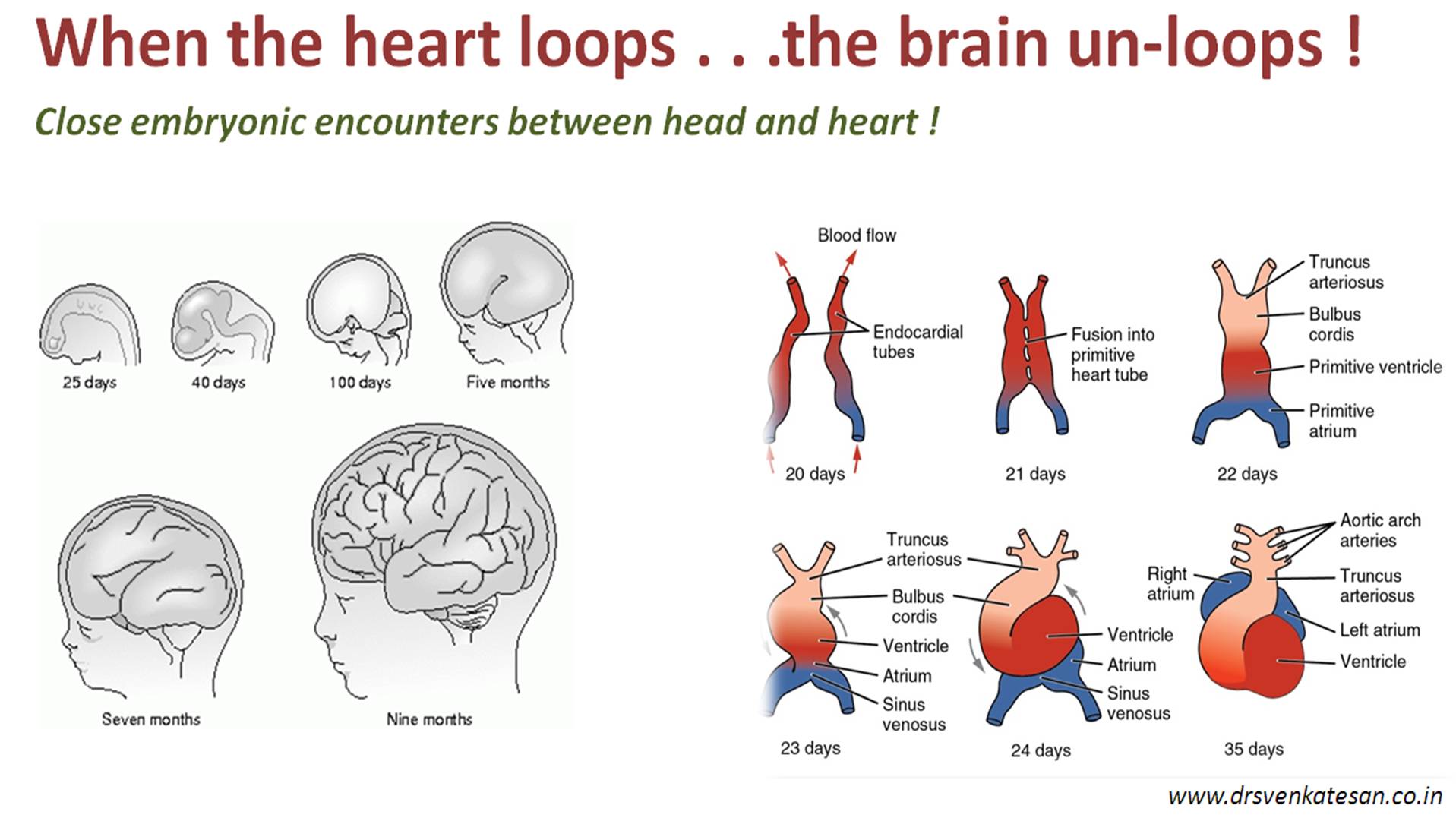
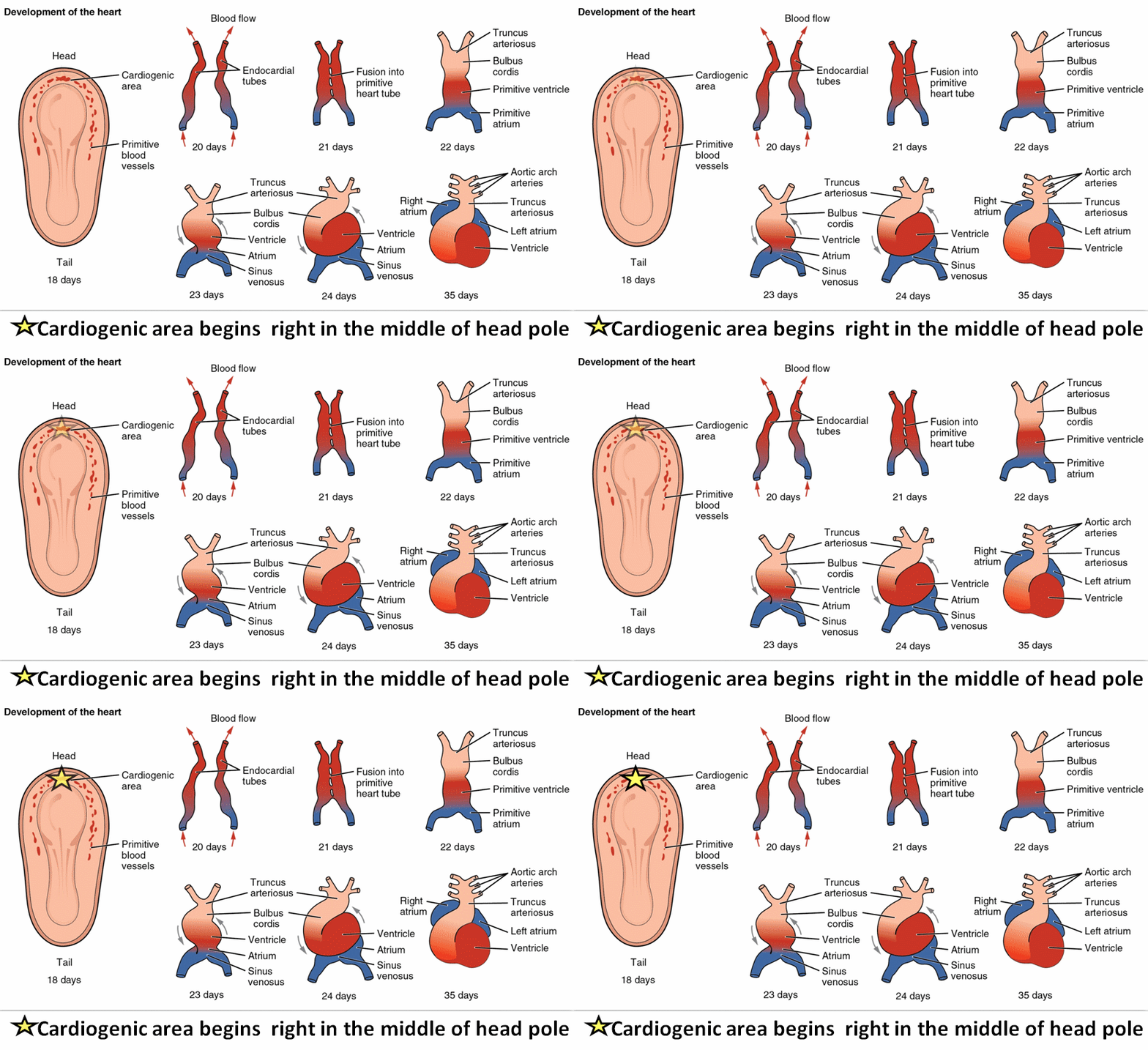
Heart by development originates from near the same spot , where the brain develop (Neuralcrest) .Hence there is no surprise to note, heart being a primary vascular organ still retain many neural connections with brain .Eyeballs with it’s extensive neural inputs can be considered as adirect extension of brain.
Oculo cardiac reflex .
When the eyeballs or the ocular muscles are manipulated or massaged slowing of heart rate can occur .This is due to a reflex called Oculo cardiac reflex mediated by vagal stimulation .This phenomenon is also referred to as Aschner phenomenon
The circuit
- Afferent _Trigeminal branch of opthalmic nerve
- Center- Medulla : Trigeminal neural signal spill over signals to Vagal nucleus
- Efferent- Vagus -SA node
Biochemical mediator -Acetyl choline
Prevention
- Adequate local anesthesia
- Retro bulbar block of ciliary ganglion
- Prompt Atropine injection
Clinical scenarios
- Opthalmic surgery : Serious bradycardia even asystole can occur as a rare complication especially in elderly and very young (Cataract /Squint surgery) .
- Cardiac events and strokes are clustered around opthalmic surgery in many elderly for some unknown reason ( OCR triggered ?)
- OCR can unmask hidden sinus node dysfunction in elderly.Routine cardiac evaluation before eye surgery may be recommended .
- Orbital fracture especially Medial orbit can elicit dangerous bradycardia (BMJ Case Rep. 2014 Apr 15;2014.)
- Rarely sudden death has been reported (Smith R (1994). “Death and the occulocardiac reflex.”. Can J Anaesth 41 (8): 760. )
- OCR for termination of SVT/AVNRT : , One can use the eyeballs to stimulate the brain stem nucleus of vagus to terminate a rapid supraventricular tachycardia (Like carotid sinus message) .Cold water immersion of eye is effective way to stimulate the vagus.(Diving reflex -Mathews 1981)
Neural control of heart how Important it is ?
Many sudden cardiac deaths are now believed to be neurogenic in origin . Though, somatic nerve supply of heart is least important except over pericardium , extensive sympathetic and parasympathetic nerve supply is present . They can now be visualized by adrenergic receptor imaging . Neuro cardiology is distinct developing field. A hyperbole: Of course one could argue , these connection has less overall significance as a person can live with an entirely new donor heart with zero neural connection with brain.
Reference
1.Lang S, Lanigan D, van der Wal M (1991). “Trigeminocardiac reflexes: maxillary and mandibular variants of the occulocardiac reflex.”. Can J Anaesth 38 (6): 757–60
2.Mathew PK (January 1981). “Diving reflex. Another method of treating paroxysmal supraventricular tachycardia”. Arch. Intern. Med. 141 (1): 22–3.
3.Borumandi F1, Rippel C, Gaggl A.BMJ Case Rep. 2014 Apr 15;2014.Orbital trauma and its impact on the heart.
Posted in Neuro cardiology | Tagged circuits for occulo cardiac reflex, diver's reflex, eye ball massage, occulo cardiac reflex, trigemianl vagal reflex | Leave a Comment »
William I am Harvey first discovered human circulatory system in the year 1628 .Published his work in “De Motu Cordis” (otherwise known as “On the Motion of the Heart and Blood”) as a 72 page booklet in Frankfurt book fair. The world of medicine changed forever , and new system of human circulation was born.
Read this now
Excerpts from Chinese classic of Internal medicine , written 2000 years before William Harvey,
All the blood is under the jurisdiction of the heart .Twelve blood vessels are deeply hidden between the muscles and cannot be seen.Only on the outer ankles are visible because there is nothing to cover. All other blood vessels that are on the surface are veins. The harmful effects of wind and rain enter the system first through the skin , being conveyed to the capillaries. When these are full , the blood goes in and turn empty into the big vessels .The blood current flows continuously in a circle and never stops
Post-amble
Of course , this in no way takes credit away from any body .William Harvey collected every data on circulation available at that time , and came with that classic De Moutu Cordis , the importance of which is undisputed. But ,history time and again tell us there are silent restless brains pondering over important concepts all over the globe .Whoever has the access to scientific facility , proves the same point , publishes first and gets attention . After all thoughts are never rewarded in human domain ! (God , does it I guess !)
Reference
1.Hume E.H Medicine in china ,old and new,American medical history 1930; 2;272-280
Posted in history of cardiology | Tagged history of cardiology, human circulation, william harvey | Leave a Comment »