Feeds:
Posts
Comments

While many of us are preoccupied with wires and balloons ,( coronary  myopia ! )  , our radiology  colleagues are making rapid strides . Let us spend some  time  to understand  how  the myocardial segments  are inflicted the  final insult . We need to realize , there is a pattern  to  this myocardial  end game of scarring and fibrosis.

MRI is the  gold standard to assess the myocardial architecture . It has a role in both assessing the anatomy , function  , perfusion and viability .

how to differentiate ischemic dcm from idiopathic dcm myocardial scar epicardial transmural

  • LV function is assessed  by cine MRI
  • Viability  stud by  delayed enhancement MRI (DEMRI , also called as  LGE- Late Gadolinum enhancement  )
  • Myocardial scar best assessed by DEMRI*
* Why do you require DEMRI to identify scar ?
One can detect scars in plain MRI but contrasts make it better .Hence delayed enhancement in by DEMRI is used  to detect scars.
Is it ischemic  DCM or Non ischemic DCM ?  ( That is the question we commonly ask  
We rely too much on CAG anatomy for this. It can be misleading. Cine MRI with DEMRI  gives the answer straightway with high degree of accuracy  .  CAG is required in all  ,  but if it is normal , or  has insignificant lesions  , the dilemma  of ischemic DCM would continue !)
**Note ,there is one   simple algorithm proposed by the author   to  differentiate  Ischemic DCM from Idiopathic DCM  without MRI – Click here to  Link
Following  scar patterns in DEMRI help us to arrive a diagnosis.
Favors Non ischemic  DCM
  • Mid myocardial scar
  • Epicardial scars
  • Global sub-endocardial scars
  • No scar(Ironically if  no delayed  hyper-enhancement is noted it is likely to be non Ischemic DCM )
Favors ischemic DCM
  1. Regional transmural scars
  2. Localised sub-endocardial  scars
* Ischemic DCM will always involve subendocardium as ischemic wave front goes from sub-endo to epicardium.
examples for Non Ischemic DCM
  • Amyloidosis (Can be restrictive as well )
  • Chagas
  • Fabrys

Why is  scar localisation and Quantification important ?

Apart from differentiating various cardiomyopathies  it has  few clinical implication .

  • Since scar indicates irreversible damage , if extensive  it will  argue  against any re-vascularisation .
  • Scar location becomes vital if we plan CRT .It will be futile  to place a CRT lead over a scar.
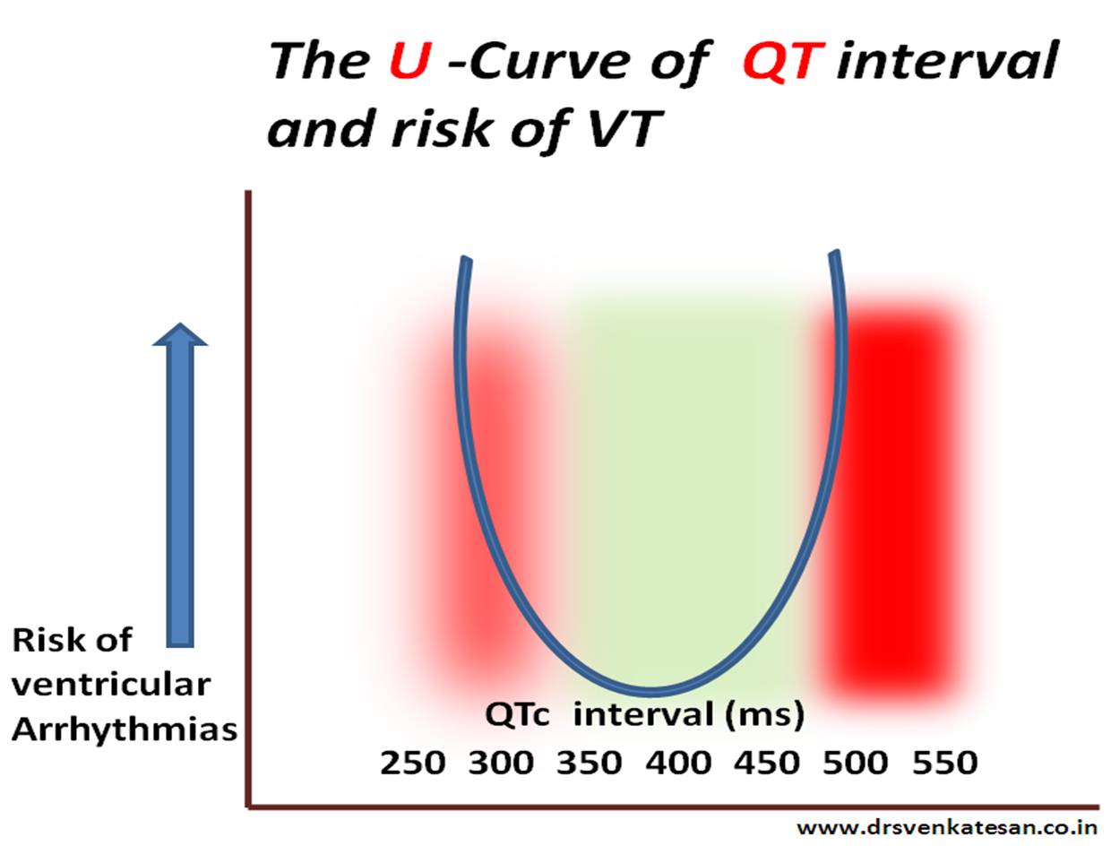
  • Scars are often  form a macro re-entrant circuits for VT .Help us localize or zeroing in VT focus.
  • Scar quantification is helpful risk stratification of patients  with HOCM .and their family.
Final message
Myocardial scar location and quantification  is the new mantra in a  patient with dilated heart with cardiac failure.
It may be more important than even a coronary angiogram .MRI  will prevail over   any of the available echocardiogram modalities to assess the scar pattern.
Reference
myocardial scarring mri

I know ,there is a VSD out there !  but I am  unable to  get the gradient across it.This situation can be quiet  common .The reasons could  be technical, anatomical or  hemodynamic.

As a rule ,  if we hear  a  pan-systolic murmur clinically ,   one must be able to catch  a good  Doppler spectrum somewhere by  echocardiography . However , If  the murmur is restricted to  early or mid to late  systole, VSD  jet is often attenuated in echocardiography .

In the  following situations ,  VSD  jets  may not  record a distinctive Doppler spectra. Invariably the velocity is low , spectrum is short,  less intense ,  lacks good shape and borders are hazy !

  • A closing  VSD
  • A Small muscular VSD
  • VSD with  Severe pulmonary hypertension
  • VSDs with muscle bundle criss crossing
  • Double chambered right ventricle (DCRV, where VSD usually drains to high pressure chamber.)
  • VSD  associated RVOT obsruction  (Note: classical TOF VSD will never generate a murmur)
  • VSD with sinus of valsalva aneurusms (  Doppler  jet  can be really  difficult to record )
  • Inlet VSDs are missed because  convectional  views of echo are perpendicular to these inlet jets.(Short axis better  )
  • Another common situation  is post STEMI VSR.Both a small apical VSD or multi tract  VSD associated with  infero posterior  STEMI   gradients are  difficult to obtain.

What is   inference ?

Doppler spectrum will help detect  small VSDs and color doppler will not miss even a tiny VSD.Doppler spectrum across VSD  is dependent many  factors other than the presence of VSD. However some large VSDs are detected better by 2D echo rather than doppler signals.

Final message

Presence of a Anatomical  VSD does not  imply it should generate a noise.The murmur as well as Doppler signals  are  primarily  determined by the pressure difference on either side of VSD. After all , one of the  largest VSD  that  we encounter

Syncope in CHB is due to unsafe escape rhythm, changing focus of VPDs,  extreme bradycardia, (<20 /minute),  pause induced VT, (Usually polymorphic and torsades is quite common .)  ultimately may end with convulsions,  ventricular fibrillation, and death.

Syncope in SND is due to extreme slowing of SA node . Sinus pauses or even arrest can happen resulting in ventricular standstill. Fortunately, a stable escape rhythm ensues more often than in CHB. (It may just be around 20 or 30/mt. still, ventricular arrhythmias are uncommon. ) This implies an important fact that stability is more important than slowness.Fatality is rare in SND.However, the mechanism of syncope in  SND is influenced by the integrity of AV conduction also. If it is severely impaired it can trigger ventricular arrhythmias as well as the escape focus becomes unstable infra hisian location.

Paradoxically, in patients with SND, an episode of palpitation due to AF  or sinus tachycardia precedes the episode of syncope. An intelligent patient may recognize this as a warning and can take lying posture after runs of palpitation.This is because of tachycardia-induced suppression of  SA node prolong the sinus node recovery time still further.

How to differentiate cardiac syncope from simple vasovagal syncope?

Cardiac syncope  is differentiated by common vaso-vagal syncope (VVS) as the latter occurs during erect posture . It may be entirely due to vascular component and hence it may simply represent hypotension without a true cardiac limb .(Vasodepressor syncope)

Hence the pulse rate and volume may take some time to recover in VVS, while Stokes  Admas of CHB  usually have a well-formed bounding pulse in the recovery phase, as the rate is low and systemic hypoxia is a consistent feature.

How is the respiration during Stokes – Adams syndrome ?

Intact. Oxygenation in the lungs goes on for time being. The pooled pulmonary blood gushes after the termination of syncope and causes  the classical flushing. Since the hypoxia causes systemic vasodilatation the flushing is more obvious.(Unlike vasovagal syncope where they are often pale)

History of stokes Adam’s syndrome Morgagni is the  one who gave credit to their  discovery

Though Morgagni first described the clinical picture of this syndrome in 1761,  It was published much later by Two Irish Physicians  Stokes, Adams. Wish this entity is referred to as Morgagni-Stokes-Adam’s syndrome

Reference

1.R. Adams. Cases of Diseases of the Heart, Accompanied with Pathological Observations. Dublin Hospital Reports, 1827, 4: 353–453.

2.W. Stokes. Observations on some cases of  permanently slow pulse. Dublin Quarterly Journal of Medical Science, 1846, 2: 73–85

qt interval animation short and long002

The stretch and strain experienced by the action potential’s left shoulder region is almost similar* in both long and short QT syndromes that trigger a VT.(* Hope this explanation makes some electrical sense !)

*Click  over the image for  high resolution

What is short QT interval ?
It is a range . Any thing less than 380 ms can be considered  short.Generally It becomes important only at < 320ms.
Relationship between QTc and risk of VT .
U curve of long and short qt syndrome

Time is muscle. This quote  became  sort of ” cardiology sermon”  in the last  few decades .Cardiologist think  they stand  on a 100 meters sprint track once a patient with STEMI arrives .This is indeed true ,  if we  agree  time is  muscle and  our urge is to reduce the door to balloon time .Please  remember ,  this rush matters  much ,  only if the patient comes through very early  when the muscle is really getting damaged . (No issues  . . . even if the fire engine comes in  slow motion if the  house is burnt fully !)

Time is muscle agreed  . . .  but  muscles are  kept alive by  factors other than time  !  So muscles can  defy time if God  is willing !

Time is one of the important components of management of STEMI.  Other things matter too !  Age , baseline co-morbidity ,  underlying extent of CAD, collateral support of IRA territory , and finally  individual variation in hypoxic damage in myocyte is (Rarely  been studied in detail.)

Door  to balloon time for a patient  who lands up within  1 hour window need  to be  much  different from a patient who comes at 10 th hour .The issue is important  because  we use a procedure which requires delicate decision-making ,(IRA-Non IRA issues etc)  the results can be  sub optimal ,  and even be hazardous in low risk STEMI . So , door to balloon time  may be a less  important  component of  time window in a patient who comes after 6 hours .This is the reason  overall outcomes are not changing in a large cohort  of rapidly performed PCI.

The presumed  absolute  relationship  between  “Time  and  muscle”  concept is  always been a suspect . This  is proven by a flawless study from  NEJM .

nejm stemi most important article

http://www.nejm.org/doi/full/10.1056/NEJMoa1208200

This study should infuse more sense to  us ,  time and again, we are  hijacked and sedated by high dose of  pseudo scientific concoction .In fact ,  indiscriminate rapid PCI may not be in  the good interest of  all  patients with STEMI ,  if it is not properly done  .Without realising this fact many developing countries are indulging in extravagance of  costly STEMI programs wasting  the exchequer.

This landmark NEJM  paper convincingly underscores a fact  that  achieving  rapid door to balloon time  is not  going to be the game changer in  conquering  the Global   STEMI  championship  . We have to take the coronary care into the streets  or to their homes as well .This is where the pre-hospital thrombolysis will  emerge in a big way in the future .

A slow and steady thrombolysis beats a fast and furious primary PCI on any given day in all uncomplicated STEMI .This we have proven for over three decades in  one of the India’s largest coronary care unit .( Where is the data man ?  Genuine experience is data . Why  we require , the act of publication to convert an experience into evidence . Often times ,  I  would feel , data is the most unscientific word in medicine . Many Truths  lack evidence , false hoods come with plenty !  For all those  scientific  homo sapiens  , please recall  70 % of ACC/AHA class 1 recommendations are backed by level C evidence ie simple opinion from  perceived experts! )

Final message

A fast and furious primary PCI may not be  the answer in all STEMI population

Thrombolysis  can be  done  with near  zero time delay , it does not require special expertise where an ambulance driver can reperfuse   a myocardium without much fuss and glamor ! He does not have to  split his hair to identify which is the IRA in a complex multivessel STEMI as well ! The streptokinase and TPA will home in  to the target site  smoothly and swiftly .

If indeed ,  time is the major factor in STEMI , we have many other ways to tame  the time . If muscle is more important than time ,  pPCI is  rarely  the answer !

Some India specific  thoughts

Is it not a shame  , we talk about primary PCI  for all  our patients  who do not even get timely Aspirin* after a STEMI! .It is something akin to what we witness every day ,  as our country folks  wield touch screen  Androids  . . . conversing  in open air toilets !

* While the importance of  Aspirin is undermined , It is different story altogether , these patients  get sorbitarate promptly whenever they get chest pain  (mis-placed and  dangerous priority ! )  prescribed by the  roaring  GPs ,  who suffer from discontinuous medical education ,  propelled  by the deeply penetrated 1000 crore oral Nitrate market .

And STEMI workshops are conducted by self-proclaimed experts  every few months in posh  7 star hotels all over India .

Anterior mitral leaflet  has a classical M  shaped motion. Infrequently , M mode echo will record a triphasic pattern .

Triphasic AML motion

The exact  answer is not known . I guess it is a normal variant.

Often  it is recorded  when there is a long  and redundant AML , especially if the M-mode cut is too close to  the tips.

Though it is not common , I have seen in few the triphasic gets converted into classical M shaped pattern if the cursor is moved slightly away from the tip of AML.

Relationship to Heart rate

Some times it appears in slow heart rate and tends to disappear with tachycardia .

Triphasic Doppler filling vs Triphasic M-Mode

We do not know yet ,  how  the  triphasic AML motion  correlate  with triphasic Doppler filling pattern which  is considered a fairly good evidence for  LV dysfunction.

The pericarditis  of acute rheumatic fever is not a true infective pericarditis.It is more of inflammation .It is primarily  T cell mediated  reaction . Neutrophils rarely take part  in this inflammation and hence  no significant  exudation . Hence , there is less  sticky and adhesive molecules inside the pericardial space .The most inflamed layer is epicardium which a nothing but visceral  pericardium .This layer lacks the tensile strength to constrict the underlying myocardium.

why rheumatic pericarditis does not go for constrictionFor constriction to occur the fibrinous  ( thick ) layer of pericardium need to be involved . In rheumatic fever  even though it is  pancarditis ,  fibrous layer is not  involved. Further the inflammatory gradient is thought to spread from within  (Unlike tuberculosis )

Note :  In chronic tuberculous pericarditis,  diffuse inflammatory process  invade from the exterior surface . Very often ,  one can not differentiate  layers. In extreme cases even myocardium and pericardium can not be separated .

Summary

The peri-cardial effusion of acute rheumatic fever

  1. Is transient ,non infective and resolving (Unlike valvular inflammation !)
  2. Less of neutrophil activation  (Less adhesion)
  3. It does not involve the thick , tensile  fibrous layer of pericardium hence lacks the contractile force .

Other lingering  questions

1.How common is tamponade  in acute rheumatic fever ?

2.How important is the mass of the effusion (Viz  a Viz  Intra pericardial pressure !) in causing tamponade ?

//

In this mean world ,most truths  exist without evidence  . . . and often  falsehoods  masquerade as truths with  overwhelming evidence !

Human  biology   has  always  been a mystery  and can express  in dramatic  ways  . While  ,  many  disorders   combine to play havoc on the body ,  few tend to  protect  each other. HT and DM can join a deadly coalition to attack the heart .Smoking  causes  extensive peripheral vascular disease ,  still  thrombo angitis of coronary arteries ( due to smoking ) is  virtually unknown. Tuberculosis does not  have the  courage to attack  the heart  valves  ,  while  it  can inflict serious injuries  all over the body . Similarly , systemic hypertension and  Rheumatic heart disease  does not  combine  well . So , it can be assumed  some unique  and hidden   protective factors  are at play  among different pathological entities and their target organs.

A  brief  account  of how COPD could be related to  CAD !  (* Mostly Imaginary !)

We know ,  COPD ,   stresses  the right  ventricle by pressure overload and in extreme situation  affects  the  LV function because of  hypoxia. It rarely impacts the coronary artery disease  . This has been  our  consistent  observation. While COPD patients often land up with LV dysfunction , investigations reveal  they are  more of a dilated cardiomyopathy and their coronary arteries are entirely normal. Diffuse atherosclerotic CAD is a rarity in  patients with  history of  bronchial asthma. Coronary micro circulation  is also observed  to be largely  intact in most people with  COPD .

We  haven’t got a call   from our pulmonology  wards  in  many decades ,  for  a true   emergency  coronary consult . Mind you ours is a  200 year old  Institution , with 3000 beds  , largest east of Suez canal !

It’ s very rare for bronchial asthma  patients  to die of  a cardiac event. Thousands of   elderly patients   throng our ER with acute severe asthma every winter  , still  extremely rare  to  precipitate an acute coronary event !

We are yet to see  critical  triple vessel disease in a patients with documented  bronchial asthma and COPD .  Even  non-critical CAD is far less  frequent  in  COPD   vis a vis  general population .  It is indeed a strange  observation  , considering both entities are rampant in the community  .

What could be  mechanism  for the perceived disconnect between COPD and CAD ?

Is it a myth ? Does it happen in all geographical zones ?  If  hypoxia is the sine qua non  of COPD  ,  one would rather  expect a close association  with CAD  , isn’t ?

One  suggestion  that  keeps  erupting  from my cortex   . It is  the  wide swinging intra thoracic pressures in COPD or  asthmatic individuals  . . .   somehow responsible . These wide swings  of pressure  are  transmitted to aortic  root . They  transform into  good coronary perfusion pressure  ,   keep the vessels  clean by pressure vacuuming effect .

We have  asked our epidemiological unit to  analyse the  25  year data from our coronary care unit  to decode the mystery .

Counterpoint

Meanwhile,  a  diagonally opposite  question was asked in UK  and found a partial proof as well . Our experience do not agree with this study  conclusions .

What is your take on the issue ?

bronchial asthma and cad

http://ije.oxfordjournals.org/content/33/4/743.full.pdf+html

Introspection

How can a opinion (rather an Imaginary essay !) based on personal observation   projected as a  scientific fact ? We need to observe , analyse and publish the data . This is what the scientific world expects us to do . Unfortunately , the journey form observation into publication has been kept  purposefully difficult . In my opinion bulk of  the international peer reviewed  medical journals with high impact factor can  convert any  junk  data  into a  scientifically palatable  recipe  !


Platelets are humble blood cells that  roam  in clusters and guard  against  any  bleeding in internal organs. Though it has natural powers to regulate itself  against aggregation at inappropriate sites , certain high risk individuals need to take these drugs to prevent cardiac event. Patients  who harbor intra-coronary foreign bodies like stents  need more intense antiplatelet  regimens.

We have variety of antiplatlet drugs. Aspirin does it  by blocking  COX. It is irreversible .Clopidgrel does this differently by inhibiting  P2 Y12 receptors .Clopidogrel has been used extensively in India. Some brands of it are many folds costly as well !

A curious encounter

A certain patient with a stable CAD,  from higher strata  of society was offended when I replaced his long term  prescription of Plavix(Clopidogrel)  with Aspirin.His major humiliation was  this  new drug costs just  50 paise ! He suggested  to me  ,  it is huge insult to him  as his driver also takes the same medication !

What does pride  do in platelet inhibition I tried to explain him  ?

He was amused !

I asked him to go elsewhere , to any star-rated , upscale health suit  nearby to fulfill his wishes !

The above event happened few years ago . I am just posting it from by diary .

Final message

In this unequal world , prestige comes to play even in illness and the drugs they take ! I wonder, how prevalent, is the issue of pride  in our patient’s mind that decide  the treatment modality  in modern day medial care !

Your comment.

The doctrine  of  modern medicine  goes  something like this   . . .

For most medical problems ,  there  would be a solution. Keep  trying . . . till you get it !

*But , just make sure that problem on hand deserves a solution in the first place !

Modern medicine continues to  remind us  every day , the much hyped solutions  often end up in new problems and  many  times worse than the original problem !

Oh ! what a  great a quote ! When I  was boasting   myself  . . .  My wife reminded  me ,  this is just  plagiarized version of  a  2000 year old Hippocratic thought !

Primum non nocere  . . . first do no harm !

But , Hippocrates’  life  was  not  contaminated  with  drug eluting stents, I pads, and  BMWs

If   Hippocrates  arrives  in   cath lab  today  by BMW which sucks  50000 Rs EMI  ,Everolimus coated  coronary  jewellery   will  definitely  tempt him !

You  can’t  simply   compare lives separated by 2000 years

. . . I told my wife !