Feeds:
Posts
Comments

Archive for the ‘myocardial disease’ Category

There are many  organic causes of mitral regurgitation. ( Ischemic , degenerative , valvular , cardiomyopathy etc.) It is not  rare for  pure  electrical events to result in valvular regurgitation.   A 70year old  man  with SHT   presented  with palpitation  and exertional dyspnea  .He was  later referred  for  Echocardiography.  Echo revealed LVH with intermittent MR and moderate LV dysfunction.
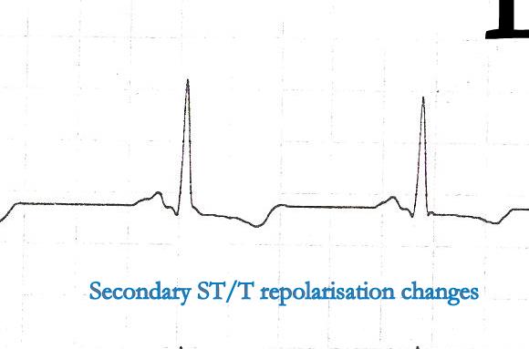
His ECG looked like

Ventricular ectopic recorded in bi-geminal rhythm

His  echocardiogram showed


His echo showed randomly timed mitral regurgitation was detected .See the Doppler MR jets below.

We know ventricles are integral  part of mitral valve apparatus  .Hence  it  wouldn’t  be a surprise to note  abnormally timed ventricular contraction  can  have a major impact  on mitral valve function.

When ventricles  prematurely begin  to contract  ( As  during  VPDs) it  interferes with  opening of mitral valve. In other words every VPD  technically imparts a  sort of  diastolic dysfunction !

VPDs occur in which part of cardiac cycle ?

VPDs  occur  either in early   or mid  diastole . Thank fully VPDs can not occur in systole . (Refractory period )

What would be the status of mitral valve at times of  VPDs?

Though it depends upon the timing of VPD ,  generally it interrupts the rapid inflow period of diastole .

In fact ,  it converts the cardiac  cycle from diastole to a partial systole or  a combination( fusion ) of diastole   and systole ! *

More MR jets are visualised than LV filling waves . Note the some of the E waves are sandwiched between two MR jets. ECG gating should have made this image more interesting .Any way , we have good MR jets to time systole nicely

* Is that a funny  imagination  ?

During   diastole ,  if  LV suddenly  begins  to contract   instead of  receiving the blood  ,  what will happen ?

VPDs are such a common arrhythmia , we  rarely  wondered  ,  it can have a dramatic  consequence  in a any  given cardiac cycle .While   the cardiologists think too  technically  their  patients observe with  shrewd  sense and tell us clearly  what  they feel  is  actually a   missed beat !

(Yeh  . . .  how simple  they describe the complex  hemo-dynamics  of  missing  diastole !)  .They also tell  us ,  next systole is felt as big thump as palpitation . (Post VPD potentiation )

Just imagine ,  if a patient  has  multiple VPDs  with  different  coupling intervals   that fall in different location of diastole  also  interspersed with sinus beats ,   how chaotic  would be the  the  mitral   filling .

This is what  is recorded in the above patient with multiple random MR jets .

Why all VPDs do  not cause MR ?

The timing is critical .We know all VPDs do  not generate a powerful contraction to cause MR. Atrial fibrillation, Prolonged PR intervals , heart blocks , critically raised LVEDP all can influence the trans mitral gradient . In fact these situation can result in  an  entity called diastolic MR that would be discussed later.

Can  VPD induced MR be  referred to  as diastolic MR ?

When VPDs  occur  in  diastole  , it  interrupts the diastole  and a new systole begins. In any  particular point of time there will be  leak into the LA  if the mitral valve is open .This is technically a new systole but in true sense it is the diastole of  the  previous beat . I wonder , whether   VPD induced MR  may be referred  to as one  form of  diastolic MR.  Of course ,  this MR can spill over to true  systole as well .

This also  makes  sense (Non !) as many of the VPDs do not open the  aortic valve ,   hence technically we can’t call the phase reset  by  all  VPDS   as a true systole !

What is the effect of VPDs  on pulmonary venous flow ?

Left atrial  cannon waves can occur that can elevate PCWP .This is the prime reason for resting or  exertional  dyspnea in these patients. Some may get a paradoxical relief  during exertion   as  exercise  suppress VPDs which are frequent at rest.

If VPDs can seriously interfere with mitral valve function , why  they are  often  considered benign  ?

VPDs are well tolerated* as long as  the  LV function is intact.  If VPDs are associated with  LV dysfunction  it  can initiate a vicious cycle of   hemodynamic deterioration .  Multiple VPDs  if left untreated can lead to progressive LV dilatation  in a  significant population .  Hence patients with  recurrent VPDS need some sort of  follow up. It  makes good medical sense to suppress VPDs in the long run. (Of course the  available anti VPD  drugs  are not very safe  !  The search for non toxic ,  ideal drug should go on !)

*”Well tolerated VPDs”   in no way  means  normal physiology.  Read a related article in my site.  “3 minutes crash course on VPDs”

Final message

VPDs  though considered  largely benign , can lead to dramatic  alterations in the  functions  of mitral valve , especially in diseased hearts.

We  must  realise  when ventricular  ectopic beats occur frequently  , it  interfere with the  both opening and closing of mitral valve.

It is really surprising  ,  the literature is  devoid of  major studies  about the  impact of  VPDs on  mitral valve  physiology . . . rather pathology !

Read Full Post »

Modern  day cardiology can do wonders. It can revive a sinking  patient in cardiogenic shock with IABP , LV  assist ,   multivessel angioplasty and bring back  life . On  the other  hand  , a young man with an infected mitral valve who is put on  intensive  antibiotic  regimen   , progressively deteriorates  throws an emboli into brain ,  raise his urea  creatinine  , cardiac   failure worsens and finally succumbs .

This is a clear case of failure  of medical therapy in infective endocarditis .  It is almost certain  surgery would have saved him .

Why  the delay ?

So the question that is been debated for so long is   “When to intervene with surgery in IE”  ?

While we show extreme  urgency for ACS , the same is not shown  with IE.This is going to change in the future .Thanks to the  EASE trial (Early surgery  in  endocarditis )  This land mark study from Korea  is likely to revolutionise  the way we will look into the  problem  of infective endocarditis. It was presented in the just concluded AHA annual scientific sessions  in Orlando

This was  our  observation  too . The issue was discussed in  the year 2008 .It reminds me ,  every  learned  thought or opinion is in fact a paper  but unfortunately modern science does not accept a  fact without evidence of a  study . Until then  it remains  as a crap !

I am glad  to note   genuine concepts will some day  get ratified . Kudos to the Korean team.It is a great study to do with  many ethical issues.

Click below to read the related article

Link to EASE Trial  http://www.theheart.org/article/1313215.do

Next question  on the cards

Should there be  a time window above which medical therapy should be   deemed (Doomed !) to have  failed  , so that the patient becomes a default candidate for surgery.

Read Full Post »

Third heart sound is a unique heart sound  because its   perfect physiology  to hear it  in the young  ,  while the same may denote  serious LV dysfunction in patients with myocardial disease.

It is a low pitched  early diastolic sound usually correspond to  the end of rapid filling phase.The mechanism of genesis of this sound has been debated for many years .(Still I think it is unsettled !)

We know genesis of intracardiac sound is contributed  by three factors

  • The blood flow
  • The valve motion
  • The myocardial contractile and  relaxyl  property

The above  three is collectively  called cardio-hemic system . When this system vibrates heart sounds are generated .In  the genesis of S3 all the three may be important . The only difference is ,  in physiological S3 the valvular and hemic component play a major role . In pathological S3 the  myocardial component has a pivotal  role .The distended LV facilitates chest wall impact during the rapid filling phase . It is now  accepted  LV S3 is  generated outside the LV  . It  was proved elegantly by Shaver et all with sound recording on either side of  LV /Chest wall.

It is to be emphasized  the mechanism of genesis of S3 is diagonally opposite in  physiology vs  pathological  S3 in some conditions . Rapid AV filling  can  rarely be  responsible for pathological  S3  associated with severe LV dysfunction , while chest wall  impact can contribute in both physiological as well as pathological S 3 .

 One can understand the complexity of genesis of  S 3  , as  there are  too many  determinants  that contribute in  varying degree of acoustics.

Factors determining the intensity of S3 is complex 

  1. Age,
  2. Atrial pressure,
  3. Rapidity of  flow across the atrio-ventricular valve,
  4.  Rate of early  diastolic relaxation 
  5.  Distensibility of the ventricle,
  6.  Blood  volume,
  7. Ventricular cavity size,
  8.  Diastolic momentum of the  heart,
  9. Degree of contact (coupling) with the chest wall, thickness
  10. Character of the chest wall
  11.  The position of the  patient.


It is ironical, pathological   S3 which is a  diastolic  sound  though ,  still  its genesis  is largely  determined by the systolic function of the heart .This mystery is partially solved as we recognise  now ,  LV S3 is equally common in  severe degrees of diastolic dysfunction. In fact ,  while we were studying the relationship  of LVS3 in DCM  , it  has strongly predicted the  presence of   severe restrictive pattern in them .


 Reference

1.Multimedia of S3

http://www.inovise.com/learn/s3causes.html

2.Importance of  S3 in cardiology NEJM 2001 article

http://www.nejm.org/doi/pdf/10.1056/NEJMoa010641

3.Chest wall impact theory of S3  by Shaver

Shaver JA, Salerni R. Auscultation of the heart. In: Hurst, ed. Heart. 8th ed. New York, NY: McGraw-Hill, Inc; 1994:291.

Read Full Post »

Sustaining a STEMI  may be a  pathological  end  point  for  coronary  artery disease. But ,  from the  management point of view it is  actually  a starting point for CAD evaluation  .Strategies to prevent further   cardiac  events   must be formulated .

How do you manage a asymptomatic  un-complicated  post  STEMI   patient*  at discharge ?

  1. Do a sub- maximal symptom limited EST and then discharge.
  2. Advised  to come back after 2 months for a  stress test or Perfusion imaging
  3. Continue  with intensive  medical management without EST or  CAG and monitor only the symptoms
  4. Advice coronary angiogram   in all and decide depending upon the lesions (Pre -discharge CAG )
  5. I am a modern day cardiologist  . This question does not arise . . .  as I do only primary angioplasty for all my cases !

( *Please note ,  this forms the bulk of  STEMI population (up to 60 %  )

Answer : Your guess is the correct answer!

Why we need to risk stratify STEMI at discharge ?

The  morality and outcome in STEMI  though appears  to be a   continuously falling  curve ,  the slope is not linear.

The classical   mortality till discharge is about 6-8  %. Between discharge and 3oth day there is 1-2 % additional mortality

At end of first year there is  further   2 % mortality. From  second year onwards there is an annual attrition rate up to 3 %.

The aim of doing  a pre-discharge  EST is to do identify  ” patient  subset ” who are destined to die  within 30 days of STEMI.  If you schedule the   EST  after 6-8  weeks  one can not prevent these two deaths out of 100 !

( Of course ,  we assume   a prompt revascularisation in those vulnerable would prevent this !).  By doing so , we can avoid the bulk of unnecessary PCIs  that  happen  with  routine CAG following STEMI.

Pre discharge EST can be done safely  within 5-7 days  with  a symptom  limited test (70 % of  THR or up to HR of  120 /mt ) This  simple test if it is negative can virtually R/O  a  critical proximal  lesion with near 100% sensitivity.

Should we  risk stratify patients  who have undergone pPCI as well ?

Most of us  would love to believe ,   once  pPCI is  done to the  patient , he  reaches  a therapeutic end  point. But  it is not the truth . It is  the degree of  LV dysfunction ,  extent of contrary coronary lesion  ,   co existing risk   factors  and  the  intensity of medical treatment  only  would  determine the long term outcome.

It is very important to  realise  the pPCI is aimed at opening the IRA  and other lesions are  often left alone. So never  believe  pPCI   per se  would confer total risk reduction following a STEMI  .  There is considerable evidence to suggest  the opposite may be true at least in high high risk pPCI  ,where  metals are   placed  in  complex ,   vulnerable thrombotic milieu.  Hence it  seems logical  to risk  stratify  all patients   after primary PCI   (In fact, this population require  more vigilance )  .

When will you advice an  EST following  pPCI ?

It is usually not needed in the immediate discharge phase in single vessel disease which  would have been  tackled during pPCI.In multi-vessel CAD , where  only the IRA was tackled during pPCI  ,the same guidelines that of  thromolysed  STEMI shall apply  .Since we know the coronary anatomy already ,  EST helps us to evaluate the hemodynamic status of non IRA lesions if  there are any  . While ,  this is a  logical debate , logics has a rare place in medicine . It is ironical ,  stress test   is rarely  done  even after 6months following pPCI  in most centers.

Final message

It is  a  pity  ,  anatomical risk stratification  has squarely beaten  the scheme of   physiological risk stratification in most cardiology centers . A pre -discharge EST* was a  good concept that gave us an idea about the coronary reserve  after the ACS.  It was a collective wisdom of cardiologists  that has hanged this useful concept.  It is still more shocking ,  to note even the  scheduled  6 week   EST is  dropped from the  post MI work up in some  institutions.

* Many would consider  ordering an early EST in STEMI is an act of bravery ! The fear seems to be genuine   and most will agree with that.  But , please remember a physiological test  (Cheapest and simple is EST or a  Nuclear perfusion )  should precede  CAG  in all  asymptomatic  post STEMI  population  whenever possible . If  EST could not be done  prior to CAG for some reason   , at least do it following the CAG . It  will have  an  important impact  on the downstream decision making  which is often an  inappropriate  PCI  !

Read Full Post »

If  we   think we have  unraveled  all the  mysteries  of   human coronary  blood flow   we are  sadly  mistaken . Most cardiac physicians spend  their  prime life  in opening the  obstructed coronary arteries  playing  a role of coronary plumber.

Like any plumber ,  it is not going to be  one time job and   our patients  would  have to hire their  services  periodically  . Many times  it turns  out to  be a 108/  911 call  as well !

Unfortunately , hem0dynamics  of  coronary blood flow  do not follow  the principle of  water flowing across  a domestic  pipeline.The most dramatic   difference  between  the  coronary blood flow and   water pipe  is ,   in the later  ,   as  the water is being ferried   across the house  ,   neither the building   nor the   pipe    contracts    (Unfortunately all our understanding , derivations and research were  based on simple physical  modules  of  hydrodynamics in a static  delivery  system )

Pressure flow relations especially in biological system is  not  simple. Since  our  foundations on principles of   blood flow  is based on this simplistic model  ,  every assumption  could be proven  wrong. This  is what  is happening now . Nothing seems to work  in a  learnt  manner.

A patient  with  100% occlusion  walks comfortably  without damaging his muscle.While an other patient  would  develop cardiomyopathy even if the occlusion is  gradual   and  incomplete  ! Hemodynamic  logic tells us blood flows from high pressure to lower pressure  zones  like a water fall !

But coronary waterfall is not a simple and smooth affair. It is not a free fall  ,  even as the water falls there are  pumpy  interruptions .When these  pumpy ride  occur  even in physiology one can imagine  the pathological states  , when  the coronary  artery is blocked ,  the myocardium is  scarred and the systemic blood pressure fluctuates .

While every  organ welcomes   the systole  ,  as they are fed  with  blood  during this time of cardiac cycle  . Heart  is only organ which sacrifices  its own blood flow during this phase  as the systolic contraction  interrupts the blood flow .

Determinants of coronary blood flow

What we learnt over the years has been too simplistic. It is not the  patency of vascular  system that matters. The coronary micro vasculature, the metabolic demand, the neuro  humoral regulation etc.  For  most cardiologists  the epicardial  patency   or stenosis remains the only relevant  issue

The reality is  much complex  to comprehend

  1. The coronary perfusion pressure
  2. Coronary flow reserve
  3. Coronary wave forms
  4. Sub endocardial vs subepicardial flow ratio
  5. Effect LVH on myocardial flow
  6. Coronary venous tone and arterial ischemia.

Now,  we have an entirely new concept which proposes (Rather proven concept !)   the  integrity of  myocardial contraction and relaxation on the coronary blood flow. This land mark paper in circulation has identified  six wave forms of coronary blood flow This include 4 positive  waves and two negative waves

Questions need to be answered 

During diastole  myocardium relaxes . Only if  the myocardium  relax   optimally  the compressive effect of systole  on coronary  coronary   micro vasculature is reversed  ,  intra coronary resistance  falls so that coronary blood flow can occur smoothly. We do not know  whether diastolic dysfunction would  affect the diastolic coronary filling waves  jeopardizing the coronary flow.

Myocardial viability is important for one more reason  , in the distribution  of   coronary blood flow .A dysfunctional muscle can not receive  and  inject  the blood  deep into  sub  endocardium (Note this becomes  important  when  revascularising   severely  dysfunctional segment )

Does myocardium has a  calf muscle analogy   and  behave like  a  powerful  intramuscular perfusing pump .

A breakthrough concept  from Davies et all in circulation .  These are not new ( Buck -Berg  ?)thought  about this decades ago .  The interest is rekindled in recent years  ,  as  complex angioplasties  following myocardial infarctions  failed  to improve outcome and relive symptoms in many .

During primary PCI ,  no- reflow  often  denotes a meaning  of  failed  PCI .The issue involved  is  hydrodynamics of intra myocardial  blood flow .The following  article partly  answers the  issue  underlying no re flow .http://circ.ahajournals.org/content/113/14/1721.full.pdf+html

Final message

Young  physicians  need to  spend  more time  in  basic  cardiac sciences . Lest, what  we  do  in cath lab blindly  will become a laughing stock  ! We have to go back to the golden years of  research in cardiac physiology  (1960 -1970s)  . Mastering coronary  angioplasty  may increase the blood  flow  up to the  myocardium ,  but pushing the blood beyond the muscle requires more sense  and effort .

A simple  hemodynamic  model based  on  physical  principles alone is a  greatest error we make in cardiac science . * Further, human heart muscle is not only influenced by the quantum of blood  it receives  but to the great extent the content of blood.The blood caries all the ill effects of  systemic diseases and  damage   the vessels and muscle .The interaction  between the  blood and  the muscle  is never  an issue in  the pure  physical labs .( Even animals misbehave !)

Read Full Post »

This seemingly straight forward question is often asked in cardiology boards.

The answer to this question is  important in the bedside as well ! Ironically ,  with  sophisticated  diagnostic modalities the complexities  has  also multiplied .

The following table attempts to simplify it. ( Mostly written with a personal knowledge and ignorance !)

Please click on the table to visualise a high resolution  image.

Read Full Post »

Tuberculosis is rampant in our country ( of course in any developing country ). Many of the patients who are referred  for cardiac  failure has history of pulmonary  tuberculosis.

In our echo lab we handle  100s of  patients every month from the regional TB sanatorium  . While the clinical presentation  mimics  COPD ,  many of them  have be severe bi-ventricular dysfunction  in echocardiography   and  bulk  of them satisfy the criteria for dilated cardiomyopathy . So what are we missing ? or guessing ?

The following paper  which  finds a  strikingly similar observation  . . .   is important for many reasons

  • It  comes for a prestigious Institute from India (PGIMS Chandigarh)
  • It strongly  suggest the link between DCM and tuberculosis
  • It also tells us both can be completely curable.

We know cardiac failure is a relentlessly progressive disease . We also know ,  certain forms  of DCM are reversible  especially infectious and toxic ones .Whenever we ask  our residents (Bitten by the western bug !)   for causes of reversible cardiomyoapthy  they promptly rattle out exotic conditions  like selenium deficiency and cobalt cardiomyopathy etc ,

  we tend to forget  “a big possibility  of   tuberculosis”  as cause for reversible cardiomyopathy.

  Students are not be blamed ,we have never taught them to think  in Indian  perspective !

Finale

DCM is a biggest  cardiac problem in our country next only to CAD. While our country men suffer , we are perennially happy to  label  them    as  idiopathic DCM   ,  even as   we  continue to  loose our  precious time  every day ,   in  ballooning dubious lesions  in  hi -tech cath labs  and help  fill the corporates coffers !

I entirely agree with the authors ,   DCM due to Tuberculosis  is  a hugely under-recognised entity  in a country with over  12o0 million people. So , youngsters  are  argued to find  answers to this   burning  issue . (* I am afraid how many of us are aware of such  an important  article published from India. )

We propose to under take a study from from Chennai  tuberculosis research center about this. Any body wishing to fund this project ?

How does tuberculosis affect heart muscle ?

Will be posted  soon .

Read Full Post »

LVH can be diagnosed with fair degree of  accuracy  by surface ECG . We have a set criteria .The Estes  scoring is  the most popular. Very rarely we have all  the classical features of LVH in a given ECG .

With the advent of echocardiography ECG diagnosis of LVH has become redundant . Still , it is essential to  build the  foundations  in cardiology  for the current generation cardiologists.

The following are the  magnified views from the above   ECG

High Voltage

High voltage QRS is a hall mark of LVH .It increases in both chest  and  limb leads .In chest leads , both R and S wave gets amplified , while in limb leads only the R wave  is taller . We have to sum up R  from lead  V 5 and S from V2  (Practically any deep S and tall R can be added . LVH is diagnosed  if  sum qrs voltage  is  >35 mm . Voltage criterias in limb leads do not require these  addition business . An  R wave amplitude > 11mm  in limb leads by itself  would indicate an LVH (In the absence of bundle blocks )

Pit falls in voltage  criteria

It is our belief    qRS voltage  would faithfully   reflect the   quantum of cardiac muscle mass ,  but in general  to equate qRS voltage  to myocardial  mass  is   a  huge error we make ! (Of course  It  may be true in  some cases  following MI )  .

The qRS  voltage is determined by   numerous  factors (Important ones are :  chest wall thickness , age , LV cavity size ,  amount of blood inside LV cavity,  heart rate , conduction delays  etc ) This is the reason a 10-year-old boy’s   ECG will  satisfy the criteria of LVH  by 100 % .Do not ever report a ECG without knowing the age of the patient .

At high heart rates R wave amplitude increases(Broddy effect) due to high conductance of blood

Chest lead always balances RV and LV forces .One can mask the other .So be ready for surprises when you find a perfectly normal ECH in bi-ventricular  hypertrophies ) A balancing act !

Mini summary : Never diagnose LVH with high voltage alone

Left axis deviation

The axis deviation is again non specific  . The LV mass shifts the mean axis to left (Beyond -15 degrees) .The axis shift would also be contributed by mild forms of LAFB . This  fascicle  which criss crosses the LVOT  easily gets injured to hemodynamic stress ( or rather insulted ) and  lose its function . So its job is  transferred to  the posterior fascicle  which  shoots  towards  anterior and superior and left , hence the  left axis deviation) .The LAFB is generally a benign defect unless it occurs in an acute fashion as a response to ischemia.

Mini summary : Never diagnose LVH on the basis of left axis alone

Left Atrial  abnormality

This need not be present in every one with LVH . It happens only  if  LVH  is associated with relaxation defect , when   it calls for  LA’s  assistance .(In other words , presence  of LAE in hypertensive  patients is  a  sure and simple way to confirm diastolic dysfunction ) . Similarly absence of  LAE (  with a   significant LVH )  is a good sign as the LV is able to tackle the hypertensive stress in solo fashion in all likely hood free from significant diastolic dysfunction.

Apart from LAE , note also the p wave encroaches good part of PR interval .

Mini summary : LAE can be very useful parameter to diagnose LVH . (Is it not ironical  to note   LAE is more reliable to diagnose LVH ! . This is because qrs morphology is unreliable as it influenced by many factors  while p wave  changes are  not subjected to such influence )

Secondary repolarization changes

We know ventricular depolarization and repolarization are interlinked phenomenon .Both  occur in  opposite directions still  , able to  record   ECG deflection  in same direction  (positive QRS/positive T)  . This is due to the fact  the epicardium and endocardium has  action potential with different velocities . At times of   LVH this epicardial  , endocardial heterogeneity in repolarization becomes void. (Note : This is a simplified statement of a complex repolarization process)

Because of this the repolarization is recorded opposite to that of depolarization .Hence we get all sorts of secondary ST /T changes. (The  term secondary is used to denote secondary to alteration  in depolarisation ).

Many times  all of the following  could   mean the same  in the bed side clinical parlance !

  • Secondary ST/T changes
  • Non specific ST/T ,
  • LV strain
  • LV systolic over load etc .

Note : Primary ST depression occurs in true ischemia without any alteration in LV Mass or conduction defect.

*** For advanced readers  only : Some of the ST depression that occur in ischemia could again be secondary changes. This  needs further reading.

Definitions

Echo is the gold standard for diagnosing LVH .There are two definitions .

  1. Based on septal thickness
  2. Based on LV mass*

LV mass > 200mg in men and 175mg in women is considered LVH . LVH based on LV mass is  ideal . But can be misleading in a dilated heart where the mass may be increased with a  relatively   thinned  out IVS .

Final message

There are numerous  ways to miss    LVH in ECG,  But the definite way  for not missing  is by echocardiogram !


Read Full Post »

Can the LV  ejection fraction change with every  heart beat  ?

EF % is one of  the  glamorous  cardiac functional indices that has  caught  the  imagination of both patients and physicians. How accurate it is ? How reproducible it is ? How many methods are available  to arrive at EF % ?

Picture courtesey http://rachel.worldpossible.org/ocw.tufts.edu Munther Homoud, M.D

 How many of us  realise  it can  potentially   change  with  every  heart beat ? *

Apart from the heart rate dependency ,  the echocardiographic error can be amplified  by

  • Difficulty in identifying  the  leading and trialling edges  of endocardium
  • Patient posture errors
  • Edge detection errors in 2D
  • Pap muscle shadowing .
  • Angle errors
  • Sub optimal echo windows  when EF is measured  in the  bed side  in critical care units
  • Mental status of performing sonographer/cardiologist  (One who chops  2D shells hurriedly and obliquely !  )

All these make this index a highly  variable parameter(  next only   to your  city temperature ! ) This happens whether you measure EF  with M Mode, 2D Simpson , 3D volumetric etc .

* The term  “beat to beat” changes may be  a little exaggerated  statement .It is used   to convey the point of   ” huge  variability” of this parameter.  It  means there can be variations of EF %  with varying heat rate.

The heart is not an Independent organ rather, it is a slave to preload and afterload !

How to overcome the limitation  of EF ?

To overcome this  error a new  parameter called myocardial performance index (MPI) which accounts for heart rate came into vogue . (Did it come really ? Ihaven’t seen a single cardiologist  do this in his clinic ) . 3D volumetrics,  velocity vector imaging , and many other innovations has been added.  Nothing  was  able to replace the EF % . Because of complexities in the newer  modalities  most cardiologists (including  the author  )  continue to romance  the  much flawed EF %  .

Simplicity  shall   reign supreme   .  .  . in spite of  inaccuracies ,  in any walk of life  !

 How does  EF  change  beat to beat ?

The answer is simple . The contractility of heart is dependent   upon the previous  diastole ,  during which heart fills. Heart is primarily an elastic organ. Whenever the  filling is  is more   ventricle is stretched  more ( diastolic filling is the stretch ) and the subsequent force of contraction is more . This is the basis of famous frank starling law.

LV filling is dependent on RV filling which in turn depend on venous return ..Venous return is a function of  vascular tone and the persons physical activity .

Apart from this  adrenergic drive make the heart contract vigorously . This is the reason ,  many patients  with  severely compromised  LV function  in ICU  , supported  with  inotropic agents  show vigorous contraction of heart .(Basis of doubutamine  stress test )

** Every one of us is aware about the huge influence  the preload  has ,  on LV contractility .  Surprisingly,  it   can also  swing  with changing  after load . This fact is often  under recognised .This is called Anrep effect .

So , imagine the scenerio . . .the heart is simply  a “squeezing- slave”  of   pre load and  after load  !  . . . And still we are happy with assessing the cardiac function ,  in isolation without giving any respect to the loading conditions.

Final message

EF ,  would rank  first among all  medical  investigations ,  that is  significantly  flawed , still  continue  to  enjoy huge popularity  ! It has little value as a  screening   test for assessing  LV function in  general  population . But ,  it  has an  important role to assess  the damage following   MI and in  the  follow up of patients with   significantly  compromised LV function.

Cardiologist are aware of this fact ,  but most non cardiologists , especially  Anesthetists  and Surgeons  revere  the  EF% with    sanctity  . This is definitely un-called for . It is the duty of the cardiologists to pass on  this  message to their colleagues in other fields.

Read Full Post »

                                                     Every day thousands of  hearts  end their life   due to terminal heart failure . Much more  lives are  confined  to their bed rooms.In refractory cardiac failure and severe LV dysfunction the only  long-term option is cardiac transplantation.

Medical therapy has reached its saturation point.  Neuro- humoral modulation shows some promise. The other modalities like cardiac resynchronisation ,LV assist devices ,  ventricular  reduction surgeries ,  restriction devices , mitral valve splinting  are  still experimental .

Simply watch this Image : Your heart will get Energy

Modern day  cardiology is trying to add life to these dying  hearts  .

There are two aims

  • To prolong survival
  • Improve functional capacity (Make them at least take care of daily activities and live a fairly independent live)

This is the purpose of the  mushrooming heart failure clinics all over the  world . These clinics , though started with  good intention , ultimately   become  feeding  centres for so many experimental  bridge modalities  , sometimes  with an  infinite wait for  a potential donor  or at the mercy of their insurance companies  . (Many time it turns out to be a  bridge to heaven as the patient fails to cross it !) .

Even though there is strict criteria for terminal  heart failure ,  in practical terms it has many issues .Temporary functional deterioration is misinterpreted   often .

Premature  dependence on LV assist devices and  indulgence  in inappropriate  mitral valve reconstructive  procedures are the currently most important pseudo cardiac interventions .( Myosplint/AV groove tying etc)   Some where along   the  academic  corridors ,   we failed to realise many patients can bridge themselves  to a  transplant (or even   self de-list  from transplant programme  )  provided we are willing to wait and take few   risks  .

It is observed exercise training  programme is awfully inadequate in most centres  who deal with late stages of cardiac failure.

The hidden link  between skeletal muscle and  cardiac muscle

Skeletal muscle  function is impaired in cardiac failure . This impairment is attributable  to both  dis-use and low cardiac output.  Proper training of these muscles can not only improve the functional capacity  but also  sets in  a positive hemodynamic cycle  that   ultimately improves cardiac function as well.

In  our  country we have data  of  thousands of patients  with severe LV dysfunction living with the much ridiculed  digoxin   ,   diuretics ,  ACEI  and minimal exercise living a comfortable life for over 10 years .  It is often said in  cardiology class rooms ,  do not whip a tired horse  as the   failed heart needs rest  .This statement  has  truth  in it even in  this  space age cardiology !

Whipping  a failing heart with electrodes in the name of CRT   could be as  bad as  whipping with inotropic agents . This is not a  personal joke ! This fact has been repeatedly  proved by various inotropic  studies in terminal heart failure(Dobutamine to be specific ) Even CRT  is a suspect .These patients walk for 30 meters  further  with  no convincing survival  benefits .(Of course it requires a ICD -Combo to prevent sudden deaths ) Zero impact in non sudden deaths ?

Can  we propose a  new therapeutic  concept to our  patients   ?

Do you  want to   live with a  low functional capacity (Restricted  life   still  happy  )    for 5 years   or live  apparently unrestricted  life   and die prematurely ?

                         In simple terms,  for all those patients with severe  grades of  heart failure   the  best advice could be . . .to  avoid the levels  of exertion that cause dyspnea / Modern gadgets  may help relieve  exertion for a short  while  , but it  can cut short your longevity * (* This is not a threatening message. This applies to near terminal stages of cardiac failure .All other minor grades of CHF are encouraged to exert up to 70 % of their limits.)

Peripheral mechanism in cardiac failure.

We know cardiac  failure is not a simple  mechanical failure of heart , it activates a complex neuro endocrine system which makes it a systemic disorder .Many of the current research is aimed at favorably modify this. It is now certain Skeletal muscle function is a  major determinant of  cardiac failure outcome and hence a therapeutic target .

If you have good muscle mass ,  good diaphragm and intercostal muscles one can  compensate the compromise inflicted by the heart to a large extent.  We know,   the entire vascular tree has a mechanical function  to do . The stiffness and compliance of aorta , other  major vessels, the muscles  through which these vessels  traverse determine the  ultimate  efficiency  of  circulation.We know  the pulse wave , as it  travels to the periphery , gets amplified. This amplification is not without any significance. It aids in muscle  blood flow . This agumnetation is missing in poorly trained cardiac failure patients. Further muscle respiration is synonymous with  functional capacity . Numerous defects (Both structural and functional )  in skeletal muscle mitochondria are reported.

This is why meticulous  exercise training  becomes an important   intervention in  cardiac failure . There are very good studies that document   muscle respiration defects  getting reverted  with  proper exercise training and  muscle  care  .  Among all muscles the   calf  and thigh muscles show great promise.   We have observed  cardiac failure patients  with good calf muscles ,  outperform others with identical ejection fraction.(Will be published shortly )

Strangely there is no comparative  studies between calf muscle  efficiency   and other available modalities  in cardiac failure .

The concept of  Venous pump vs  Arterial pump

Skeletal muscle mass acts not only as venous pump  it also has a modulating  effect on the arterial pulse transmission .A good venous  pump will activate  vascular  tone . In congestive heart failure  a the RV filling pressure is raised,  blood tends  to  move sluggishly  in right heart chambers .  A proper venous tone  can alleviate this . Well trained  calf muscle  can exactly do this  by a controlled elevation  of  IVC pressure at times of exertion . 

 Dyspnea  of muscular  origin (Peripheral dyspnea)

The symptomatology of cardiac failure has an intimate  realtionship with skeletal muscle integrity  !

Lactate in blood and  hypoxia  in   exercising muscles  can trigger   non hemodynamic dyspnea . Further , there is strong reason to believe  the sensation of dyspnea   is perceived at the chest muscle level  (By muscle spindle length/tension   mismatch ) .It is not known whether lower limb  muscles can generate a feeling of dyspnea  !

But , one thing is certain   by altering the tone of the muscle  spindle and the  optimising the  stretch signals the peripheral component of cardiac dyspnea can be significantly neutralised . This  is what  happens in well-trained   cardiac  failure patients .

How to train the skeletal muscles ? ( In to heart friendly  muscle )

  • Passive stretch
  • Simple 6 minute walking three times a day will help .
  • Muscle massage and toning
  • Drugs like Trimetazidine may improve muscle metabolism by better ATP utilisation
  • Diligent use of diuretics (Excess diuretic can make your muscle exhausted )
  • Chest exercise for improving intercostal muscle function


Final message

Skeletal muscle training  in cardiac  failure  could be as important as  the  digoxins  , diurteics   and ACEI .When a 300 grams of heart muscle is struggling  , God  is willing to  help  it with huge muscle mass that lies elsewhere , we should read the silent  signals of nature . Many cardiac failure patients  realise this and live  a happy live without artificial assistance .This applies  in all grades of cardiac failure .

For  all those physicians  out there in modern hospitals who treat cardiac  failure , spend at least  few minutes  for prescribing a good exercise  program with a specific  mention about calf muscle function  . After all , it  may turn out be the most efficient  RV/LV assist device !

References

                                                                     http://content.onlinejacc.org/cgi/content/abstract/30/7/1758

 http://www.uptodate.com/contents/skeletal-muscle-dysfunction-and-exercise-intolerance-in-heart-failure

Read Full Post »

« Newer Posts - Older Posts »