Feeds:
Posts
Comments

Archive for the ‘pericardial disease’ Category

Pericardial effusion is often detected in patients with Infective endocarditis. Incidence can be as high as 25% . Most often it is mild, can be moderate in few.

Mechanism

  1. Sympathetic effusion in response to endocardial infection. It’s never more than minimal. (Evidence ? it’s only an assumption)
  2. IE related cardiac failure (Raised systemic venous pressure to which pericardial veins drain)
  3. Local sepsis, Abcess formation tracks to pericardial space through transmural lymphatics
  4. Fungal , granulomatous , Tuberculous IE (Rare) Here IE and PE  share the same pathology
  5. Part of systemic sepsis activated Immune mechanism (Polyseroists)
  6. Renal Involvement of IE-Renal failure
  7. Postoperative pericardial effusion in Prosthetic valve IE (Common, often loculated)

Clinical Implication

  • If the pericardial effusion is more than mild, it often denotes worse outcome. This implies more extensive infection or a marker of extracardiac causes of effusion like renal dysfunction.
  • Effusion may predispose to local dissemination of infection and ends up as peri-annular abscess is whether it is a cause or effect of effusion remains to be understood.It is often exudate as one would expect, but transudative  effusions also occur and would indicate more benign course.
  • The sterility of pericardial fluid has not been proven. Culture studies are rarely done from effusions associated with IE.
  • Pericardial effusions appear more often seen in IE of right heart valves. They turn out to be  IV drug abusers.
  • Contained rupture of an abscess needs to be differentiated from effusion

Can we give steroids for PE associated with IE?

Steroids can rapidly plug the inflammatory pores in the from the pericardial surface.It may also prevent future constriction. Currently, routine steroid therapy is not advised in infective pathology . If the infection is confirmed and is being taken care of by antimicrobial therapy there could be a role for steroids with user discretion.

Final message

During the echocardiographic evaluation of IE, the presence of pericardial effusion should be specifically looked for. These patients should be flagged and will require monitoring as the prognosis of PE complicating IE is a concern unless proved benign.

Reference

Two studies one from Spain and other from Egypt looked into this issue specifically.
 

Read Full Post »

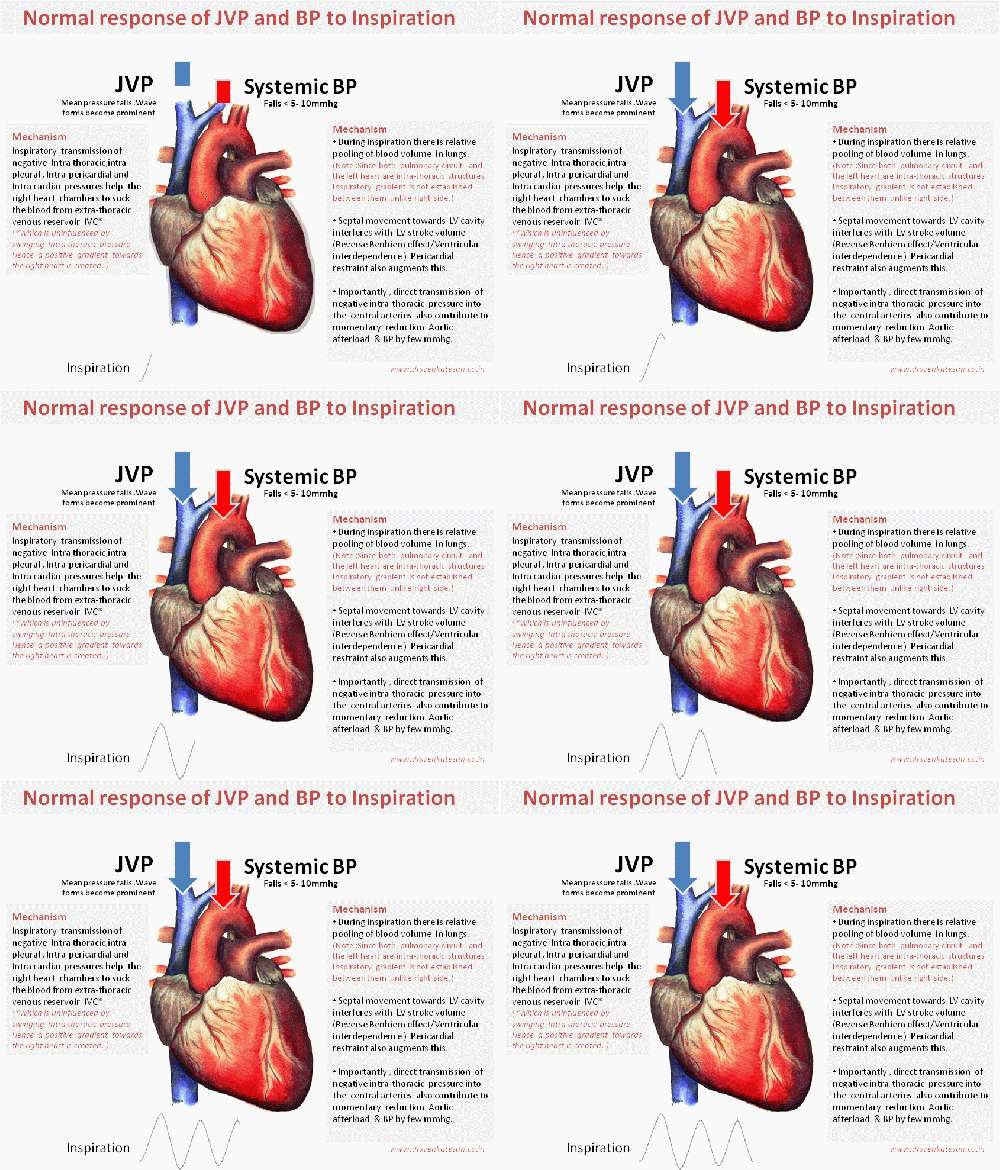
effect of inspiration on jvp and bp pulsus paradoxus bernhiem effect ventricular interdependence

Image  modified  from  http://www.anatomygallery.info

That’s  normal . . . what happens during pathological states ?

There are important diseases  that  restricts entry of blood into right heart chambers. They can occur either in an acute  (Tamponade) or in chronic  fashion like constrictive pericarditis  and restrictive cardiomyopathy.These entities  show distinctive impact on JVP and systemic pulse.

The two pathognomonic signs are Kussmaul sign and pulsus paradoxus* that go hand in hand in most  situations.Inappropriate elevation of JVP with inspiration is termed as Kussmaul sign , while exaggerated fall in systemic BP with inspiration is called Pulsus paradoxus.The later is the  arterial counter part of  Kussmaul sign in JVP .However, there can be dissociation between these two signs occasionally.

* Pulsus paradoxus is a term originally  used by Kussmaul when he noted heart sounds were  retained while pulse dissappeared  in  patients with cardiac  tamponade .Later we realised the loss of pulse was linked to inspiratory cycle  of respiration. To make  this sign objective  sphygmomanometery  criteria was formulated which measured the difference between inspiratory  and expiratory korotkoff’s  sounds .

Coming up next 

Why Kussmaul sign  is often absent in Tamponade while  its arterial counterpart pulsus  paradoxus may still be conspicuous ?

Read Full Post »

The pericarditis  of acute rheumatic fever is not a true infective pericarditis.It is more of inflammation .It is primarily  T cell mediated  reaction . Neutrophils rarely take part  in this inflammation and hence  no significant  exudation . Hence , there is less  sticky and adhesive molecules inside the pericardial space .The most inflamed layer is epicardium which a nothing but visceral  pericardium .This layer lacks the tensile strength to constrict the underlying myocardium.

why rheumatic pericarditis does not go for constrictionFor constriction to occur the fibrinous  ( thick ) layer of pericardium need to be involved . In rheumatic fever  even though it is  pancarditis ,  fibrous layer is not  involved. Further the inflammatory gradient is thought to spread from within  (Unlike tuberculosis )

Note :  In chronic tuberculous pericarditis,  diffuse inflammatory process  invade from the exterior surface . Very often ,  one can not differentiate  layers. In extreme cases even myocardium and pericardium can not be separated .

Summary

The peri-cardial effusion of acute rheumatic fever

  1. Is transient ,non infective and resolving (Unlike valvular inflammation !)
  2. Less of neutrophil activation  (Less adhesion)
  3. It does not involve the thick , tensile  fibrous layer of pericardium hence lacks the contractile force .

Other lingering  questions

1.How common is tamponade  in acute rheumatic fever ?

2.How important is the mass of the effusion (Viz  a Viz  Intra pericardial pressure !) in causing tamponade ?

//

Read Full Post »

Hypothyroidism is  a classical example of  generalised edema formation .  The mechanism of which is extensively studied  .Still we are  not clear  about  it .

Content of edema fluid

  • Mucopolysacharides -Hyalinorunic acid
  • Albumin

Mechanism

  • Albumin leak into interstitial and extra cellular space  due to capillary dysfunction.
  • Reduced lymph clearance – probably due to poor lymphatic tone  .

Why hypothyroid  edema  does not pit on pressure ?

For pitting to occur tissue should be compressed and retain the elasticity .This means  the fluid can escape into the cell when mechanically compressed and comes back when the  elasticity of skin  brings  it back to its original status. For this to occur the skin and subcutaneous matrix should be normal in texture.

In cardiac failure and renal  failure   edema is   primarily due to  altered hydrostatic forces and skin  is intrinsically normal .So  some amount of pitting  is retained . In hypothyroidsim and lymphedema  where there is  an intrinsic  pathology  of the  skin   pitting  is rare.

Why constriction and  cardiac tamponade are rare with hypothyroid pericardial effusion ?

Like the generalised slow response of hypothyroid individuals  the effusion is  also very slow forming and is rarely large so tamponade is rare .

The hypothyroid infiltrates which  collects within  the pericardial space it rarely infilitrates  the fibrinous pericardium (The thick outer shell ) .Unless fibrinous pericardium is inflamed or infiltrated constriction is exceedingly rare .Simple epicardial infiltration may cause some restrictive  physiology but not constriction .

Reference

Lancet. 1975 Mar 8;1(7906):564-6.Effusions into body cavities in hypothyroidism.  http://www.ncbi.nlm.nih.gov/pubmed/47029

Read Full Post »

Ventricular septal rupture is a major mechanical complication of STEMI . Excruciating  chest pain ,  is the sine qua non of  any myocardial tear , dissection and rupture . It is surprising ,   VSR  following STEMI  is rarely a painful event . I can recall number of  such events  , when a  stable   patient with persistent ST elevation  in the  coronary care unit ,   wakes up next morning  with a systolic murmur.And echo reveals a septal defect promptly.

Three  reasons  can be  proposed  for relatively  pain free rupture of IVS in STEMI.

  1. Typically  VSR  occurs in 3rd or 4 th day of infarct . By this time myocardium  can be as  soft as an ice cream ! . There is not much stress and strain at the site. The necrotic  debri just gives way to spikes of   LV systolic pressure .
  2. For rupture to occur there   must be  transmural infarct  .The pain nerve terminals also die in the process .
  3. Further , it is a cavity to cavity rupture  (LV to RV ) . Direct pericardial  stretch  does not occur .

* Ventricular free wall tear   is a near fatal event is extremely painful .This  often occurs  in the first 24 hours when  the nerve terminals are  alive . The free wall rupture is more of  a  tear in the plane of  myocardium . The  pericardial  (epicardium)  layer has  rich   somatic  nerve supply .

In summary

Early  myocardial  tear   involving the epicardial  surface can be severely  painful  .  Late giving way  of softened  , necrotic  often  hemorrhagic muscle ( especially in the IVS ) is less painful or totally painless.

Coming soon   . . .

By the    . . .  what happens  to  pieces of  septal myocardium as it  gives way  and enter the right ventricle   ?

Read Full Post »

A  patient who presents with predominantly right  heart failure  is  an interesting clinical challenge . Constrictive pericarditis (CP)  remains  a popular diagnosis in this setting. However  in the bed side clinical  examination (and in cardiology Board exams )  the following  differential diagnoses are  to be  considered .( And ruled out one by one)

  1. Restrictive cardiomyopathy* especially Right  sided .In India endo myocardial fibrosis tops the list
  2. Primary Tricuspid valve disease( Tricuspid stenosis / Carcinoid etc)
  3. Chronic cor-pulmonale in terminal RV failure
  4. Silent Mitral stenosis with right heart failure
  5. Ebstein anomaly
  6. Severe forms of valvular pulmonary stenosis with RV dysfunction
  7. SVC obstruction
  8. Cirrhosis of liver
  9. Porto pulmonary hypertension

( The list is not complete , readers may contribute )

Bed side clues

  • Remember  a deep “y” descent  is  the bed side counter part of   Square root sign  recorded by  invasive RV pressure study
  • Similarly , pericardial knock is the auditory   equivalent (You hear the square root !  . . .yes  )as the ventricle thuds the rigid thickened pericardial shell in very early diastole !)
  • Pulsus paradoxus and kussmal sign can occur in both CP and RCM.
  • If a good LV apex , is  palpated it  goes against CP .
  • Please be reminded , even restrictive cardiomyopathy  will ultimately dilate their chamber pre-terminal and clinical features may be confounded with that of DCM.
  • Silent heart would suggest CP.
  • AV valve regurgitation would favor RCM
  • Features of  Pulmonary hypertension will help confirm Mitral valve disease , Cor pulmonale,
  • Deep  “y”descents  are against  any form of  Tricuspid stenosis.
  • Opening snap of mitral valve is to be distinguished from pericardial knock.( Opening snap high pitched  and occur later than   pericardial knock in diastole   , best heard in expiration )
  • Cirrhosis liver with hypo- proteinimic   fluid retention is  a traditionally close mimicker  .It  may be ruled out by the careful history taking as exertional dyspnea is an exception , if  at all , it is a very late event  in cirrhosis.
  • The issue gets further weird   as chronic constriction can lead on to chronic congestive liver and cardiac cirrhosis .
  • Severe  forms of constriction can invade the myocardium and result in features of myocardial dysfunction .It is more common than we recognise.

How to confirm ?

Following should be performed in that order

  • ECG
  • X -Ray
  • Echocardiogram
  • CT scan
  • MRI

*Cath study is no longer done (Only for academic purpose )

Final message

Even in this era of sophisticated  medical  imaging  , clinical examination  remains the key . One should  realise the importance  of  meticulous  clinical history  ,  sequential examination and interpretation .It  will   “rule out  or rule in”  majority of  cardiac disorders .

The hi tech imaging  modalities should be used only to confirm , risk stratify and  plan management . If you skip the clinical  part , one  may still arrive at a correct  diagnosis  but there is  high chances of erring in  management.

(Cardiac pearls lie in the bed side not in cath labs !   Here is  one such pearl  . Not every constriction  require surgery !

Please note about 20 % of constrictive pericarditis are  transient !)

Read Full Post »

Constrictive pericarditis is a well known mechanical disorder  of heart that occurs due to the  compression  by thickened pericardium .Constrictive pericarditis is  the classical cause for  severe diastolic dysfunction.

We know , lungs are   prone for restrictive disorders due to chest wall , skeletal  disorders. Does the heart get mechanically restricted in extreme obesity ?

Not really , one may reason out . Chest wall fat can have little effect on cardiac function but when excess fat accumulates within the layers of heart , it is indeed possible for  the  fatty layer to impede mechanical filling of heart. This may be considered rare as of now , but many times it is not recognised ,  as most of the dyspnea in morbid obesity is attributed to some other known factors.

Dyspnea in obestity  can  due to

  • Pulmonary hypoventilation
  • Increased  MVO2 due to elevated cardiac mass
  • Diastolic dysfunction of  LV/RV
  • Increased demand  due to  excess BMI.

Image courtesey : http://www.onlinejacc.org

Now, we have evidence for  altered RV hemodynamics due to compressing effect  of epicardial fat pad. It may be due to   simple mechanical effect  of epicardial  fat over the distensiblity of RV or occasionally  LV. (The distribution of epicardial fat is mainly over the right ventricle or septal areas.)

This   paper  from Korean circulatory journal  succinctly describes this new possibility .

http://www.ncbi.nlm.nih.gov/pmc/articles/PMC2771803/pdf/kcj-39-116.pdf

Final message

Bed side cardiology  continues to  bring surprises  , it  never fails to fascinate us   !

Heart  is  a dynamic organ ,  has a potential to get restricted by  any  layer that  surrounds  it. Constriction by pericardium got huge attention so far .We need to realise , the epicardium which is  a part of pericardium has a variable fat depot  . It  can take a different avatar  in an occasional obese individual  and   exert  important hemodynamic impact.

Excess fat is excess load on heart . . .  we have  to unload it

It is possible , sucking out the  epicardial  fat in morbid obesity can bring important relief to  those patients with unexplained dyspnea . We  need to  explore this possibility.

Read Full Post »

A 50-year-old man was referred to us with suspected  angina. Here is his ECG.

Epicardial fat : One more cause for Low voltage QRS

He was an obese man weighing 105 Kgs. He was put on a tread mill  .It  was convincingly  negative .
The echo cardiogram revealed a prominent epicardial pad of  fat measuring 6mm throughout the anterior surface.He had  normal valves and normal myocardial function.It was concluded the low voltage and poor R waves , and T wave inversion was due to the thick epicardial fat.

ECG -Fat correlation

The lack of R wave progression  is attributable  to the insulation effect of fat .Chest wall fat rarely dampen the electricity .Epicardial fat does it more.T wave inversion may not be  due to dampening effect of fat  .We think epicardial fat when adherent to true pericardial surface of the heart it alters  the epicardial  action potential  .It is possible  electrical  neutralisation by the fatty infiltration of epicardium  reverses the direction  of repolarisation  towards the epicardium .

Other ECG manifestation of thick  epicardial  fat

  • Poor R wave progression
  • Anterior Q waves
  • T wave inversion in ;leads v1 to v4 or V5

Final message
Epicardial fat deposits can have clinically  important influence on the surface ECG recording .
Simple chest wall obesity causes only diminutive  R wave . If fat encircles epicardium it has high chances of  producing repolarisation   abnormalities  in the form of T wave inversion or flattish   ST segment.

Read Full Post »

WPW syndrome remains as  a   fascinating ECG entity ,  ever since it was described by Wolf , Parkinson and White in the year 1930.It is  primarily a  disorder of cardiac embryology . Heart is an organ made up of  tissues from mesoderm and neuro ectoderm.The muscle which comes from mesenchyme has to be incorporated with specialized conducting system. This is a complex  process .It is determined by the bio-genetic forces. When errors happen in the embryonal  tissue  flow  congenital anomalies occur.

In  WPW this  error   happens  exclusively in the conduction  tissue movement  . Normally the specialized conducting system    pierces  the  entire  AV ring and connect atrium  and ventricle  .Later ,   it regresses in  all areas  except in the AV nodal zone  . When  It  fails  to regress ,  these  remnants of  conductive  tissue act as AV accessory tissue  and create electrical  short circuiting .This is the reason , all these pathways are located in the close vicinity of AV ring.

Accessory pathway shows   varying conduction velocity , but generally devoid of  decremental conduction properties .  The presence of such pathways make these individuals prone for variety of cardiac arrhythmias .It can range from  simple AVRT  to  malignant antidromic  AVRTs  that can end up in  VT /VF.

Resecting  these  pathways surgically was once popular.  Effective blocking  of  the pathways with  drugs  is a good option. Currently ,  it is possible to  locate and  ablate  most of these  pathways   successfully.

Even though there are many protocols to locate accessory pathway the one that is very popular is  simple   Type A and type B  WPW , which locates the pathway either in the  left  or  right  ventricle  respectively.

Huge data base  has been accumulated over the past 80 years  regarding WPW syndrome,  still   many questions are unanswered.  One of the important clinical issue is  multiple  accessory pathways , scattered  at  random  across the  tissue planes of atrium and ventricle  .

The other issue is intermittent pre-excitation and shuffling  of path ways during tachycardia  .

It is very rare to see a patient who manifests both Type A and type B pattern during sinus rhythm .Here is an  article from  unexpected  quarters  , Colombo Sri-Lanka in the year 1972  candidly  describes a patient with classical  combination  of  Type A and  B  WPW . It is great to see such an interesting  observation in the pre  EP/Echo era from a remote island nation.

Now , let us ponder over  these questions

    1. Can a pre-excitation  happen simultaneously in both right and left free wall pathway ?
    2. How will the ECG look like  when impulse travels over multiple pathway ?
    3. When dual pre-excitation combines   with  normal AV  conduction  ,     will  it not make  a  triple AV pathway ?
    4. How does a supra-ventricular impulse decide ,  which pathway it is going to travel  when confronted with a choice of  three or  four pathways ?
    5. How do you plan ablation for such a patient  ?

Read Full Post »

Heart has three layers

  • Epicardium
  • Myocardium
  • Endocardium
  • Epicardium is same as visceral pericardium . If pericardial inflammation dissusely occurs ,  it is bound to injure  the epicardium and subepicardium .

    Does  the  troponins  located deep inside the  myocardium  ?

No .It  can  even be present just few microns below the visceral pericardium  . Hence  severe forms of pericarditis can elevate the troponin levels without any issues .

Is troponin  release related to ST elevation ?

 Ideally  most forms of  pericarditis can be termed as epicarditis. The mechanism of ST elvation in pericarditis is actually a sign of  epicardial injury.In fact ,  there is no easy way to  differentiate  a  slice of epicardial infarct from an   inflammatory pericarditis accurately .

Is there any form of pericarditis which invlove only parietal pericardium ?

We do not know as yet ,  about  existance of such  an entity. It is distinctly possible. However , if present it is unlikely to result in significant  ST elevation in ECG.

In pericarditis ,  troponin release is due to inflammation  or necrosis  ?

Both are possible .Even transient wall motion defects are reported in isolated pericarditis.

What is myopericarditis ?

It is  a general term used  to indicate the above situation . In practical terms Pericarditis + Troponin positivity can be termed as myopericardits. It is well known pericardits can extend to endo- myocardium but it is rare other way  around( ie endocarditis extending to pericardium )

What is pancarditis ?

It is the carditis  involving all three layers of heart ,Cassically occura in rheumatic fever. Fulminant carditis is known to raise the troponin to significant levels.

Does troponin elevation  in pericarditis  occur in all  ?

We are yet to collect adequate data about this .  Diffuse , extensive pericarditis ,gross ST elevation ,  and associated pericardial effusion  correlate with troponin.

Crazy questions in pericardiology

What is the pericardial blood supply ? Is there  an entity called ischemic pericarditis ?

Final message

Do not ever underestimate the  importance of  pericardium  whenever you encounter unexplained ST elevation in ECG.

Reference

Here is an article which has   meticulously studied this issue

Read Full Post »

Older Posts »