Feeds:
Posts
Comments

Archive for the ‘cardiology -Therapeutics’ Category

Even though multiple mechanisms operate  the major mechanism is due to augmentation and  diversion of blood flow towards sub endocardial region* which is  main area of ischemia in most patients with Angina .
subendocardial-blood-flow
*Beta blockers  does this by smoothing   the  myocardial contractility there by  reducing  trans-myocardial gradient. The coronary arterial perforators which traverse from epicardium to endocardium gets less squeezed and promotes sub endocardial perfusion. 
Link to  a related article from this site

Read Full Post »

This  is the story of a 55 year old  women ,  who was received  in our CCU  with a  dramatic STEMI (ECG looked like an action potential ) ,  LV  S 3  and  hypotension.    It was impending cardiogenic shock.Since we do not have full fledged primary PCI  program  , thrombolysis was planned. She had  cardiac arrest   immediately after  starting streptokinase infusion . She  was  promptly shocked  and  revived .  The ECG changes rapidly  reversed(ECG -3) . Every other  hemodynamic parameter got stabilised as well . To our surprise   ( few hours later ) this patient  was  so comfortable , sat up on her bed ,  demanded a discharge . (Which was refused of course !)  One week  later coronary angiogram was done, a near complete recannalisation of RCA was documented.

ECG 1 on arrival
Inferior MI 2  

ECG -2 Developed cardiac arrest  10 minutes  after  starting the Streptokinase Infusion

primary VF 2

ECG -3 .Taken few minutes following   the VF

inferior MI evolved 2


Acute myocardial infarction (STEMI)  kills more than a million life every year . Majority of death  happens within an hour of onset of symptoms. Ventricular fibrillation  is the arrhythmia of death. Why this occurs  only in  few , while  many are  immune to it ?

God keeps  this secret  close to his chest ,  how and why  he selects   candidates for this arrhythmia !

Scientists are still  far away  in finding the truth . But , one thing  is obvious .The  moment   coronary artery is totally occluded  , the heart begins a fight  and try  to  get rid of this obstruction . In the process ,  it  goes into convulsion (VF)  with a foolish belief  , it  can shrug of the thrombotic insult . Death often   ensues if  not intervened . (Very rarely  VF can be a non sustained one  and patient survives cardiac arrest !)

VF  as  a electrical  response  to  reperfusion injury .

Often times ,  we witness patients  to  go  for  VF  very early following thrombolysis . The  thrombus in situ is an irritant , it  triggers the inherent fibrinolytic system (Natural TPA included) If it is successful  it opens the occlusion ( atleast partially )  and salvages the myocardium .If the fate is against  the patient , very early reperfusion of IRA triggers  VF  .  If this occurs at home   survival  is  low .If  the VF occur at hospital the probability of survival is near 100 % .

               The  intensity of  natural lytic mechanism  is the major determinant  of   early reperfusion . Ironically  the same  factor   determines  occurrence of the deadly  VF .

I would believe  , the STEMI patients  who die early (even before reaching  the hospital ) are (un) blessed with a  fighting  heart  ! Ironically , the lazy hearts  reach the hospital  alive ! (slow &  steady win the race !) .  Of course , reperfusion  injury is not the only mechanism of VF . Other common suspect is  left main STEMI .

Link to related video “Ignorance based  cardiology ”

https://www.youtube.com/watch?v=J9DH6Vr04es

Final message

While , VF  is  referred  to as arrhythmia  of death , it may  in-fact , represent  a common form  of  reperfusion arrhythmia in  the setting of  STEMI !  .  . .  Hence , it can  Initiate  a new lease of life in  many   lucky ones !  I hope the title of this article  makes sense  !

Read Full Post »

           Do not ever under estimate  the importance of  TIMI 1 flow .  It can save a  major chunk of myocardium !   A late TIMI 3  flow   . . . is far inferior . . .  to  an early TIMI 1 flow . * Even a trickle  of  flow (Ooze )   can keep the myocardium  alive .  This point we have realised very late. Thus came the   pharmaco Invasive strategy for  all STEMI  who have no immediate access to cath lab ! (please note 90 % of STEMI belong to this group )

pharmaco invasive strategy for stemi002

For a high resolution Image  click below

pharmaco invasive strategy in stemi

* Even a trickle (Ooze )   blood flow can keep the myocardium  alive .

Read Full Post »

Thrombus laden plaque  is sine qua-non of UA/NSTEMI . That’s what we  have been taught  !  right ?  It may be  true in many  situations , but please remember there is another concept  called  demand ischemia , where in there is  no active thrombus ,  still resting  angina may occur due to  increasing heart rate etc.

I just wanted to test how far this concept is understood ,  by the  fellows in our coronary care unit . Following  is  story of a patient who arrived at CCU with  angina  at rest .  I showed  this   ECG asked them the  management .

positive est and unstable angina

History was  purposefully blinded . 5/6 cardiologists wanted to admit the patient either in CCU or rush to cath lab.  Heparin/ Fondaparuinux was prescribed by all. Tirofiabn was suggested by few.It is a  high risk UA with left main disease some one  mumbled .

I silently listened to them and  revealed the history . This patient  has just finished the  exercise stress test , it was terminated as he had angina at peak exercise. and was  reported as  positive . A date was fixed for elective   coronary angiogram. 10 minutes later ECG totally normalised  , and the patient went home (Boarding a crowded Chennai  city bus )

The fellows realised the importance of history . In fact no body asked for it ?  I felt  bad  as  all my fellows failed in this test That reflects bad teaching on my part !

What is the mechanism of ST depression here ?

  • Fresh thrombus ?
  • Mechanical occlusion ?
  • High  heart rate ?
  • Combination of high rate and probable flow limiting lesion .

(Severe forms of  stable angina can occur at rest . So do not equate all rest angina as true  unstable angina !)

Final message

Do not label an ECG straightaway  as acute coronary syndrome when there  is  baseline  tachycardia and ST depression . Spare few minutes and apply your mind !

If  a combination of ST depression  and angina  can be taken  synonyms with UA  every EST positive fellow should be labeled as UA and admitted in CCU. Please remember any tachycardia with a fixed tight lesion will  mimic UA . Further ,  since there is no thrombus here  and there is absolutely  no role for heparin.

Read Full Post »

95 % of hypertension is designated as primary HT .What does it mean ?  It means  95 % of times  we do not   know what  exactly is the cause for raised blood pressure  . Simply stated  . . . it  reflects 95% ignorance .

So what is secondary hypertension ?

Secondary HT is the one,  in which we have specific reason for the raised BP.  The most important cause is Renal  ,  endocrine etc.

When will you suspect renal HT ?

https://drsvenkatesan.wordpress.com/2010/09/01/when-will-you-suspect-reno-vascular-hypertension/

How is secondary HT different ?

  • Occur at relatively at young age (<45)
  • It is generally more severe.
  • Diastolic BP is proportionately  higher ,
  • End organ damage is more.
  • It is very unlikely primary HT to present as acute LVF.  One rule of thumb is ,  if diastolic blo0d pressure is > 120 never diagnose  primary HT . Some amount of renal component is very likely.

Is stress related HT a form of secondary HT ?

https://drsvenkatesan.wordpress.com/2013/04/26/why-stress-obesity-related-hypertension-is-not-considered-as-secondary-hypertension/

Read Full Post »

Heart disease was  once considered as   rich man’s disease  . . .  It’s no longer  true .  We in India ,  are witnessing an epidemic of CAD . The reasons are  varied  . Apart from  conventional factors ,   social   factors  like changing demographic pattern  ,  life style , ethnic  risk  like  south  Asian metabolic profile are responsible .

While  Rheumatic heart disease (RHD ) continues to be a huge burden , CAD  is  the number one  cause for cardiovascular morbidity and mortality .

CAD affect the poor and rich with equal vengeance . The later is better equipped financially to tackle it . Of course ,  it has resulted in maximum inappropriate interventions. The poor (or borderline poor ) have  no  other  option  but  to knock the doors of Government  hospitals. It is heartening to note, various state  Governments   are  gradually involving insurance schemes.

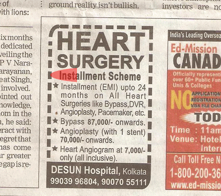
Still , many struggle to find the required  finance for  a major cardiac intervention. It roughly costs 100,000 rupees  for PTCA  .While  PCI is required in all symptomatic ,  critical coronary occlusions , still . . .  majority of the  CAD in general population  do not require it . There are 675 cath labs in India performing 180000 angioplasties every year  on an average of   15000  PCI per month ( 500 /day )  This is grossly inadequate . We  have huge potential

What is the hurdle ?

  • Expertise ?
  • Hard ware ?
  • Awareness ?

No  . . . it is all about  financial resources

Recently I stumbled upon an  advertisement on Times of India

cardiology ad ptca

Disclaimer: This article does not in any way defame any hospital that offers the scheme.It just want to debate the concept.

Hospitals  want  to  market the procedure . Convert angiograms  to angioplasties . That’s   corporate boardroom mantra  . And one fine day ,  bankers and medical doctor sat together and brought a brilliant idea.

Why not do the procedure  on credit and  push the patient  life long  into a financial debt !

Wonderful idea  . . . many thought .Thus came the financing scheme for  cardiac procedures.

Final message

Financing a poor patient  with good intention is welcome. But, there is big caveat .In a vast country   with high  illiteracy , inappropriate  procedures   may be thrusted upon  on   the  poor  souls.

After thought

Now ,  our patients   have  one more  risk parameter to  assess   ” Number of remaining EMI( Equal monthly instalment )  and incidence of stent thrombosis”   “Accumulated  interest  and angina”   What a wonderful way to provide cardiac care !

I can recall a  patient who sold his livestock  (his sole income source ) for undergoing a open heart surgery and lost his life as well in the process  leaving the family stranded !

Solution

The only solution is  to  provide  a strictly regulated Govt sponsored  insurance scheme.  High tech procedures should be  continuously and meticulously  audited for cost effectiveness .


Read Full Post »

In HCM every myocyte is  genetically made  defective . Myofibrils are in disarray every where . Still , can we identify some vulnerable zones that acts as arrhythmic  focus ? If that is possible , we  have a opportunity to abate that focus .

In HOCM  , which is the most stressed area ? LVOT ?  Septum, ? When we say stress , it can mean either mechanical or electrical .

VENTRICULAR TACHYCARDIA 002

Does electrical instability involve the same zone as mechanical stress ?

How often VT originate from LVOT in HCM ?  For this we have good clinical model _, the patients who underwent alcohol septal ablation.

What happens to the incidence of VT  post septal  ablation  ?

“It is reported  post septal ablation the incidence of SCD  becomes  equal to general population” (Read the paper below )

If that is true , it is obvious the  arrhythmic  focus is also ablated along with LVOT myocardium .

Outcome of HOCM after alcohol septal ablation

Though many studies claim  so !  It  fails to convince us  .  HOCM is a diffuse disease of  myocardium.  Even a cluster of myocyte disarray  with fibrosis   can be a future focus  irrespective of it’s location .

However ,  it is always possible relieving the mechanical stress of the LV can definitely reduce the likelihood of an electrical event .(Even if the arrhythmic focus is intact elsewhere !)

* We know RVOT is  developmentally arrhythmia prone zone . We also know HCM involves RVOT (After all ,  IVS  is legally shared by both ventricles !  ) . Some of  the monomorphic  VTs with LBBB morphology may originate from RVOT in HCM .

Management of recurrent VT in HOCM

  • Drugs (Amiodarone/ Calclum blockers/ Beta blockers/Disopyramide)
  • ICD- (Probably mainstay  )
  • Very rarely ablation (If localised focus is well documented )

Reference

1.A case report for successful ablation of  VT in HOCM   http://www.ncbi.nlm.nih.gov/pubmed/9255687

2.http://www.ncbi.nlm.nih.gov/pubmed/23076968

//

Read Full Post »

Why should mitral  annulus gets calcified ? .  Degenerative  calcification can be benign in  elderly .  If it occurs prematurely (say < 55 years )   there is enough reasons to worry .  This may represent a systemic vascular inflammation and  is considered a surrogate marker for athero- vascular -sclerosis .  A study from Cidar Sinai  , Los angels  has well documented the link way back in 2003  !

mitral annular calcification mac cad link

This is a  large study involving  17 735 patients (who were investigated for symptoms of CAD )   were screened.

The incidence  of MAC was high (As expected !)

  • 35% > 65 years
  • 5 %  < 65 years
Angiography  revealed more surprises .
  • The incidence of angiographic  CAD among those who had MAC and no MAC   was  88% v68% respectively ,( p = 0.0004),
  • Left main coronary artery disease  was (14% 4%, p = 0.009)
  • Triple vessel disease  was (54% v33%, p = 0.002).
mitral annular calcification  www_drsvenkatesan_co_in

Image source  S.Atar ,  Heart 2003 : 89, 161-164

Conclusion
This study concluded ,  CAD is more aggressive in patients with MAC. It can  also be  an independent  predictor of  high risk CAD .
Further Implications  of MAC
  1. MAC is more common in women, especially diabetics .
  2. Degenerative Mitral regurgitation  is common ,rarely  mitral stenosis
  3. Recurrent VPDs and even  trouble some mitral annular VT is possible
  4. Extensive calcific lesions in coronary  artery is also reported with MAC.
Link between Stroke and MAC .
This was well proven by this paper  published in  NEJM in 1992.
MITRAL ANNULAR CALCIFICATION AND STROKE NEJM  EMELIA BENJAMIN 1992

Read Full Post »


Those who answered  “Yes” ,  can leave this article . Those who answered  “No” read further .

* Logic would tell us myocardial revascularisation should correct  stress induced ischemia and it  should disappear promptly  . This does not happen in all cases  real world  ! That is  why medicine is  different  from mathematical science .

Some of the  reasons for  persistence of stress positivity even after an apparently successful PCI are  . . .

  1. Incomplete  correction of ischemia. (Ideally  to be referred as failed PCI )
  2. Error in Identifying culprit 9Angina related artery ) .Common feature of poorly worked up  multivessel CAD.
  3. Re-stenosis /Re-occlusion
  4. Doing very early stress test without giving time for revascularisation to work *
  5. Rapid progression of non culprit lesions .(Sub -optimal medical management )
  6. Chronic N0-Reflow phenomenon  surrounding  area of infarct .(Especially in  PCI of CTOs)
  7. Dyskinetic  or grossly remodeled ventricular segments  can result in non ischemic positive EST response (ST drag **)
  8. Associated systemic conditions especially  Anemia/ SHT & LVH -(False positive )
  9. Many diabetic patients may  continue to show stress ischemia due to  small vessel disease.
  10. A  patient with  syndrome X  characters  can have incidental  epicardial lesion as well . In such a patient EST will always be positive .

* Optimal time to do  EST  for assessing the  efficacy of  PCI/CABG is not established .Six months may be the reasonable point .If done within 2- 3 months it may  end  up  in embarrassment for the Interventionist . (So only it is kept at 6 months , this also help us  greatly  as  we can always blame it on poor life style control and progression of  the disease !)

** No reference  for this  , a  personal observation .We know  Q leads following MI ,  will show ST elevation during stress test especially if the segments are dyskinetic  . In leads diagonally opposite to q leads ,  ST depression is observed . This may not be  a evidence for true  ischemia . It probably represents   ST drag due to mechanical stretch .

Read Full Post »

bifurcation angle

  • At any branch point three angles are possible .True bifurcation angle is formed between LAD and LCX .
  • The angle between LM and LAD or LM and LCX can also be important in specific situations ,especially when we encounter short left mains and Medina 1,1,0 lesions .
  • Major bifurcation angle can  occur in mid  segments  as well ,  between LAD / major Diagonal  , LCX and OM.
  • Logic would tell us the  left main  bifurcation  angle is relatively fixed by the anatomical AV and IV grooves. Still early course of LAD and LCX can be out of grooves.
  • Further ,the bifurcation angle is imparted some amount of dynamism by cardiac cycle . It can vary between 80 -120 degrees (LAD/LCX).
  • Most importantly various  angiographic views can alter the true angle (by illusion ) in dramatic fashion . RAO caudal view appear ideal to measure it. (LAO caudal make every bifurcation angle obtuse !)
  • Acute angled bifurcations are prone for stent related mechanical issues both during deployment and in the long term outcome . (When two stent technique is used) This is because ,  acute  angled bifurcations has a tendency to drift the carina , and  encroach  the lumen  which can create new  turbulence . Of course final kissing balloon is expected to reduce this hemodynamic side effect at least on paper !

Read Full Post »

« Newer Posts - Older Posts »