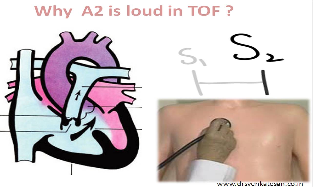
Second heart sound in TOF is often single and loud . It is often best heard in left 2nd intercostal space as well as on right 2nd space.
This is primarily due
Presence of pulmonary stenosis and resultant reduced pulmonary blood flow makes P2 soft or absent .Hence A2 becomes loud by default.
In tune with any cono -truncal anomaly , aortic root is anteriorly malposed in TOF. This brings the aorta closer to chest wall (Nullifying the aquastic insulation of main pulmonary trunk ) and results in a booming aortic second heart sound.
Increased flow across aortic valve . In cyanotic heart disease with reduced pulmonary blood flow aortic flow is augmented and may even result in dilatation of aorta . A large aorta with increased flow is perfect setting for generating a loud A2 . It is common to hear a aortic ejection click as well in these situations .
When you hear a single second heart sound at the base of heart , how do you recognise it to be A2 or P2 ?
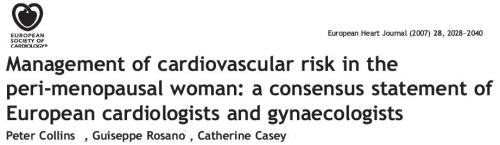
Gynecologists do have interactions with cardiologists frequently in their day to day practice.In fact , in any big hospitals cardiologist consult invariably happen every day . In our institute fellows visit the maternity ward almost daily to give opinion about a cardiac issues . These are mainly emergencies like breathless rheumatic heart patient in labor , A DVT to R/O pulmonary embolism, women with prosthetic valve waiting for delivery, and a women with LV dysfunction posted for hysterectomy etc.
While it is common for our Gynec colleagues to call us in emergencies , and we do have a cardiac clinic every week , it is rare to discuss broad based practice issues. There is little inter departmental brain storming sessions.
The beauty of this document lies in the succinct practice points written in every page .
In India , even though premier bodies like cardiological society of India exits it rarely considers bringing about such guidelines in collaboration with other scientific bodies . ( To be more precise . . .they do not have their own guidelines either ! )
I believe , FOGSI (Federation of Obstetrics and gynecologists society of India ) is doing a much better job and they have created exclusive guidelines in O & G.
Q : Beta blockers reduce blood pressure mainly through
Reduction in Heart rate
Reduction in cardiac output
Negative Inotropic action
Vascular sensitization to circulating catecholamines
Blocks Renin secretion and reduce vascular tone.
Answer :5 (May be 4 as well !)
Our understanding of beta blocker’s action in SHT has changed considerably over the years .The negative inotropic action on the myocardium attributed for BP reduction , is no longer considered important . Now we know , beta blockers can reduce peripheral vascular resistance significantly.(There were days , we presumed the opposite to be true , iewhen beta blockers are blocked , alpha action will overshoot to cause excess vascular resistance ! ) This is more of perceived fear. This concept was never proved convincingly even in the dreaded Prinzmetal angina* where beta blockers are relatively contraindicated for fear of aggravating vasospasm.
*Note : This is may still be valid in selected few who show a tendency for Raynaud phenomenon especially in peripheral vascular system.
Additional factors influencing beta blockers in SHT
Suppression of central adrenergic drive , modulation of brain stem vasomotor centre are aslo considered vital . This action is linearly related to the ability of beta blockers to cross the blood brain barrier which is more with lipophilic drugs like metoprolol.
The role of beta blocker in isolated systolic hypertension in elderly is unique.Here it reduces the myocardial dp/dt (ie contractility ) and hence help them prevent systolic spikes of pressure and the resultant stroke.
The newer vasodilating beta blockers like Nebivolol, (Nitric oxide mediated ?) and Carvidilol may have additional advantage in controlling BP.
It needs to be appreciated , beta blockers combine well with diureticslike hydrochlorthaizide .This makes it easier to control severe forms of HT especially volume dependent ones in both young and elderly. (SHEP trial )
Final message
The modification of vascular response to catecholamines is the single most important mechanism of reduction of blood pressure.
This may be a direct consequence of 1. Blockade of vascular adrenergic receptors . 2 Indirectly through suppression of Rennin secretion.
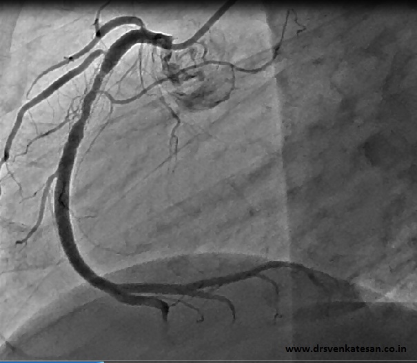
We know primary PCI is a race against time both for the patient and his physician.
What is the upper limit for this unique race where the stakes are high and it involves human lives and big corporate warfares ?
6 hours
12 hours
24 hours
36 hours
54 hours
Time does not matter . You can do a PCI as late as possible as long as patient has sufficient insurance coverage and we have the expertise
Answer : 2
Please note there is only one exception . Cardiogenic shock has been given a extended lease of time window (Which can be technically up to 54 hours ) . PCI can be performed if the onset of shock is within 36 hours of STEMI and to be performed within 18 hours after the onset ! )
* Even though we have a well set criteria for re-perfusion which bans primary PCI to be performed after 12 hours , cardiologists have enough technicalities to overcome this hurdle and keep doing the futile pPCI well after 12hours.
How they are able to indulge in these futilities without any ethical issue ?
The answer is very simple. Instead of calling it as primary PCI they refer to it as delayed PCI or rescue PCI ! Strict time specific guidelines are only for primary PCI . By changing the terminologies they make a mockery of the concept of time window which is vital for any intervention for STEMI !
Success of primary PCI is defined by different yardsticks by different cardiologists , indifferent institutions ! But , for the patient and his family, getting discharged alive with out symptoms is a huge success !
They do not bother even if they are charged Rs 3-4 lakh for their stay , as they believe surviving a heart attack is a God’s grace rendered thorough the hands of the doctor.
But we know the real success lies elsewhere. Cardiologist’s perception of success of pPCI should be based on scientific concepts. Unfortunately many physicians continue to think like their patients . This tendency to get self gratification with a patient’s frame of mind is common and needs introspection .
This is esepcailly true for primary PCI . It came with big fanfare a decade ago . Soon ,many cardiologists developed a habit of criticizing the practice thrombolysis for STEMI .If primary PCI is such a superior modality every patient should be prevented from significant myocardial damage following STEMI .
Primary PCI may be the the most logical method still , for reverting the STEMI process . But “A properly performed primary PCI as a concept ” lies mainly on paper , not been replicated in real world for various reasons.
Please remember , a successful primary PCI
Is notrestoring TIMI 3 flow in IRA
Is not relieving the angina
Is not discharging a patient in stable condition
Even if . . . we accomplish each one of the above . . . if the patient carries home anything equal to , or more than moderate LV dysfunction , primary PCI has deemed to have failed.
Final message
What is the reality check ? In one of the preliminary analysis out of 20 randomly selected patients who have undergone STEMI* within 12 hours , significant LV dysfunction was present in 12 patients making it pPCI only 40 % successful in real world .( Which would struggle to beat the outcome of promptly administered fibrinolysis )
* Primary PCI done in state of the art institutes .
“STEMI is Acute MI” , and “Acute MI is STEMI” ! This is how we have been taught over the years.
But STEMI can present in any one of the following odd ways
1. Posterior MI with ST depression in V1 to V3 ( Manytimes mistaken for unstable angina , and reperfusion not considered !)
2.Only with tall T waves (TEMI)
3. Left bundle branch block
4. Present as ventricular tachycardia. Untill VT it is reverted ST elevation will not manifest.
5. When a STEMI presents with complete heart block ST elevation may not manifest.
6.Atypical ECGs and subtle ECG changes are especially common in elderly, diabetic and in patients with LVH.
7. Finally, it is often quoted in text books acute MI occurs with normal ECG in up to 10 % . This is a high estimate .We belive acute MI with normal ECG is very rare presentation < 1% . That too in the very early stages of evolving MI.
As of now only condition No 1 and 3 are approved for thrombolysis or PCI. The unfortunate few who fall in other categories will continue to lose their muscle in the golden hour without any intervention as the guidelines has not addressed these issues.
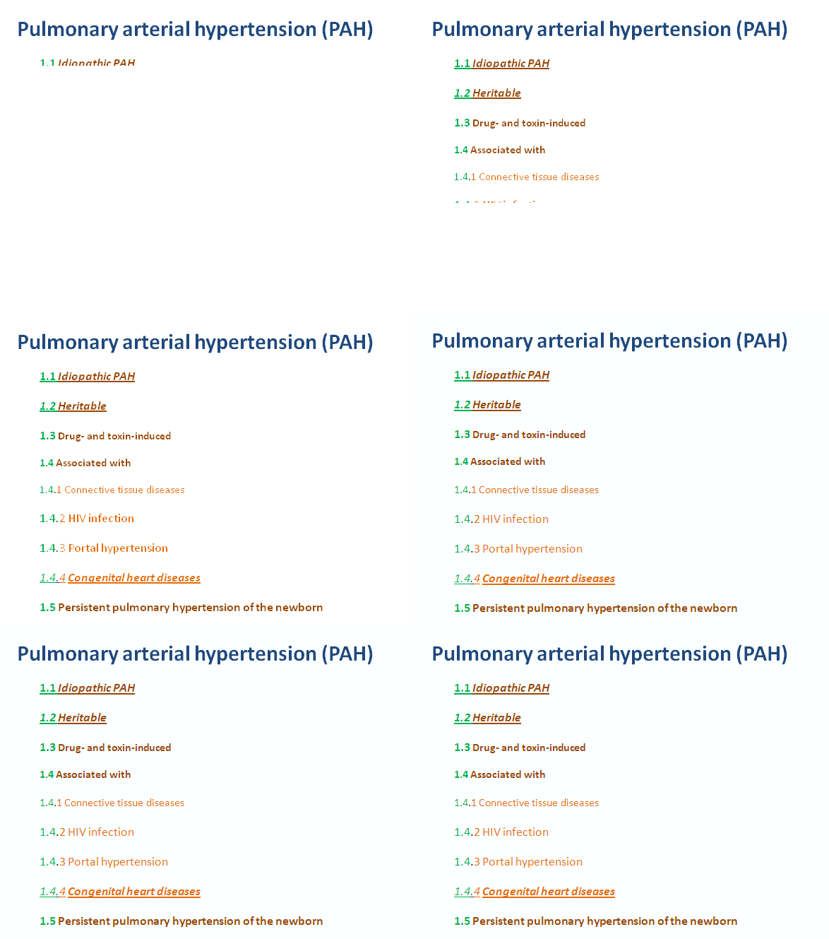
Pulmonary hypertension (PH)is a very common clinical problem in cardiology.The classification of PH has been little complex. Now we have a fairly clear scheme formulated in 2008 in a small beach side county of California called Dana point .
I have tried to simplify it without affecting the core
Click on the image if slide is not displaying
Category 1 is again divided into 5 categories
1.1 /1.2/1.3/1.4/1.5
Other categories( Category 2 to 5)
* Note there is a special category called 1 ‘ for pulmonary veno-occlusive disease .This should be distinguished from CTEPH
Summary
Remember 99 % all pulmonary hypertension will be constituted by the following seven entities .
Idiopathic PAH 1.1
Familial PAH 1.2
Connective tissue disease 1.4.1
Congenital heart disease 1.4.4
Left sided valvular /Myocardial heart disease 2.3.3
Secondary to COPD 3.1
CTEPH 4
In India (probably worldwide ) the commonest cause for PH is 2.3.3
Before answering the above question , there need to be a correction to the question itself . PDA is persistence of ductus arteriosus . In pulmonary atresia ductus itself is not formed .So the question should ideally be Why ductus is absent in pulmonary atresia with VSD !
Ductus is formed from the dorsal portion of left 6th arch .The sixth arch also gives raise to right and left pulmonary artery.This can happen only if everything from aortic arch and pulmonary artery development is normal
Ductus has to connect aorta with left pulmonary artery , when pulmonary artery itself is poorly developed or not developed how can the ductus connect to LPA ?
Pulmonary atresia is essentially a defect in the development of pulmonary arterial tree (Please note pulmonary valve and MPA is formed from different structures in different times and it would get docked with developing LPA,RPA and subsequently to the rest of the pulmonary tree .)
In fact , one of the terminologies used for pulmonary atresia with VSD is , total anomalous pulmonary arterial connection(TAPAC) .In fetal life, blood flow in ductus is from RV to pulmonary artery and then to descending aorta through the ductus . When RV is disconnected with pulmonary artery( Rather there is no pulmonary artery ) ductus can not be formed for both anatomical and physiological reasons . Some consider the left 6th arch in these patients would become a poorly identifiable minor Aorto pulmonary channel .
A Link to 3D vedio of aortopulmonary collateral
Embryology of major aorto pulmonary collaterals.
The lung perfusion in patients with pulmonary atresia is important only after birth, as fetal lung is largely non functional.
In patients with pulmonary atresia with intact IVS this becomes critical and usually death ensues unless intervened.
If VSD is present it allows the baby to survive as the lung gets perfused by major or minor aortopulmonary collaterals.
These collaterals can range between extensive and sparse. Hence the symptoms can also vary from volume overload /cardiac failure to severe oiligemia and recurrent hypoxic spells.*
MAPCOS , if present can connect directly the aorta to hilar pulmonary artery or indirectly from the branches of aorta (subclavian /LIMA/RIMA etc) . These arteries supply with or without a central confluence . It may enter the lung through the hilum or away from hilum .The MAPCOS can be located anywhere from the arch of aorta to descending thoracic aorta.It is very rare in the ascending Aorta .
*The natural history directly depends on extent of aorto pulmonary collaterals and its anatomical patency .
Final message
Embryologically both the major arteries of thorax Aorta and Pulmonary artery have the same parent structure namely the dorsal aorta and its six arches.Hence there is no surprise , when these embryological divisions and fusions goes awry , pulmonary artery fails to get carved out from the dorsal aorta in the normal fashion .The randomly formed pulmonary arteries continue to have link with the parent -dorsal aorta .These are manifested in various ways as major aorto pulmonary collaterals .
(It is to be noted in pulmonary atresia , VSD is an offshoot developmental defect . Embryologically VSD is not linked to the primary defect of pulmonary artery development . This is the reason many would consider PA with VSD as a distinct entity with that of TOF (Which is a cono truncal anomaly) This also explains the lack of MAPCOS in true TOF .
When know statins have revolutionised the management of coronary artery disease , why we hesitate to say the same thing for cerebro vascular disease ?
Is it because
Atherosclerosis is different in cerebrovascular disease than coronary artery disease
or Blood pressure control becomes more important in preventing stroke than lipids?
Unlike MI , Cerebro vascular system have variety of insults.
Embolic stroke
Thrombotic stroke
Ischemic stroke
Hemorrhagic stroke
Low flow – Water shed infarcts
Hemorrhagic , embolic and low flow mediated myocardial infarction are very rare and denova hemorrhagic MI is non existent.This is due to the fact , coronary autoregulation is vastly different from cerebral vessels. Carotid plaque stabilisation is a major mechanism by which statins can prevent stroke . Still , statin therapy( however aggressive) has less impact in the incidence of stroke than MI.
This meta analysis claims statin to be very effective in prevention of ischemic stroke.
We have observed ,simple statin administration without concomitant BP reduction has absolutely no effect on stroke prevention, while in coronary prevention even if the blood pressure optimisation is less than the desired levels, significant number of MI are still prevented.
The contents of the this blog is being published as Kindle E book , as per the request of many of the readers. Every article will continue to be open source in this site. Again I shall reiterate the book format is not aimed at any commercial intent. It is only to facilitate learning in a single book format Here is the link to book https://amzn.in/d/euhL5vu
Click below to see who is watching this website live !
This site will never aim for profit. Still ,this donation link is added at the request of few visitors who wanted to contribute and of-course that will help make it sustainable .
Please Note
The author acknowledges all the queries posted by the readers and wishes to answer them .Due to logistic reasons only few could be responded. Inconvenience caused is regretted.