Feeds:
Posts
Comments

A patient with  chest pain   is being rushed   into the ER  of  a medium sized cardiac facility in an urban county of India .The fellow briefs the senior consultant cardiologist about the arrival of  this  extensive anterior  STEMI  over phone.  The  consultant  enquires  about the “symptom to door time” and  was little concerned  when the fellow said ” it is only  2 hours sir”  .The fellow was amused with the consultant’s  reaction.

The consultant arrived in 15 minutes  and  began  the all important  discussion with the  patient  and his spouse  .(Meanwhile tablet clopidogrel and Aspirin was  loaded  per orally . Note : Heparin is not given yet )

Cardiologist : It is a  massive heart attack . One of your coronary artery  got  blocked suddenly , I have to remove the block at the earliest . There are  two ways of doing it .  One is fibrinolysis which lyses the clot .It can open up  your vessel  , though incompletely but would prevent myocardial damage  at the earliest .

The other one is PCI ,  which if performed rapidly  will completely  open up the vessel in question , what  we call TIMI 3 flow ,  But it has to be done within one hour.

Patient : Which is better doctor for me ?

Cardiologist : My cath lab  logistics  does not allow me to do a  PCI within the stipulated  time ,   still  I wish  to perform   a primary angioplasty  as i am not a believer  in thrombolysis !

So , even though you are  eligible for both modes of re- perfusion, in the strict sense doing a  PCI  which will  ultimately be  delayed  beyond the recommended time window  is technically contraindicated “

(Please  be notified : Currently , if the delay is during the procedure due to some technical issues after starting the procedure  or if the delay is at  the patient   level in arranging the required finance or insurance clearance it can be condoned without any ethical issues )

Patients spouse : Doctor please save my  husband . You  start  the fibrinolysis  every minute is risky isn’t doctor , and do the PCI once its ready doctor .

No . . . Fibrinolysis  does not combine well with PCI  in fact it will worsen the situation .   Thats what FINNESSE  study says . We can do only one of them . . . not both .

The patient and spouse (Terribly confused  by now )

Cardiologist:  By the way , what is  your insurance limit  sir ?

Patients spouse : Its 4 lakhs

Cardiologist : OK , that should be suffice  90 out of 100 times . Any way keep another lakh ready in case I need IABP.
Patient : My pain is worsening doctor at least relieve  my pain till this debate  is over !

(A patient’s relative  browses his  i phone  and argues  the  cardiologist   to administer streptokinase at the earliest . Suddenly  the patient family lobbies for fibrinolytic mode  empowered by the i phone guy )

One of them shouts “Please doctor you do something either take him inside cath lab or  start a fibrinolyitc therapy ”

How did the cardiologist  fight his way through ? ( He gulps a cup of coffee and  starts a fresh discussion)


Cardiologist : 
I am sorry , you have come too early  for PCI .  Guidelines do not allow me to choose PCI in the first hour  as our  anticipated delay for PCI is more than 3 hours . I wish you arrived after  the 3 hour window .

“Of course you are on time for thrombolysis”  which  unfortunately I am against !

Now, I am going to wait ( You may think it is a waste of time i call it as patient preparation time )  .This waiting period incidentally  allows us to cross  three  hour  time  window ,  then the issue of not lysing  does n’t creep in at all . A PCI done after 3 hours is not a race against time , while a PCI done early is like  a power play in cricket .

I do not know whether this delay which is happening  right now –  Intentional /Unintentional,  Scientific / unscientific reasons  would  damage your heart or not !

Since you have an extensive MI , I earnestly believe  with all my wisdom and knowledge, you will do well with primary PCI  even if  i do it  little late .  Please allow me to violate the standard criteria in the interest of your heart .
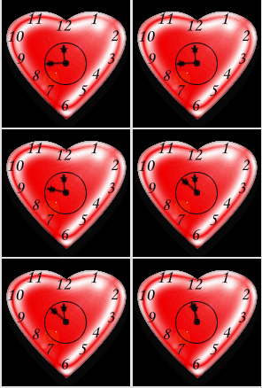
Cardiologist : I wonder , if you had arrived little late we wouldn’t be discussing this terrible conversation at all .Your myocardium  will also feel thrilled for getting a better mode of re-perfusion. You put me in an awkward situation by coming early !

Patient : But  . . . doctor every one  tells me ,  one should reach the hospital  after a heart attack  at the earliest  is it not ?   Please believe me doctor ,  I  made  extraordinary  efforts to  arrive early to your hospital , but you have put me on hold doctor !

Cardiologist :I agree  . . . but you won’t understand the modern jargons  in  interventional  cardiology ,  some times (Or is  it many times !) we will be doing the diagonally opposite to what  is   preached  in text books  .  Both intentional and unintentional delays are common  in the emergency cardiac  care . Do not bother about it   . . . science will take care of it !

Patient : You mean  . . . you are going to waste the golden hour in STEMI by simply arranging for cath lab staffs and machinery .

Cardiologist : You got the point right ! Just sit back enjoy the  flight !

Patient : Your wish is my wish doctor . Please handle with care doc !

* Please note this is an imaginary conversation ( Most often happens in silent mode !) in many of the cath labs which do not have 24 hour service. In a country  with  100 crore population like India ,  less than a dozen cathlabs  work round the clock . Guess  how often such a  situation would come in day to  day cardiology practice.
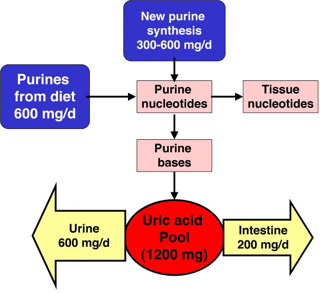


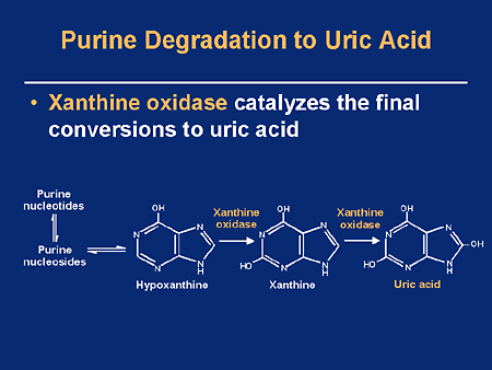
Uric acid is a  metabolite of purine metabolism.Purine is dynamically present in  every  active multiplying cell .Uric acid is formed when Xanthine oxidase  acts on   Xanthine and hypoxanthine  which are products of purine . The estimation of serum uric acid level can give us a rough estimate of cell metabolism and turn over. We need to  understand there is a dietary source for purines as well.

UA is mainly  excreted in urine . Normal levels of UA is   3-6mg in women and can be 1 mg higher  in men

Biological actions of uric acid

UA is a physiological molecule . It is more of an byproduct  and  devoid of any unique  action. Hence , most physicians still   believe it to be an unwanted  dangerous toxic molecule. What we fail to realise is ,  uric acid is a strong natural reducing agent .Hence it acts as an antioxidant .(Comparable to Ascorbic acid Vit C !)

Some believe excess uric acid is  a natural metabolic weapon against cellular degeneration . In fact , hypo- urecemia has a well known  association with multiple sclerosis and augmenting UA  is known to improve multiple sclerosis.

However, the problem with this  physiological molecule  is ,  we do not know yet,  when the  levels become pathological .We know uric acid in excess can lead to  urate stones  in kidney and Gout in joint . Does these crystals have any effect on coronary and cerebral circulation ?

Is uric acid a marker of inflammation and cell turn over ?

Yes it is.  What ESR  means to  inflammation , uric acid means for cell turnover . Since Inflammation induces white cell turnover  uric acid  level  becomes   a marker of inflammation as well .

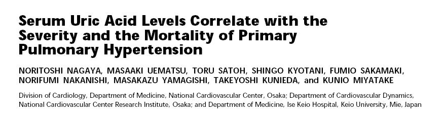
Uric acid in  excess  is a  marker of vascular  damage as  atherosclerosis  is an inflammatory process  , especially  with  pulmonary endothelial damage. So , in patients  with primary pulmonary  vascular diseases  like PPH , uric acid levels   may indicate  the progression or regression of PAH.

Some studies have correlated right atrial pressure with uric acid levels.

Uric acid and hypoxic states

Uric acid formation is more in hypoxic states as hypoxia depletes ATP and adenine metabolism is promoted and more inosine and  Xanthines  are produced . Uric acid can be a simple  marker  of increased oxidation stress of human biological system.

No surprise  to note   pulmonary hypertension  an important hypoxic state  increase uric acid levels .

Why uric acid is rarely considered as a useful diagnostic marker in cardiology  ?

  • The major  reason is it is an old molecule and has  lost its  flavor .
  • The name is not exotic (Like BNP, Di dimer etc)
  • Finally it is  a  cheap investigation and hence  lacks the required glamor.

Technical limitations

  • Uric acid levels are non specific (Like any other  modern  molecules  Tropinin , CRP etc! ) No one  would like  to compare uric acid vs hs CRP one to one as a marker of inflammation in vascular  disease.
  • UA  levels depend on kidney function .
  • Dietary influence can be significant (Especially meat, Liver Beans, Cauliflower etc)

Knowing the  basal level of uric acid  in a given patient ,   help us  monitor the net cell turnover during medical   management of chronic illness.

UA’s Clinical utility in cardiology practice

Importance of UA in PAH   is well recognised now  . Most studies on PAH  use it as a marker  or even  to define a therapeutic endpoint  But , please remember  elevated uric acid is a  simple  index of elevated  cell turnover and oxidative stress and it mainly represent  the effect of  pathology rather than a pathology itself.

So , attempting to reduce uric acid levels   with drugs like  Allopurinol may not  improve the  vascular function as one would wish ! The only indication for  reducing uric acid level   is  when the levels   become  too much and it starts depositing   in body.

Final message

Uric acid is a useful bio marker for  vascular function. It can indicate  the  quantum of  inflammatory , metabolic  and cell turnover of any progressive vascular  disease. With serial measurements  it definitely helps us in monitoring   cardiovascular disease  especially pulmonary hypertension  as  lung tissue is major source of this molecule . Now , uric acid  is used  for prognosticating  cardiac failure also.

Reference

http://qjmed.oxfordjournals.org/content/93/11/707.full.pdf+html


http://ajrccm.atsjournals.org/cgi/reprint/160/2/487?ijkey=dc24281a22fcf54ed27ac4466393abd691047408

http://cel.webofknowledge.com

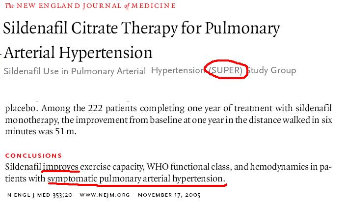
Sildenafil was first approved for pulmonary hypertension  in 2006  after a much hyped study called “SUPER” published in New  England journal of medicine.

This study population  included only  idiopathic pulmonary  hypertension and secondary  pulmonary hypertension due to pulmonary vascular disease like scleroderma  etc .The exclusion criteria were not clearly described in this paper ,  but it was clear PAH due to COPD was not included.

Please note , the title and conclusion are misleading .The real conclusion should be read as sildenefail may be useful some of the patients with in idiopathic PAH and secondary PAH sue to connective tissue disorders.

Further ,  the conclusion  of  SUPER study gave considerable  room for   misinterpretation (Intentional ? ) .The term PAH was used in too casual and generalised  manner. Many of the physicians  started  using sildenafil for every case of PAH including COPD.                 

COPD is a disorder of  airway , while sildenafil is a drug which acts on  the vascular  system (Vascular smooth muscle )  Apply your own logic  ,how  effective sildenafil  would be in  COPD patients .

As on today there is no good  evidence to suggest sildenafil will be useful in COPD. (Few studies  suggested it may be useful in fibrosed  lung)

Still , no body can prevent a physician  from testing this drug in otherwise refractory COPD as  we have a convenient   semantics called  ” off label  indication ”

So, the answer to the title  question still  eludes the majority    . . . even though many of us  know  the answer !

Ans : You   can use it  . . . but your patient   may not  really benefit ,  of course it satisfies  the physician and the drug company.

Reference

An editorial from Indian journal of chest diseases debates the issue

http://medind.nic.in/iae/t08/i4/iaet08i4p317.pdf

http://www.nejm.org/doi/pdf/10.1056/NEJMoa050010

http://www.nejm.org/doi/pdf/10.1056/NEJMc053442

The prosthetic cardiac valve which saved so many lives  in the later half of 20th century is no more. A valve which is not only  known for its excellent durability  but also  devoid  of sudden  mechanical  occlusion , unfairly  lost its place  by the rival  mechanical  bileaft valves. (Supposedly superior hemodynamics !)

Here is a nice video  from Indian surgeon  which  describes the hardware of  Starr Edwards valve .

Please read a related article from my site

Who killed Starr Edwards valve ?

Heart is a dynamic organ . It can alter  its force of contraction with every beat  according to the needs.Generally it responds to  length  of  previous  diastole.This is famously called frank starling law , ie the force of contraction is directly proportional to the end diastolic fiber length. So changing diastolic  duration as in atria fibrillation classically result in varying amplitude of LV contraction and pulse volume.

However , the commonest cause for  pulsus alternans  is  due to  severe left ventricular systolic dysfunction .There has  always been a suspicion about the existance of  beat to beat variation in  diastolic function as  well.  We have recently observed a  new* explanation for pulsus alternans .We know AV inflow is subjected to respiratory swings . Non  respiratory swings in mitral and tricuspid valves are rarely described. This pattern is now increasingly recognised.

These  non respiratory swings in the mitral inflow doppler pattern  is seen in  some of the  patients with hypertension and LVH.This  probably confirms the existence of  beat to beat variability of diastolic function . This phenomenon is relatively a new observation . Such pattern are common in patients who have had a recent hypertensive failure .


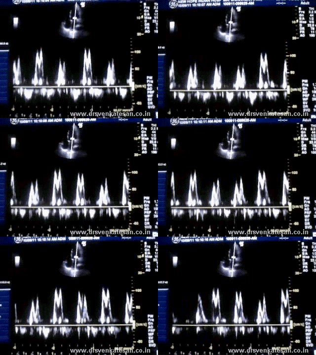
Here is a doppler of mitral inflow recorded from a patient with hypertension with LVH .

This is the doppler mitral inflow profile of a patient with Hypertension, LVH and class 2 dyspnea .Note the non respiratory swings in both "e" and "a" velocity

It is proposed  to  define  a new class of diastolic dysfunction that can be referred to as diastolic  mitral inflow  alternans .This phenomenon probably indicates a more severe grade of diastolic dysfunction.At the molecular level this is related to  undulating flux  in the calcium uptake from cytoplasm into SERCA .There is one more possible explanation for diastolic alternans  -Left atrial  dysfunction .

Occasionally one can visualise  a chaotic pattern of  diastolic filling waves  (e=a e>a a> e )  Such patterns are thought  to be markers of impending acute diastolic shutdown .

Further  analysis of  this  mitral doppler inflow pattern will be reported  later.

Reference

* Though we observed this for the first time , this is not a new phenomenon .There are few reports available in the literature.

http://www.sciencedirect.com/science/article/pii/S0735109785800358


http://www.sciencedirect.com/science/article/pii/S0894731706012818

Bicuspid aortic valve (BCAV) is  one of the common  congenital abnormality of heart . Incidence can be 1-2% of population . It can result in premature aortic degeneration with Aortic stenosis/Aortic regurgitation or both .

The normal development and arrangement of three cusps is altered ( rather interrupted )  during fetal life.

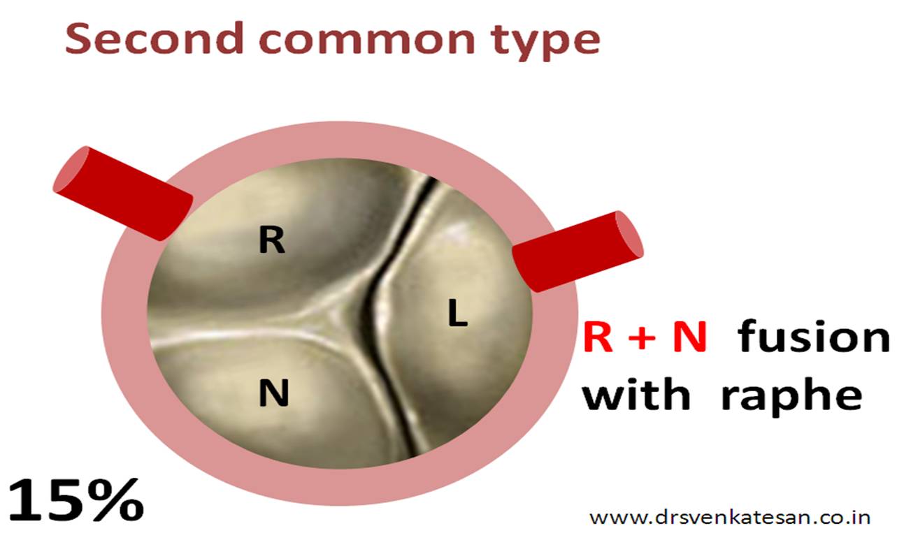
There can be two ways BCAV can occur. One is due to the   fusion* of two leaflets to covert a  tricuspid  valve into  bicuspid  ,  and the other is    two cusps develop de novo .The former has a raphe , while the later has no raphe.

The  fusion* occurs between  either

  • Right and left (R +L)
  • Right and  non coronary cusp  (R +N )
  • or Left and Non coronary cusp (L +N)

(* The fusion is embryological , not acquired )

Most often the  fusion is due to lack of division in the valve  analgen .Hence a raphe (A conjoint remnant) is noticed .

90% of BCAV has raphe  ,only 10 % lack raphe . Aortic root is also structurally abnormal in many .(Little clinical sequale though !)

Coronary artery origin anomalies  are more common with  BCAV. We also know co-arctation of aorta has a embryological link with BCAV.

The commonest type of BCAV is

The most hemodyanmic stressed BCAV is R +N type fortunately it is rare

R + N fusion is a high risk BCAV as degeneration occur fast

The least common type is

The coronary artery origin anomalies are common

BCAV in the absence of raphe is classified separately (This constitutes 10 % of all BCAV)

The nomenclature  is

  • Antero posterior (Common type ) AP
  • Lateral (L )

What is  the pathological significance of raphe ?

Many  believe presence of raphe accelerates degeneration as leaflets have  rough surfaces . Still , BCAV with raphe has less coronary anomalies and aortic root pathology .

Presence of raphe  indicate relatively  a minor embryological defect  , as the fault is in the failure to divide after the formation of analgen , while BCAV without raphe  imply  lack of development of analgen itself . This is expressed in the coronary sinus anatomy and aortic root dimension and orientation .

So currently it is  welcome  to spot  a raphe in the patient point of view  .Echo cardiogram is notoriously  unreliable to diagnose raphe. Once degeneration process sets  in ,  it is almost impossible to recognize  the  presence or absence of raphe .

* Please note ,tricsupid aortic valve with eccentric leaflet closure  shares  a close pathological relationship with BCAV. Premature degeneration ,  (AR more common than AS here ) .This entity will be discussed separately later.


Image courtesey

Part of the Image (The valve) is adopted from Yale university Image Bank .

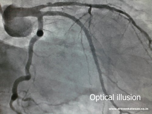
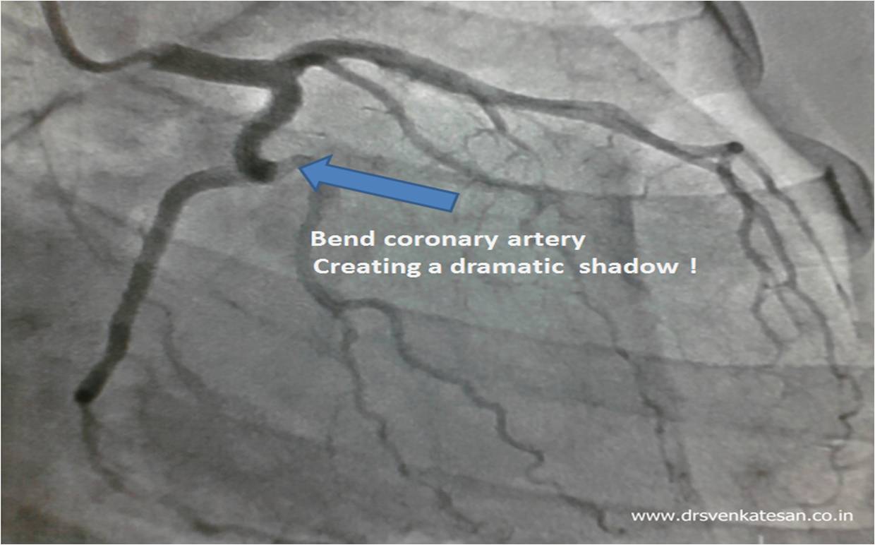
Coronary angiogram is  a videographic snap shot of  moving targets. Coronary arteries are dynamic  , tortuous  vessels of varying dimensions .Normal vessels  sometimes appear as an  illusion of lesions and tight lesions may appear innocuous at times. So the  rule is never rely on a single view before reporting.

This is an  angiogram of a patient , which  one of my fellows referred to as  “a black pearl  inside the LCX !

The same patient,s angiogram showing  origin of OM1

Final message

Beware of radiological artifacts in various angiograms. It can lead to erroneous interpretation and interventions !

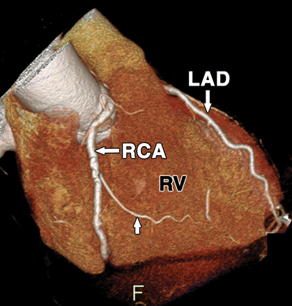
It is a well known anatomical fact  the grooves in the heart

  • Right and left  Atrio ventricular grooves.
  • Anterior  and posterior  inter-ventricular   grooves.

sincerely carry the  main stem of right and left coronary artery . It can be compared to a train running   with dedication   on its track .

Image source : AJR June 2007 vol. 188 no. 6 1665-1674

But God has not  enforced  strict rules   especially in human biology . The coronary arteries in few individuals   wander away  from the grooves.

This is very common  for Left circumflex  , followed by  RCA.  It is  relatively  for LAD to jump out of  its groove.(Except in rare Dual LAD system)

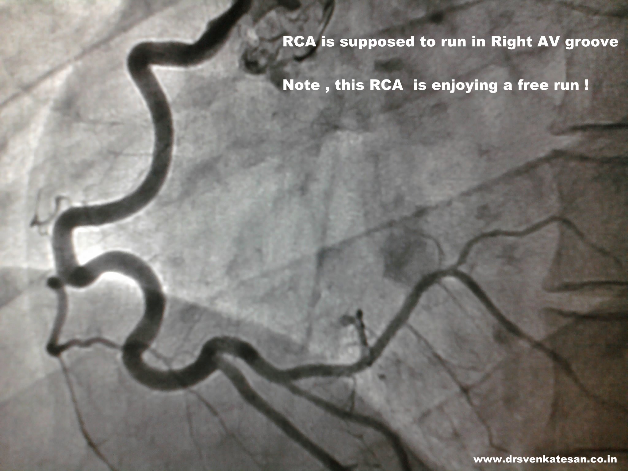
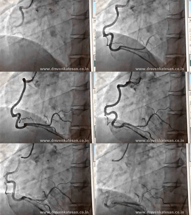
Here is a right coronary artery which enjoys its journey outside the right AV groove for   a good distance , only  rejoin the  groove at the crux.

Importance of  “Non- Grooval” coronary artery

It is true large diagonals and OMs ,ramus do   run without any  special tracks  .  But ,  it becomes ab  entirely different issue  when  the main stem of RCA,LCX or LAD itself   derail from its groove. There is no  fixing agents or vascular sheaths  that keep the coronary arteries within the groove. Surgeons  tell us circumflex is  often absent in its groove.

Note the stress and strain on the mid RCA : What will happen if that segemnt requires a stent ?

Hemodynamic implication of such free flowing non grooval coronary arteries is not been studied much .It is also observed  these   coronary  arteries   show  a  tortuous  course.This is  important   for  the  interventionist  as stenting  these segments is fraught with excess mobility and  tortuosity induced crimping .

Final message

The prevalence  of such drifting coronary arteries from its  groove  can be much more prevalent than we would  believe . The anatomical and physiological , surgical  issues need to be explored.

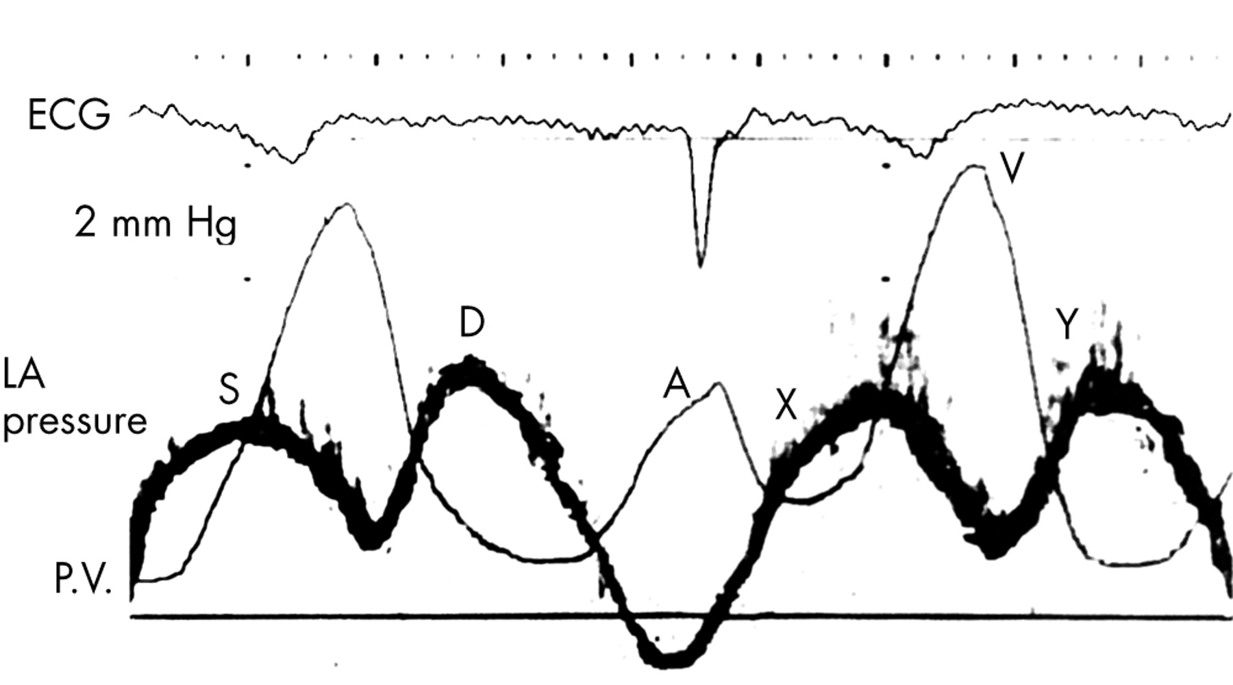
We know the atrial pressure  wave forms vary between right and left  atrium .In the right atrium “a” waves are  prominent and taller than “v” waves, while the reverse is true in left atrium .

Typical filling pattern of Right side chambers .Note The tall A waves . Source : http://www.ncbi.nlm.nih.gov/books/NBK2213/

Note the left atrial a waves are diminutive and v waves are tall .The dark black  wave is pulmonary venous waves. Source :http://heart.bmj.com/content/89/2/231.full

The  reasons for  tall  left atrial v waves are

  1. V waves are passive atrial  filling waves and  are timed  during ventricular systole .Left atrium is relatively  thick *,stiff , less compliant chamber .( Compliance : Rate of raise of pressure per unit change in volume .)
  2. Apart from relative thinness,* right atrial volume is more , hence  it can  accommodate more volume without raising its pressure .
  3. The left atrium is decompressed by  relatively stiff  pulmonary veins  with a mean pressure of 8 mmhg ,  can not adequately  dampen the   refluxing tides of  v waves , while the low pressure vena cava  of RA  dampen the right atrial v waves with ease  .
  4. Further ,the adjoining  systemic  left ventricle  ,  adds up to the stiffness of  left atrial   filling .

(*Thickness of RA -2mm,  LA -3mm )

Related article .

What is left atrial pressure volume Loop ?

http://www.wellsphere.com/heart-health-article/left-atrial-pressure-volume-loop/1208152

Ventricular tachycardia can be classified in a variety of ways. Monomorphic VT  and polymorphic VT  is one such classification based on VT morphology.Polymorphic VT  generally conveys a meaning of origin from multiple focus .But in reality  bulk of the polymorphic VT originate from a single focus .

How does a single focus have a multiple QRS  morphologies ?

This is possible because ,  even though VT arises from a single focus , the route it takes to exit  from the myocardium is different and hence they inscribe different QRS morphology for each  beat. It is also possible ,  as the conduction time varies with each  exit route  the VT  becomes  irregular. This  phenomenon   is called  polymorphic VT.

It is assumed the VT focus  is often located in the sub endocardium and breakthrough occur in the epicardial side as  we record  the activity  in the surface ECG. Electrophysiology of VT is not that simple ,  focus  of VT can be anywhere ,  the focus can be single or multiple and exit pathways  can also be multiple it and  it may even exit into endocardial cavity . Please note , even  a single  electrically abnormal cell  shall  act as a  focus . To confuse us further ,  some of  VTs  may not exit at all , extinguishes before reaching the surface.

It is  a  difficult  job to fix  a given  polymorphic VT  to arise   from a single focus or from multiple focus  .  Multifocal VT  can be  diagnosed  with  confidence only  after a through electro physiological  study.Clinically few clues are there. Electrolytic disorders and ischemia  are usually  multifocal,  while scars VT gives single focus .Other famous  example  of polymorphic VT are Torsedes de pointes .

Polymorphic VTs are  usually hemodynamically unstable but it is not a rule. Surprisingly ,some of the polymorphic  VTs are well tolerated . This is especially common in  multi focal polymorphic  VTs  which are hemodynamically and electrically  better off  . Ironically ,   presence of  multiple focus may be  a blessing  as they    compete with each other ,  in the process  pulling  down the other focus from triggering a VF . It  is possible  one focus acts like a natural anti tachycardia pacing for a  VT from another focus.

Ventricular fibrillation can be termed as an extreme form of irregular polymorphic VT as the wavefront breaks into innumerable fragments  each exiting the myocardium at will in a random fashion bringing ventricles to a standstill.

Final message

The term multifocal polymorphic  VT  is generally been abandoned at the bedside  as distinguishing  it from single focal  polymorphic VT  is a difficult task*.Still  the concept of  multifocal VT  is alive and  kicking in the EP labs ,  giving sleepless nights to  our Electro-cardiologists!

*Please note , multi focal VPDs can be recognised  with ease by different coupling intervals, but  it is difficult to identify during a  run of VT .