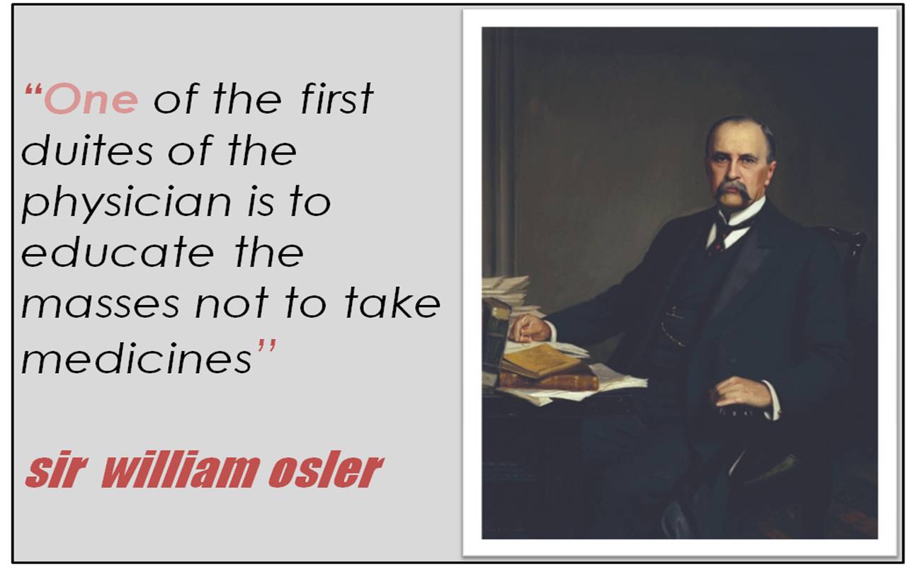
Posted in medical quotes | Tagged educate the masses not to take medicines, favorite medical quotes, sir william osler, william osler | Leave a Comment »
No one would have believed a century ago when electricity was first dosvovered for the mankind by Benjamin Franklin with glorious purpose , would now be used as a drug for treating life threatening heart ailments !
Yes , electricity is a drug by definition.
It is administered percutaneously by focusing a beam of current into the heart.
- There is a dose , shape , energy and direction for this drug.
- Paddle size determine the energy.
- Paddle location determine the direction of current vector.
- Dose is selected by the physician.
Mechanism of DC shock / Defibrillation *
When heart suddenly behaves abnormally and start generating its own electricity and sends it through abnormal channels other than its natural paths , it becomes a dangerous arrhythmia .This propagation of wave front can occur in multiple directions in a chaotic manner , resulting in VT/VF and imminent death.
Like an air to air missile ,this abnormal wave front can be tackled only by an another electrical wave front . Nothing else will work.
* The difference between DC shock and defibrillation is only technical. If one gives a synchronised shock ( with qrs complex ) it becomes DC shock .If not , it is defibrillation
The success of defibrillation depends on many factors .
The following are most important.
- The critical myocardial mass must be depolarized by the current delivered.Sufficient amount of sodium channels /less of calcium currents need be activated for this to happen .(JACC 2008)
- The direction and the angle of current entry with reference to advancing end of abnormal wave front. is also important .
- Distance between the paddles.(Antero posterior paddles more effective than Apex /Sternal pads )
- Energy level (seems to be less important ! )
Two shock forms are used
- Monophasic shocks
- Biphasic shocks
A biphasic DC shock has replaced the traditional mono phasic sine wave shocks in most machines.
What is the fundamental difference between the two ?
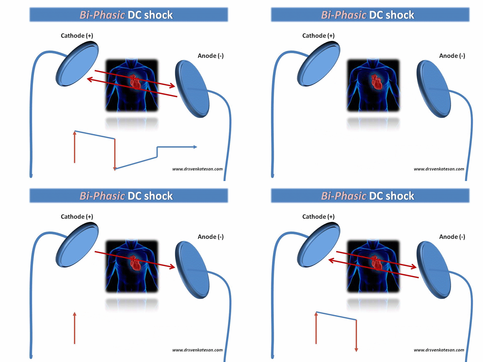
- In bi phasic shocks , the current traverses the myocardium twice .
- So, it has a second chance to interrupt the critical tachycardia circuit , if the first one fails. In other words, biphasic shocks are technically equivalent to “two sequential low energy shocks” delivered in opposite polarity . This change in direction happens in micro seconds .
- The shape of biphasic DC current wave form can be a truncated sine wave or square wave .The maximum energy of DC shock in biphasic mode is 200 joules (In Monophasic it is 360joules) . All AEDs, ICDs, now use bi phasic shocks to conserve energy .
Final message
A biphasic shock waveform has a proven advantage . It has greater efficacy ( because it traverses the heart twice ) , requires fewer shocks with low delivered energy and hence less myocardial and dermal injury.
References
Even though there is general acceptance of superiority of bi phasic shocks , it is still considered by some , that there is no great difference in the overall outcome .
http://circ.ahajournals.org/cgi/content/full/94/10/2507
Bi phasic shocks in atrial fibrillation
Posted in Cardiology - Electrophysiology -Pacemaker, cardiology -ECG, Uncategorized | Tagged biphasic shock, current as a drug, dc shock, defibrillation, electricity as a drug, electricity is a drug, energy levels in dc shock, monophasic vs biphasic dc shock, what is biphasic defibrillation, what is biphasic shock | 1 Comment »
It is often said medical professionals lack engineering sense and vice versa. The filed of bio medical engineering is is not a new one . It is there for over 50 years .The gap is narrowing very fast.
We are in the era of developing hybrid imaging , where a PET and CT come together. Raman spectroscopy is sending live images of tissue histology from the coronary arteries .
A journal exclusively catering to the cardiovascular enginnering is new development . Let us thank to the unique initiative from Purdue university .
Posted in cardiology journal club, cardiology journals | Tagged cardio vascular engineering, journal of cardiovascualr enginnering, loose cuff hypertension | Leave a Comment »
Blood pressure measurement , probably is the commonest investigation done in our patients in the entire field of medicine . It is such a common thing , both physicians and patients fail to perceive it as an investigation . (It indeed is !)
Even though BP is considered as a clinical sign , measuring it requires a device called sphygmomanometer . The BP apparatus has to be properly calibrated with the mercury , the tubing, the bladder , inflation balloon etc need to be perfect.
The following fallacies are noted in the measurement of blood pressure . Some of them are rampant* !
Patient
- Posture of recording
- Anxiety -White coat /Gender
Device
- Cuff width/Length
- Arm circumference
Ocular errors
It is surprising , such an important tool has a scale of 2mm markings which is prone for parallax errors of light with mercury column undulating .
Physician factors*
- Hasty cuff syndrome , Rapid deflation .
- Absent minded recording – Failure to note phase 4 to phase 5 due to inattention
- Failure to hear phase 4 muffling (Aging medico -Auditory insufficiency !)
It is not at all surprising to note, two BP readings rarely match , even if it is recorded by the same person with same machine at the same time !
There are many articles that describe in detail , how to record blood pressure properly. But this article from a relatively unknown journal from Purdue university , tells us most scientifically , what has been taken for granted by the medical community for so long .
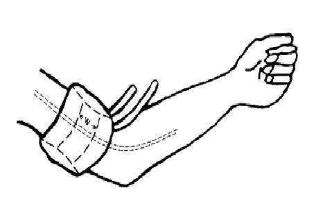
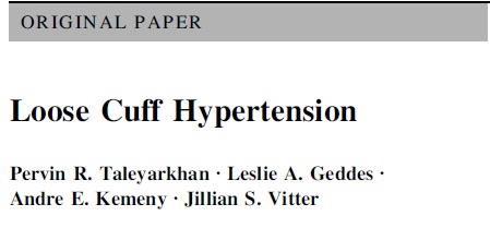
Loose cuff hypertension (Link to the journal of Cardiovascular engineering )
How much stiffness is to be applied in the arm for optimal pressure recording ?
What is the incidence of hypertension due to loose cuff ?
Final message
The BP apparatus , though appears as an innocuous machine , the readings that emerge from it determines , how millions of our fellow human beings are going to be labeled ! ( High pressured humans , slaves to anti hypertensive drug marketeers for rest of their life ) .
So , realise how important it is , to measure the blood pressure properly ! Never be casual . . . with this machine .
Experience has taught us , while it is very easy to name an individual wrongly as hypertensive , it often needs Herculean efforts to remove this medical tag from their neck . The reasonings are many .( Academic , non academic and patient factors included )
Finally , in this funny planet it is a personal observation ( Or is it an imagination ?) some men and women tend to enjoy , being referred to as high pressured ! Loose cuff or tight cuff , it simply do not bother them !
Posted in Cardiology - Clinical, Cardiology hypertension, Uncategorized | Tagged cuff length, cuff width, fallacies of blood pressure measurement, how to measure blood pressure, korotkoff sounds, loose cuff hypertension, phase 4 vs 5, riva rocci, sphygomanometer, systemic hypertension | Leave a Comment »
Cardiology as a specialty has grown faster than any other field in medicine. From the days of Da -vinci and William Harvey , the urge to understand the intricacies of anatomy and physiology of this organ that sustain the rhythm of life , life was tremendous .
Heart was a gift of life by God to the man kind
- Few men dreamed about it.
- Few were simply fascinated by it .
- Some exploited it in the name of science .
- Only few spent the entire life for it , explored it passionately . . . truly and genuinely .
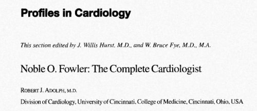
One such person , we should all celebrate is Noble O Fowler From Cincinnati USA. This unassuming ,(In contrast to some of the current hyped up achievers !) has kindled thirst in the subject to many youngsters .
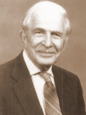
His remarkable achievement included
- The pioneering thoughts about pre-infarction angina (Now labeled as unstable angina)
- Pericardial physiology and pathology
- A overall approach to cardiac patient with shrewed physiological and pathological sense.
His book cardiac diagnosis was a exclusively authored by him is still considered as unique as his life.
Some how this book never got published beyond the 1980 s.
I personally dedicate this little service to cardiology literature to the legacy of Noble O Fowler.
A tribute by his Collegue Robert J Adokph
Posted in Uncategorized | Tagged complete cardiologist, great cardiologists, noble o fowler | 1 Comment »
Cadiac resynchrnonisation (CRT) therapy , is the most famed as well as ridiculed treatment modality for refractory failure . It is facing a real tough time for survival now .(At least in class 4 CHF.)
Confident and authentic data are emerging now , that CRT should not be used in advanced heart failure .(This is in total contrast with the original concept , when CRT was introduced nearly a decade ago ! more of class 3 and 4 were enrolled ) . Bad outcomes are expected in advanced CHF. This is something similar to whipping the tired horse concept which found inotropes to increase the mortality in severe heart failure .
The article in the current issue of circulation shows no mercy to CRT in advanced CHF
http://circ.ahajournals.org/cgi/content/abstract/CIRCULATIONAHA.110.956011v1
So what is the answer to the ailing CRT industry ?
Go and catch class 1 and class 2 CHF population* .You will get plenty , of course it got ratified by MADIT -CRT trial .
* It is attractively called prevention of cardiac failure
Posted in Cardiology-Coronary artery disese | Tagged cardiac resynchronisation therapy, care chf, circulation crt, class 4 chf and crt, companion, CRT, crt d, crt in calss 4 nyha, knock out punch for crt, madit crt, miracle | Leave a Comment »
For todays cardiologists traveling in time machine may be made as an essential assignment . It seems there is more to learn from history than the core medical science.
How the knowledge evolved ? . How our ancestors toiled ? Where are the good old medical inspirations ?
How can we enjoy the fruits of success without knowing the tree of it’s origin ?
What you are going to leave for the future man kind ?
Learn how a dream heart team led by Christian Barnard created history in the year 1967 in a remote southern hemisphere town ( To be precise Cape town ) , South africa .Click the link or over the image .Courtesey of Life magazine
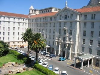
Groote schuur hospital where Christian Barnard made history
Thanks to the Life magazine for providing these stunning pictures to the present generation
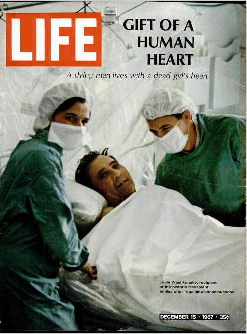
 Christian Barnard and team after the historic surgery .Groote schuur hospital .Cape town
Let us salute the men of past .We shall take an oath to strive harder and harder and the least , trivial achievements are not glorified . .
Posted in history of cardiology | Tagged cape town, christian barnard, heart transplantation | 2 Comments »
A female child aged 14 was referred for progressive breathlessness and abdominal distension

Abnormally dilated right atrium with significant pericardial effusion .www.drsvenkatesan.com
Can you guess the diagnosis ?

Apart form RA ,RV dilatation , the RV apex is seen filled with coarse treabeculations.This is believed to be a type of non compaction http://www.drsvenkatesan.com
Still difficult to conclude ? Look at the following Image.
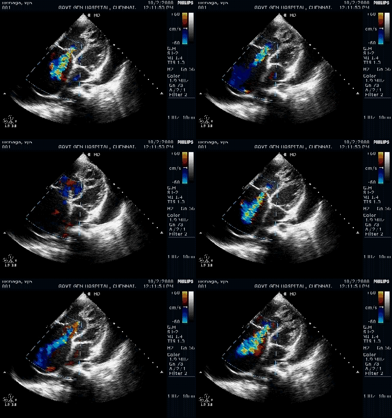
Tricuspid regurgitation is significant . http://www.drsvenkatesan.com
If you have thought . . .
- ASD with TR
- Severe PAH/COPD
- RV cardiomyopathy
All are acceptable differential diagnosis
But the real diagnosis is none of the above .
Need more time . . . the following Doppler tracing will settle the issue !

Doppler velocity in RVOT at 88mmhg. http://www.drsvenkatean.com
The final diagnosis was . . .
- Severe valvular pulmonary stenosis
- Marked RV,RA dilatation
- Acquired non compaction of right ventricle
- TR -Moderate
- Pericardial effusion -Moderate
- This patient also had dilated IVC, Hepatic veins that lead to clinical ascites.
Here , RV functional assessment becomes vital , but it is difficult many times. A simple clue is , as the RV is able to generate 88mmhg pressure it implies , the contractility should be near normal .
RV EF %, RV Dp/Dt , Tricuspid annular motion by tissue Doppler are additional measures. Cine MRI can be a useful investigation prior to intervention.
Final message
- VPS is a common acyanotic disease. Most are benign and milder forms are the rule.
- Dysplastic valves preclude balloon valvotomy. (In late stages little difference between dysplastic / non dysplastic VPS is noted )
- Severe progressive VPS , like in this patient needs immediate balloon dilatation or surgery.
- Long term outcome is excellent except in advances cases where irreversible RV dysfunction sets in.
Posted in Echo library and gallery, echocardiography, Uncategorized | Tagged dr s venkatesan, drsvenkatesan, echo clippings, echo clips, echo for diagnosis, echo quiz, echo spot diagnosis, echocardiography videos, interesting echo images, non compacted rv, pulmonary balloon valvotomy, ra rv dilatation, rare echo cases, rv dp/dt, rv dysfunction, sever vps, stunning echocardiography, valvular pulmonary stenosis, venkatesan s | 3 Comments »
A young women with Rheumatic heart disease .
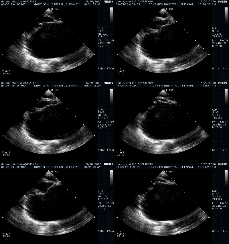
Giant left atrium in rheumatic MS . http://www.drsvenkatesan.com

Mitral regurgitation is significant .www.drsvenkatesan.com
When do you call a left atrium as giant ? When it is referred to as Aneurysmal dilatation ?
It is all semantics. Whenever LA becomes more than 6 cm , at least in two diameters many prefer to call it giant .
In India , 6 cm LA is such a common finding , we have kept a cut off at an arbitrary 9 cm .
What factors determine a LA to dilate like a balloon ?
The exact mechanism is not known.It could be the intrinsic weakness of LA wall , as very few with RHD develop this. Many LAs resists dilatation even in the midst of extreme LA pressure. But , it is a well-known fact , mitral regurgitation provokes greater LA dilatation than MS alone .This implies volumetrics play a major role than pressure dynamics in determining LA size. Acuteness of hemodynamic insult is inversely proportional to LA size.
By the way, what is the purpose of recognizing the LA as Aneurysmal ?
- In plain X -ray chest , LA may form the right heart border over shooting the RA.
- When LA becomes huge , there is a chance for mechanical complications like dysphagia, phrenic nerve , bronchial compression etc .
- Giant LA invariably increases the chance of LA clot.
Electro-physiological Issues
- Atrial fibrillation , a usual accompaniment of giant LA , is often refractory . There is no purpose to convert to sinus rhythm . In fact , one should not attempt this. There was a time when surgical incisions ,corridors , mazes were quiet popular.Now it is believed all these are adding further injury to the ill-fated LA .Electro-physiologists should be restrained . Pulmonary vein ablation should never be attempted in such cases as the focus of AF is elsewhere .
Implication in cath lab
During PTMC LA size can be an issue as the plane of IAS is distorted and make things difficult for septal puncture . Further the balloon , guidewire may often slip back into RA .
Implication for the surgeon.
For the surgeon the implication could be more. As a cardiologist I can’t comment about that .One thing we have observed is when LA becomes huge , the size of mitral annulus is too fictitious and funnily enough we have recorded up to 6 cm of mitral annulus . No valve is available for this size . We learnt from the surgeons , large LA rarely pose a problem as they suture the much smaller valve in a larger annulus .(Which makes the task that easier )
Does the LA size regress after surgery ?
In many it does regress , in as many it doesn’t. We have seen giant LAs continuing to trouble the patient even after a successful mitral valve replacement.
Posted in Echo library and gallery, echocardiography | Tagged giant left atrium, left atrial aneurysm, mitral regurgitation, rheumatic heart disease | 1 Comment »
A classical echo of a common AV canal .

Common AV canal . http://www.drsvenkatesan.com
Note the free floating common AV leaflet
An ASD
A VSD
With all chambers interconnected it is a free for all circulation . The blood seems to get mixed at the level of common AV orifice . Even though one expects severe cyanosis in common AV canal , the intensity of which is primarily determined

Common AV canal with free for all shunting . http://www.drsvenkatesan.com
the net blood flow to lungs which is dictated by the pulmonary vascular resistance or the RVOT obstruction. This patient had no RVOT obstruction but had severe pulmonary arterial hypertension. In spite of raised PVR , some amount of volume over load of lungs occur.
How to assess the operability ?
Posted in cardiology -congenital heart disease, Uncategorized | Tagged common av canal, endocardial cushion defect, inlet vsd, ostium primum asd, ostium primum vsd, single av valve | Leave a Comment »
Categories
-

-
The contents of the this blog is being published as Kindle E book , as per the request of many of the readers. Every article will continue to be open source in this site. Again I shall reiterate the book format is not aimed at any commercial intent. It is only to facilitate learning in a single book format Here is the link to book
https://amzn.in/d/euhL5vu Archives
- November 2026 (2)
- October 2026 (6)
- September 2026 (9)
- August 2026 (8)
- July 2026 (6)
- June 2026 (9)
- May 2026 (6)
- April 2026 (11)
- March 2026 (10)
- February 2026 (8)
- January 2026 (8)
- December 2025 (11)
- November 2025 (7)
- October 2025 (8)
- September 2025 (7)
- August 2025 (9)
- July 2025 (10)
- June 2025 (8)
- May 2025 (9)
- April 2025 (7)
- March 2025 (10)
- February 2025 (4)
- January 2025 (9)
- December 2024 (11)
- November 2024 (8)
- October 2024 (10)
- September 2024 (5)
- August 2024 (5)
- July 2024 (6)
- June 2024 (5)
- May 2024 (4)
- April 2024 (7)
- March 2024 (4)
- February 2024 (8)
- January 2024 (6)
- December 2023 (8)
- November 2023 (13)
- October 2023 (14)
- September 2023 (5)
- August 2023 (6)
- July 2023 (10)
- June 2023 (5)
- May 2023 (5)
- April 2023 (4)
- March 2023 (5)
- February 2023 (2)
- January 2023 (7)
- December 2022 (3)
- November 2022 (5)
- October 2022 (5)
- September 2022 (4)
- August 2022 (3)
- July 2022 (9)
- June 2022 (2)
- May 2022 (1)
- April 2022 (2)
- March 2022 (1)
- February 2022 (3)
- January 2022 (7)
- December 2021 (3)
- November 2021 (5)
- October 2021 (8)
- September 2021 (4)
- August 2021 (6)
- July 2021 (6)
- June 2021 (7)
- May 2021 (5)
- April 2021 (4)
- March 2021 (3)
- February 2021 (6)
- January 2021 (8)
- December 2020 (4)
- November 2020 (5)
- October 2020 (7)
- September 2020 (7)
- August 2020 (10)
- July 2020 (6)
- June 2020 (9)
- May 2020 (9)
- April 2020 (5)
- March 2020 (7)
- February 2020 (3)
- January 2020 (4)
- December 2019 (4)
- November 2019 (6)
- October 2019 (3)
- September 2019 (6)
- August 2019 (3)
- July 2019 (1)
- June 2019 (3)
- May 2019 (2)
- April 2019 (2)
- March 2019 (2)
- February 2019 (4)
- January 2019 (2)
- December 2018 (2)
- November 2018 (2)
- October 2018 (2)
- September 2018 (1)
- August 2018 (2)
- July 2018 (3)
- June 2018 (1)
- May 2018 (3)
- April 2018 (1)
- March 2018 (3)
- February 2018 (3)
- January 2018 (1)
- December 2017 (3)
- November 2017 (3)
- October 2017 (3)
- September 2017 (2)
- August 2017 (2)
- July 2017 (2)
- June 2017 (2)
- May 2017 (4)
- April 2017 (3)
- March 2017 (3)
- February 2017 (5)
- January 2017 (3)
- December 2016 (2)
- November 2016 (5)
- October 2016 (4)
- September 2016 (3)
- August 2016 (5)
- July 2016 (3)
- June 2016 (4)
- May 2016 (3)
- April 2016 (6)
- March 2016 (4)
- February 2016 (3)
- January 2016 (5)
- December 2015 (6)
- November 2015 (5)
- October 2015 (8)
- September 2015 (2)
- August 2015 (5)
- July 2015 (7)
- June 2015 (4)
- May 2015 (6)
- April 2015 (5)
- March 2015 (7)
- February 2015 (15)
- January 2015 (8)
- December 2014 (5)
- November 2014 (9)
- October 2014 (7)
- September 2014 (9)
- August 2014 (5)
- July 2014 (11)
- June 2014 (5)
- May 2014 (4)
- April 2014 (5)
- March 2014 (8)
- February 2014 (8)
- January 2014 (5)
- December 2013 (7)
- November 2013 (7)
- October 2013 (14)
- September 2013 (12)
- August 2013 (15)
- July 2013 (15)
- June 2013 (15)
- May 2013 (15)
- April 2013 (15)
- March 2013 (15)
- February 2013 (15)
- January 2013 (15)
- December 2012 (15)
- November 2012 (15)
- October 2012 (15)
- September 2012 (15)
- August 2012 (15)
- July 2012 (15)
- June 2012 (15)
- May 2012 (15)
- April 2012 (15)
- March 2012 (15)
- February 2012 (15)
- January 2012 (15)
- December 2011 (15)
- November 2011 (17)
- October 2011 (17)
- September 2011 (17)
- August 2011 (21)
- July 2011 (20)
- June 2011 (17)
- May 2011 (15)
- April 2011 (17)
- March 2011 (25)
- February 2011 (20)
- January 2011 (20)
- December 2010 (18)
- November 2010 (21)
- October 2010 (21)
- September 2010 (25)
- August 2010 (20)
- July 2010 (10)
- June 2010 (11)
- May 2010 (19)
- April 2010 (16)
- March 2010 (14)
- February 2010 (22)
- January 2010 (18)
- December 2009 (20)
- November 2009 (20)
- October 2009 (3)
- September 2009 (21)
- August 2009 (19)
- July 2009 (12)
- June 2009 (12)
- May 2009 (11)
- April 2009 (15)
- March 2009 (21)
- February 2009 (4)
- January 2009 (12)
- December 2008 (13)
- November 2008 (9)
- October 2008 (22)
- September 2008 (20)
- August 2008 (16)
- July 2008 (14)
- June 2008 (7)
Blog Stats
- 6,769,294 hits
Please give your feed back .
Click below to see who is watching this website live !

- This site will never aim for profit. Still ,this donation link is added at the request of few visitors who wanted to contribute and of-course that will help make it sustainable .

Please Note